. 2013 Aug;173(2):195-206.
doi: 10.1111/cei.12111.

Mesenchymal stem cells (MSCs) from scleroderma patients (SSc) preserve their immunomodulatory properties although senescent and normally induce T regulatory cells (Tregs) with a functional phenotype: implications for cellular-based therapy

Affiliations

Mesenchymal stem cells (MSCs) from scleroderma patients (SSc) preserve their immunomodulatory properties although senescent and normally induce T regulatory cells (Tregs) with a functional phenotype: implications for cellular-based therapy

P Cipriani et al. Clin Exp Immunol. 2013 Aug.

Abstract

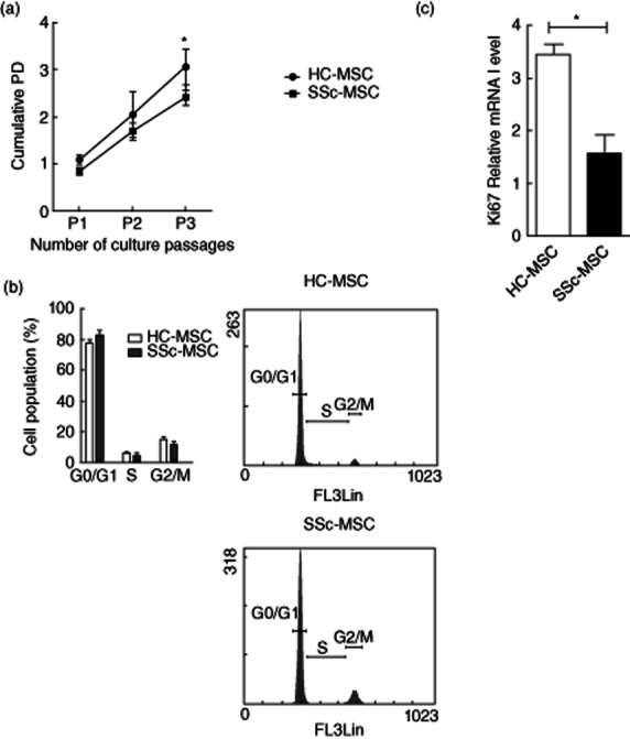
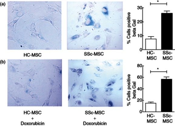
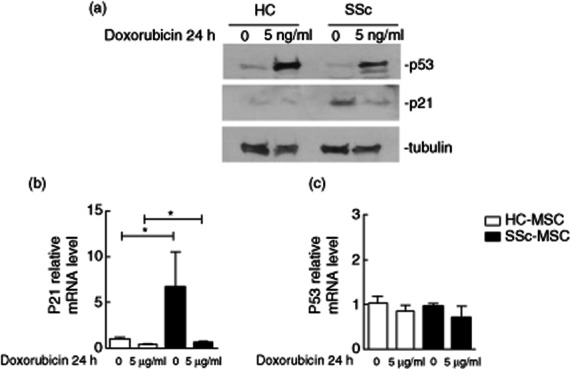
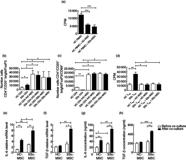
Systemic sclerosis (SSc) is a chronic disease, with early activation of the immune system. The aim of our work was to address how SSc-mesenchymal stem cells (MSCs), although senescent, might preserve specific immunomodulatory abilities during SSc. MSCs were obtained from 10 SSc patients and 10 healthy controls (HC). Senescence was evaluated by assessing cell cycle, β-galactosidase (β-Gal) activity, p21 and p53 expression; doxorubicin was used as acute senescence stimulus to evaluate their ability to react in stressed conditions. Immunomodulatory abilities were studied co-culturing MSCs with peripheral blood mononuclear cells (PBMCs) and CD4(+) cells, in order to establish both their ability to block proliferation in mixed lymphocyte reaction and in regulatory T cells (Tregs) induction. SSc-MSC showed an increase of senescence biomarkers. Eighty per cent of MSCs were in G0-G1 phase, without significant differences between SSc and HC. SSc-MSCs showed an increased positive β-Gal staining and higher p21 transcript level compared to HC cells. After doxorubicin, β-Gal staining increased significantly in SSc-MSCs. On the contrary, doxorubicin abolished p21 activation and elicited p53 induction both in SSc- and HC-MSCs. Interleukin (IL)-6 and transforming growth factor (TGF)-β-related transcripts and their protein levels were significantly higher in SSc-MSCs. The latter maintained their immunosuppressive effect on lymphocyte proliferation and induced a functionally regulatory phenotype on T cells, increasing surface expression of CD69 and restoring the regulatory function which is impaired in SSc. Increased activation of the IL-6 pathway observed in our cells might represent an adaptive mechanism to senescence, but preserving some specific cellular functions, including immunosuppression.

Keywords: immunomodulatory abilities; mesenchymal stem cells; senescence; systemic sclerosis.


Figures

Fig. 1
Fig. 1
Cell proliferation and cell cycle. (a) Growth kinetic curves of cultured healthy controls (HC)– and systemic sclerosis (SSc)–mesenchymal stem cells (MSCs) during in-vitro expansion from the first to the third passages. The growth rate was expressed as the mean value of cumulative population-doubling (PD) levels and calculated according the formula: log2 [(the number of collected cells)/(the number of seeded cells)]. HC–MSCs showed a significant higher PD than SSc–MSCs. (b) Flow cytometric assessment of the MSC cell-cycle, using propidium iodide (PI). No significant difference was observed in the percentage of cells in G0/G1, S and G2/M phases, between HC– and SSc–MSCs. Histograms are representative of cell cycle analysis of three different experiments. (c) mRNA level of Ki67 proliferation marker of HC– and SSc–MSCs. The values are expressed as mean ± standard deviation (s.d.) (*P < 0·05).
Fig. 2
Fig. 2
β-Galactosidase (β-Gal) activity. (a) Systemic sclerosis (SSc)–mesenchymal stem cells (MSCs) showed a significant increase in the percentage of β-Gal-positive cells in respect to healthy controls (HC) cells. β-Gal-stained cells showed flat and enlarged morphology. (b) Doxorubicin (5 μg/ml) treatment increased the percentage of β-Gal-positive cells in both HC and SSc cells. β-Gal-positive cells in SSc–MSCs were increased significantly when compared to HC–MSC. Percentage of β-Gal-positive cells were quantified and showed in histograms. The values are expressed as mean ± standard deviation (s.d.) (*P < 0·05).
Fig. 3
Fig. 3
p21 and p53 expression in mesenchymal stem cells (MSCs). (a) The expression levels of senescence-associated proteins were analysed by Western blotting. At basal state, no difference was observed in p53 expression between healthy controls (HC)– and systemic sclerosis (SSc)–MSCs; p21 showed increased expression in SSc–MSCs with respect to HC cells. After doxorubicin, p53 protein levels increased in both HC– and SSc–MSCs; HC–MSCs did not show an increase in p21 expression level; a slight increase of p21 protein level was found in SSc–MSCs. Anti-tubulin antibody was used as a control for equal loading of proteins. The pictures are representative of three independent experiments. (b) p21 and (c) p53 mRNA relative quantification before and after doxorubicin treatment. The values are expressed as mean ± standard deviation (s.d.) (*P < 0·05).
Fig. 4
Fig. 4
The immunomodulatory properties and cytokine profile of mesenchymal stem cells (MSCs). (a) Effect of MSCs on healthy controls (HC)–peripheral blood monuclear cell (PBMC) proliferation. Both HC– and systemic sclerosis (SSc)–MSCs suppressed significantly the phytohaemagglutinin (PHA)-induced proliferation of PBMCs without significant differences. (b) Number of CD4+CD25+brightforkhead box protein 3 (FoxP3) cells within CD4+ lymphocytes. The number of regulatory T cells (Tregs) was increased significantly in circulating SSc–CD4+ cells when compared to HC. After co-culture with MSCs the number of Tregs was increased significantly in both autologous and heterologous co-cultures. (c) Number of CD4+CD25+brightFoxP3CD69+ cells within CD4+ lymphocytes. The number of circulating CD69+ cells was significantly lower in SSc when compared to HC. After co-cultures with MSCs, CD69 surface expression was increased in both autologous and heterologous co-cultures. (d) Immunosuppressive function of Tregs. Circulating SSc–Treg inhibition of healthy CD4+ cell proliferation was impaired. After co-culture with HC– and SSc–MSCs, the induced SSc–Tregs inhibited CD4+ cell proliferation significantly. (e) Interleukin (IL)-6 and (f) transforming growth factor (TGF)-β mRNA expression. IL-6 gene expression was increased in SSc–MSCs before and after co-culture with PBMCs. Before co-culturing with PBMCs, HC– and SSc–MSCs did not show any difference in TGF-β expression. TGF-β expression increased in SSc–MSCs co-cultured with PBMCs when compared with HC cells. (g) IL-6 and (h) TGF-β enzyme-linked immunosorbent assay (ELISA). IL-6 production is increased in the supernatants of SSc–MSCs cultured alone and co-cultured with PBMCs when compared to HC–MSCs. At basal state HC– and SSc–MSCs showed no difference in TGF-β supernatant protein levels; after co-culture with PBMCs, SSc–MSCs displayed a significantly greater TFG-β protein level. The values are expressed as mean ± standard deviation (s.d.) (*P < 0·05; **P < 0·005; ***P < 0·0005).

References

    1. Caplan AI. Why are MSCs therapeutic? New data: new insight. J Pathol. 2009;217:318–324. - PMC - PubMed
    1. Pittenger MF, Mackay AM, Beck SC, et al. Multilineage potential of adult human mesenchymal stem cells. Science. 1999;284:143–147. - PubMed
    1. Le Blanc K, Frassoni F, Ball L, et al. Developmental Committee of the European Group for Blood and Marrow transplantation. Mesenchymal stem cells for treatment of steroid-resistant, severe, acute graft-versus-host disease: a Phase II study. Lancet. 2008;371:1579–1586. - PubMed
    1. Dominici M, Le Blanc K, Mueller I, et al. Minimal criteria for defining multipotent mesenchymal stromal cells. The International Society for Cellular Therapy position statement. Cytotherapy. 2006;8:315–317. - PubMed
    1. Oswald J, Boxberger S, Jørgensen B, et al. Mesenchymal stem cells can be differentiated into endothelial cells in vitro. Stem Cells. 2004;22:377–384. - PubMed

Publication types

MeSH terms