. 2013 Jul;27(7):2829-44.
doi: 10.1096/fj.13-227868. Epub 2013 Apr 22.

Differential type 1 interferon-regulated gene expression in the brain during AIDS: interactions with viral diversity and neurovirulence

Affiliations

Differential type 1 interferon-regulated gene expression in the brain during AIDS: interactions with viral diversity and neurovirulence

Maria J Polyak et al. FASEB J. 2013 Jul.

Abstract

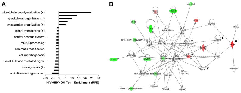
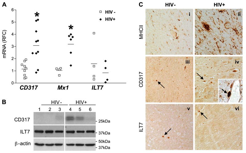
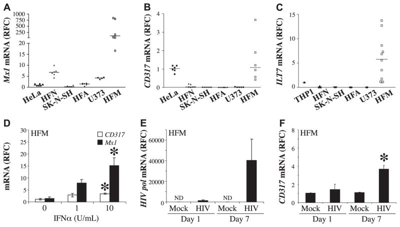
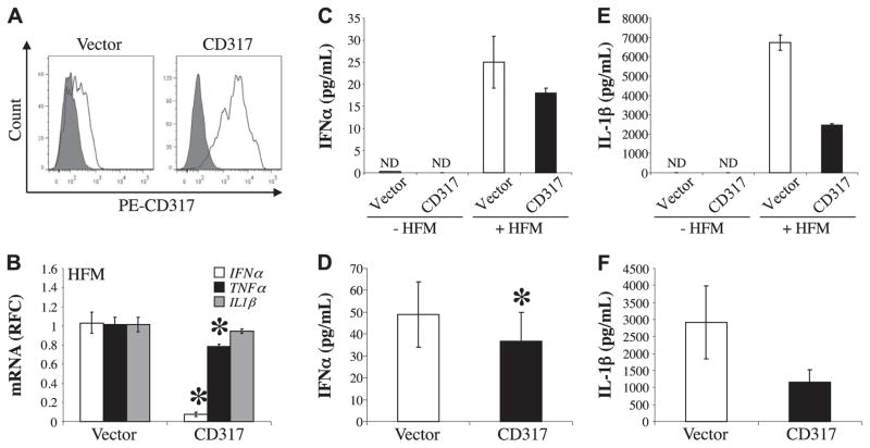
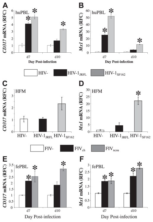
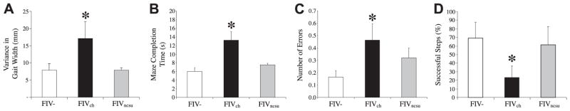
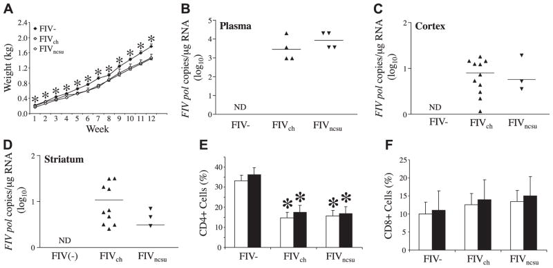
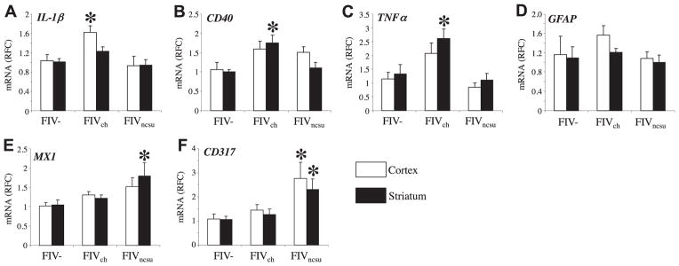
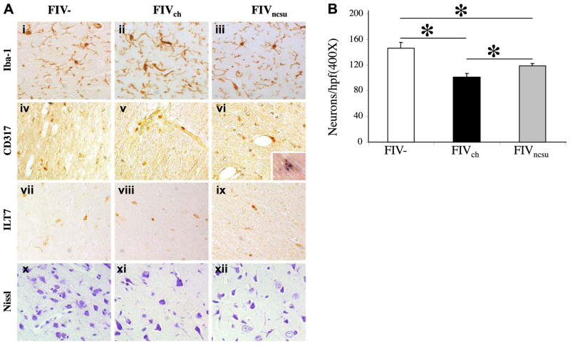
The lentiviruses, human and feline immunodeficiency viruses (HIV-1 and FIV, respectively), infect the brain and cause neurovirulence, evident as neuronal injury, inflammation, and neurobehavioral abnormalities with diminished survival. Herein, different lentivirus infections in conjunction with neural cell viability were investigated, concentrating on type 1 interferon-regulated pathways. Transcriptomic network analyses showed a preponderance of genes involved in type 1 interferon signaling, which was verified by increased expression of the type 1 interferon-associated genes, Mx1 and CD317, in brains from HIV-infected persons (P<0.05). Leukocytes infected with different strains of FIV or HIV-1 showed differential Mx1 and CD317 expression (P<0.05). In vivo studies of animals infected with the FIV strains, FIV(ch) or FIV(ncsu), revealed that FIV(ch)-infected animals displayed deficits in memory and motor speed compared with the FIV(ncsu)- and mock-infected groups (P<0.05). TNF-α, IL-1β, and CD40 expression was increased in the brains of FIV(ch)-infected animals; conversely, Mx1 and CD317 transcript levels were increased in the brains of FIV(ncsu)-infected animals, principally in microglia (P<0.05). Gliosis and neuronal loss were evident among FIV(ch)-infected animals compared with mock- and FIV(ncsu)-infected animals (P<0.05). Lentiviral infections induce type 1 interferon-regulated gene expression in microglia in a viral diversity-dependent manner, representing a mechanism by which immune responses might be exploited to limit neurovirulence.

Keywords: BST-2; CD317; FIV; HIV-1; microglia; tetherin; type 1 interferon.


Figures

Figure 1
Figure 1
Neuronal maintenance genes are suppressed with IFN signaling pathway induction in the brain during HIV/AIDS. A) GO analysis revealed significant overrepresentation (P<0.05; Benjamini’s test) of several functional categories of genes among the transcript tags increased or decreased by ≥4-fold in HIV+ compared with HIV cerebral white matter specimens. Data are represented as relative-fold enrichment (RFE). B) Network pathway analyses based on brain tag abundances displayed differential expression of specific genes in brains from HIV+ and HIV persons, disclosing either decreased (green) or increased (red) expression, or having established associations with the input reference dataset (white; IPA 31). Asterisks indicate genes associated with IFN signaling.
Figure 2
Figure 2
Type 1 IFN-related genes are increased in cerebral white matter myeloid cells in HIV/AIDS. A) Among HIV+ and HIV white matter specimens, CD317 and Mx1 transcript levels were increased in HIV+ brains, while ILT7 transcript abundance was similar across groups. Values are represented as average RFC compared with HIV. B) Cerebral white matter tissue samples were analyzed under reducing conditions by Western blot using mouse anti-CD317 and rabbit anti-LILRA4 (ILT7) antibodies followed by appropriate secondary antibodies, revealing that CD317 immunoreactivity was higher in the HIV-1+ brains, but ILT7 was similar in both groups. Membranes were stripped and reprobed for β-actin to confirm equal loading of samples. C) HIV and HIV-1+ paraffin-embedded white matter brain sections were immunolabeled with antibodies to MHC II (i, ii), CD317 (iii, iv), and ILT7 (v, vi). HIV-1+ brain sections displayed increased MHC II and CD317 immunoreactivity compared with HIV brain sections. Arrows indicate immunostained myeloid cells. Inset: HIV-1+ tissue sections disclosed colocalization of MHC II (DAB; brown) and CD317 (BCIP substrate; blue). Arrowhead indicates double-stained myeloid cell. All images captured at ×200 view. *P < 0.05; Kruskal-Wallis test.
Figure 3
Figure 3
Primary human microglial cells constitutively express Mx1, CD317, and ILT7; CD317 expression is induced by IFNα or HIV infection. A–C) Mx1 (A), CD317 (B), and ILT7 (C) transcript levels were measured by real-time RT-PCR from human cultured cells (Hela, THP1, HFN, SK-N-SH, HFA, U373, and HFM), revealing high constitutive expression of all genes in HFM cells. D) HFM cells incubated with IFNα at different concentrations showed induction of CD317 and Mx1 transcripts. E) HFM cells incubated with either medium (mock) or HIV-1SF162 showed that HIV-1 pol transcript was detected at d 1 and 7 postinfection. F) CD317 transcripts were detected at both d 1 and 7 in mock- and HIV-infected HFM cells, but there was a significant increase in HIV-infected cells at d 7. Values were derived from ≥3 biological replicates; and transcript values are represented as RFC. ND, not detected. *P < 0.05; Bonferroni’s test.
Figure 4
Figure 4
Expression of immune mediators by human microglia is modulated by exposure to CD317. A) BHK cells transiently expressing either pCDNA 3.0 (vector) or pCDNA 3.1 CD317 (CD317) were incubated with preimmune rabbit serum (gray) or with rabbit anti-CD317 serum (white) followed with PE-conjugated secondary antibody and then analyzed by flow cytometry. B) HFM cells (3×10⁵/sample) were incubated with BHK transiently expressing vector or CD317 for 48 h. Human IFN-α, TNF-α, and IL-1β transcript levels were measured by real-time RT-PCR. Values were normalized to human GAPDH to eliminate the contribution from the BHK and are represented as average RFC compared with HFM cells cocultured with BHK transfected with empty vector. C) Representative IFN-α protein levels from the supernatants of BHK cells transiently transfected with empty vector (vector) or pCDNA3.1 CD317 (CD317) either cultured alone (−HFM) or cocultured with HFM cells (+HFM) were measured by ELISA. D) Averaged IFN-α protein detected in the supernatants of HFM cells cocultured with BHK cells transfected with empty vector or construct containing CD317 from 5 independent experiments show significantly suppressed IFN-α levels. E) Representative IL-1β protein levels from BHK ± HFM cells transiently transfected with empty vector or pCDNA3.1 CD317, as determined by ELISA. F) Average IL-1β levels detected in supernatants from 4 independent experiments. ND, not detected. *P < 0.05; Student’s t test.
Figure 5
Figure 5
CD317 induction is dependent of lentivirus diversity. huPBLs were mock (HIV) or HIV infected with one of two strains of HIV-1 (HIV-1JRFL and HIV-1SF162) at matched input titers in triplicate. Cells were collected at d 7 and 10 postinfection and solubilized with TRIzol, and cDNA was prepared. A, B) CD317 (A) and Mx1 (B) transcripts were measured by real-time RT-PCR, showing that HIV-1JRFL consistently induced CD317 and Mx1. C, D) Likewise, mock- or HIV-infected HFM cells showed increased CD317 (C) and Mx1 (D) transcript levels in the cells infected with HIV-1JRFL. E, F) Similarly, fePBLs were mock infected, FIV, or infected with one of two strains of FIV (FIVch and FIVncsu) at matched input titers, revealing that CD317 (E) and Mx1 (F) transcripts were increased in the cells infected with FIVncsu. Values represent average relative RFC compared with HIV (AD) or FIV (E, F). *P < 0.05; Dunnett’s test.
Figure 6
Figure 6
FIVch-infected animals exhibit greater neurobehavioral deficits compared with FIVncsu- or mock-infected animals. Neurobehavioral and neurocognitive parameters of 12-wk-old FIV (n=7), FIVch (n=10), and FIVncsu (n=5) animals were evaluated. A) Gait function was determined by analyzing the width of inked footprints of cats walking across a suspended plank. Distance between the right and left paw placement was measured, and variance in width was calculated showing increased width in the FIVch-infected animals. B, C) Motor speed and memory ability were measured using a modified T-maze; time duration (B) as well as the number of errors (C) made during completion of the maze were recorded, revealing that the FIVch-infected animals were slower and made more mistakes. D) Object-memory test measured spatial object memory; animals were required to step over a 6-cm moveable barrier with their forelimbs to reach a food reward. Their ability to recall the height and position of the barrier was monitored using reflective dots placed on the outside of the cat’s hindlimbs. The number of successful attempts was recorded as a percentage, with the FIVch animals exhibiting worsened performance. *P < 0.05; Dunnett’s test.
Figure 7
Figure 7
In vivo viral load and CD4 T cell suppression are independent of FIV diversity. Animals were infected with either FIVch or FIVncsu 1 d postpartum. A) Weights were recorded on a weekly basis for 12 wk, revealing less weight gain among the FIV-infected animals. B–D) Viral load in plasma (B), cerebral cortex (C), and striatum (D) was measured by semiquantitative real-time RT-PCR using primers for FIV pol, disclosing similar viral copy numbers for each virus, although levels were higher in plasma than in brain. Virus quantity was determined on comparison with a FIV pol standard curve. E, F) At 8 and 12 wk, PBMCs were isolated from blood, and T-cell subpopulations were identified by flow cytometry. Percentages of CD4 (E) and CD8 (F) T cells of total PBMCs are indicated, showing similar levels of CD4 T-cell suppression in both FIV-infected groups. Open bars, 8 wk; solid bars, 12 wk. *P < 0.05; Dunnett’s test.
Figure 8
Figure 8
In vivo induction of immune genes is FIV diversity dependent. Host gene expression from 12-wk-old uninfected (mock, n=6) and FIV (FIVch, n=12; FIVncsu, n=7)-infected animals was determined. IL-1β (A), CD40 (B), TNF-α (C), GFAP (D), Mx1 (E), and CD317 (F) transcript levels were determined by real-time RT-PCR. FIVch-infected animals showed increased CD40 (A), TNF-α (B), and IL-1β (C) levels, while FIVncsu-infected animals displayed increased Mx1 and CD317 levels in cerebral cortex and/or striatum. Results are expressed as average RFC as compared with mock treatment. *P < 0.05; Dunnett’s test.
Figure 9
Figure 9
In vivo neuronal viability is FIV diversity dependent. A) FIV, FIVch-infected, and FIVncsu-infected paraffin-embedded tissue sections were immunostained with antibodies against Iba1 (myeloid cell activation marker; i–iii), CD317 (iv–vi), and ILT7 (vii–ix), as well as Nissl stained (x–xii). Inset: FIV+ tissue stained for MAC387 (DAB; brown) and CD317 (BCIP substrate; blue), ×600 view. All images except the inset were captured at ×200 view. B) Neuronal density was determined by counting Nissl-stained images of the cerebral cortical regions of FIV (n=4), FIVch-infected (n=5), and FIVncsu-infected (n=6) sections. Average number of neurons in multiple fields per animal in each group is shown. *P < 0.05; Dunnett’s test.

Similar articles

Cited by

References

    1. Clements JE, Zink MC. Molecular biology and pathogenesis of animal lentivirus infections. Clin Microbiol Rev. 1996;9:100–117. - PMC - PubMed
    1. Zink MC, Laast VA, Helke KL, Brice AK, Barber SA, Clements JE, Mankowski JL. From mice to macaques–animal models of HIV nervous system disease. Curr HIV Res. 2006;4:293–305. - PubMed
    1. Power C. Retroviral diseases of the nervous system: pathogenic host response or viral gene-mediated neurovirulence? Trends Neurosci. 2001;24:162–169. - PubMed
    1. Gonzalez-Scarano F, Martin-Garcia J. The neuropathogenesis of AIDS. Nat Rev Immunol. 2005;5:69–81. - PubMed
    1. Medzhitov R, Schneider DS, Soares MP. Disease tolerance as a defense strategy. Science. 2012;335:936–941. - PMC - PubMed

Publication types

MeSH terms

Grants and funding