. 2013 Jun 10;319(10):1398-408.
doi: 10.1016/j.yexcr.2013.04.010. Epub 2013 Apr 19.

IL-4 inhibition of IL-1 induced Matrix metalloproteinase-3 (MMP-3) expression in human fibroblasts involves decreased AP-1 activation via negative crosstalk involving of Jun N-terminal kinase (JNK)

Affiliations

IL-4 inhibition of IL-1 induced Matrix metalloproteinase-3 (MMP-3) expression in human fibroblasts involves decreased AP-1 activation via negative crosstalk involving of Jun N-terminal kinase (JNK)

Mariah Chambers et al. Exp Cell Res. .

Abstract

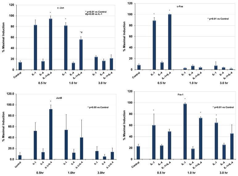
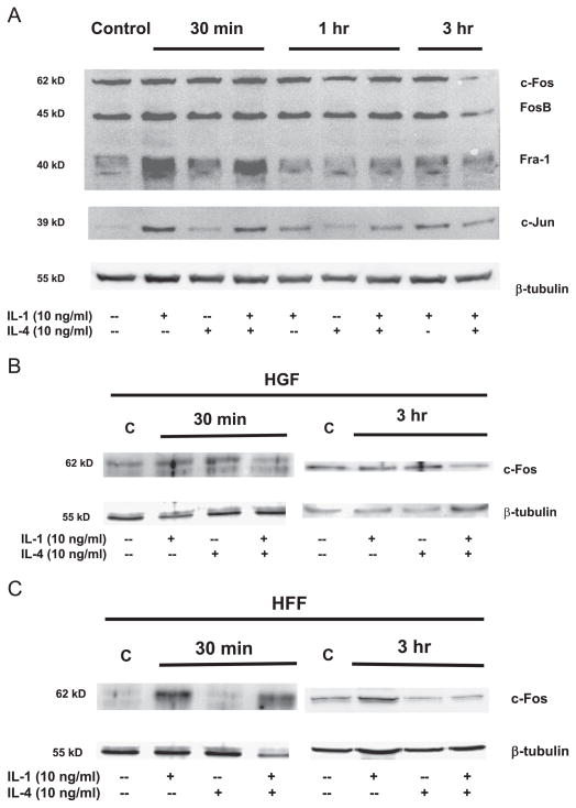
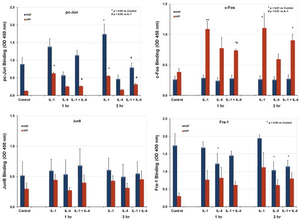
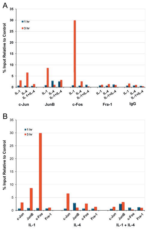
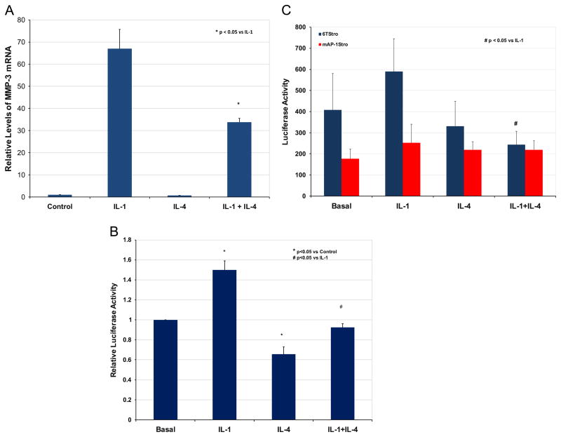
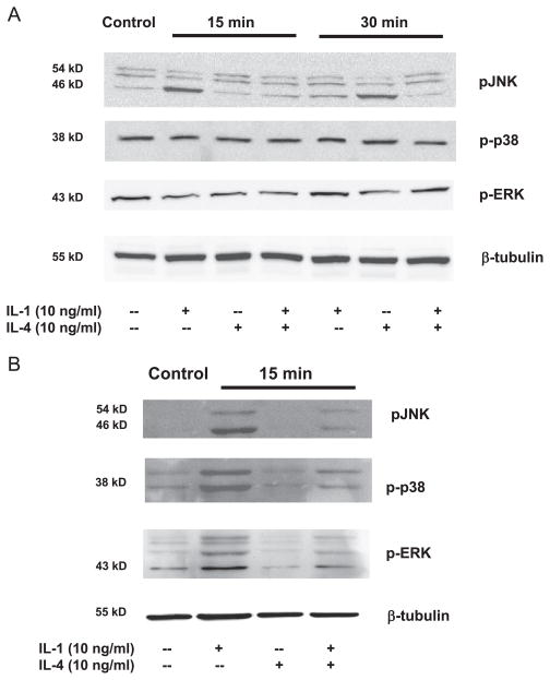
Matrix metalloproteinase-3 (MMP-3) over-expression is associated with tissue destruction in the context of chronic inflammation. Previous studies showed that IL-4 inhibits induction of MMP-3 by IL-1β, and suggested that AP-1 might be involved. Here we show that IL-1 induced binding of transcription factor AP-1 to the MMP-3 promoter consists primarily of c-Jun, JunB, and c-Fos and that binding of c-Jun and c-Fos is inhibited by the combination of cytokines while binding of Jun B is not. Mutation of the AP-1 site in the MMP-3 promoter decreased the ability of IL-4 to inhibit its transcription in transfected MG-63 cells. Western blotting showed that both cytokines activate Jun N-terminal kinase (JNK), but with somewhat different kinetics, and that activation of JNK by both cytokines individually is inhibited by the combination. These results indicate that IL-4 inhibition of MMP-3 expression is associated with reduction of IL-1 induced binding of active forms of the AP-1 dimer, while less active JunB-containing dimers remain, and suggest that these changes are associated with decreased activation of JNK.


Figures

Fig. 1
Fig. 1
Effects of IL-1 and IL-4 on Jun and Fos mRNA expression HGF cell cultures were serum-deprived for 16 h prior to the addition of 10 ng IL-1β/ml and/or 10 ng/ml IL-4. Total RNA was harvested from control (untreated) cells and from cells treated for 0.5, 1 or 3 h with cytokine(s). c-Jun, JunB, c-Fos, and Fra-1 mRNA levels were quantitated by real-time PCR and normalized to levels of GAPDH mRNA. The graph represents data from three independent experiments with three different HGF cultures derived from samples from three different individuals, average +/− SEM. Statistical significance was assessed by ANOVA followed by Tukey’s post-hoc test.
Fig. 2
Fig. 2
Effect of IL-1 and IL-4 on Jun and Fos protein expression (A) HGF cell cultures were serum-deprived for 16 h prior to the addition of 10 ng IL-1β/ml and/or 10 ng/ml IL-4. Whole cell protein lysates were obtained from control (untreated) cells and from cells treated for 30 min, 1 h or 3 h with cytokine(s). Twenty μg of each lysate was separated on 10% SDS-PAGE and transferred to a nylon membrane. Proteins were detected with anti-c-Fos antibody, which also recognizes Fra-1 and FosB, as well as anti-c-Jun and β-tubulin antibodies, followed by chemiluminescence. (B and C) HGF and HFF cell cultures were serum-deprived for 16 h prior to the addition of 10 ng IL-1β/ml and/or 10 ng/ml IL-4. Nuclear extracts were isolated from control (untreated) cells and from cells treated for 30 min or 3 h with cytokine(s). Twenty-five μg of each extract was separated on 10% SDS-PAGE and transferred to a nylon membrane. Proteins were detected with anti-c-Fos antibody specific for c-Fos, as well anti-β-tubulin antibody, followed by chemiluminescence.
Fig. 3
Fig. 3
Effect of IL-4 on IL-1 induced binding of AP-1 family proteins to a consensus AP-1 site HGF or HFF cultures were serum-deprived for 16 h and then incubated for 0.5, 1 or 3 h with IL-1 (10 ng/ml) and/or IL-4 (10 ng/ml). Control cells were incubated without cytokine addition. AP-1 DNA binding activity was determined using the TransAM AP-1 Family ELISA-based binding assay from Active Motif and 20 μg nuclear extract. For HGF, data shown are from three independent experiments with three different HGF cultures derived from samples from three different individuals, average +/− SEM. Statistical significance was assessed by ANOVA followed by Tukey’s post-hoc test.
Fig. 4
Fig. 4
Effect of IL-4 on IL-1 induced binding of AP-1 proteins to the endogenous MMP-3 promoter HFF cells were incubated in the presence and absence of IL-1 and/or IL-4 for 1 or 3 h. ChIP assays were performed using the SimpleChip Enzymatic Chromatin IP Kit from Cell Signaling and Technology according to the manufacturer’s suggested protocol. Antibodies to c-Jun, c-Fos, JunB, JunD, and Fra-1 and IgG were used for immunoprecipitation. Quantitiation of the immunoprecipitated DNA was performed by quantitative RT PCR using primers specific for the region of the human MMP-3 promoter containing the AP-1 site (-231 to -101). Results are expressed as percentage of input relative to the untreated control.
Fig. 5
Fig. 5
Effect of mutation of the AP-1 site on IL-4 inhibition of transcription from the MMP-3 promoter (A) Total RNA was harvested from control (untreated) MG-63 cells and from cells treated for 12 h with 10 ng IL-1β/ml and/or 10 ng/ml IL-4. MMP-3 mRNA levels were quantitated by real-time PCR and normalized to levels of GAPDH mRNA. The graph represents data from three independent experiments, average +/− SEM. (B) MG-63 cells were transfected with a luciferase reporter plasmid containing 2.3 kB of the wild-type human MMP-3 promoter (WT), or with a similar plasmid containing a mutation in the AP-1 site at −70 (mAP-1), or (C) an AP-1 reporter plasmid, along with SVβgal as a control for transfection efficiency. Cells were replated and allowed to rest for 24 h prior to addition of IL-1 (10 ng/ml) or IL-1+IL-4 (10 ng/ml). Cells were harvested after 12 h. Normalized luciferase numbers were expressed as % inhibition of the IL-1+IL-4 combination relative to IL-1 alone. Statistical significance was assessed using Student’s T-test.
Fig. 6
Fig. 6
Effect of IL-1 and IL-4 on levels of activated forms of MAPKs. (A) HGF or (B) HFF cell cultures were serum-deprived for 16 h prior to the addition of 10 ng IL-1β/ml and/or 10 ng/ml IL-4. Whole cell protein lysates were obtained from control (untreated) cells and from cells treated for indicated times with cytokine(s). Twenty μg of each lysate was separated on 10% SDS-PAGE, and transferred to nylon membrane. Proteins were detected with antibodies directed against the activated (phosphorylated) forms of JNK, ERK and p38 MAPK, as well as β-tubulin.

Similar articles

Cited by

References

    1. Honig J, Rordorf-Adam C, Siegmund C, Wiedemann W, Erard F, et al. Increased interleukin-1 beta (IL-1 beta) concentration in gingival tissue from periodontitis patients. J Periodontal Res. 1989;24:362–367. - PubMed
    1. Stashenko P, Fujiyoshi P, Obernesser MS, Prostak L, Haffajee AD, Socransky SS, et al. Levels of interleukin 1 beta in tissue from sites of active periodontal disease. J Clin Periodontol. 1991;18:548–554. - PubMed
    1. Cheung NT, Dawes PT, Poulton KV, Ollier WE, Taylor DJ, Mattey DL, et al. High serum levels of pro-matrix metalloproteinase-3 are associated with greater radiographic damage and the presence of the shared epitope in patients with rheumatoid arthritis. J Rheumatol. 2000;27:882–887. - PubMed
    1. van den Berg WB, Bresnihan B, et al. Pathogenesis of joint damage in rheumatoid arthritis: evidence of a dominant role for interleukin-I. Baillieres Best Pract Res Clin Rheumatol. 1999;13:577–597. - PubMed
    1. Shapira L, van Dyke TE, Hart TC, et al. A localized absence of interleukin-4 triggers periodontal disease activity: a novel hypothesis. Med Hypotheses. 1992;39:319–322. - PubMed

Publication types

MeSH terms