. 2013 Jun;21(6):1160-8.
doi: 10.1038/mt.2013.67. Epub 2013 Apr 23.

Mature T-cell lymphomagenesis induced by retroviral insertional activation of Janus kinase 1

Affiliations

Mature T-cell lymphomagenesis induced by retroviral insertional activation of Janus kinase 1

Tim Heinrich et al. Mol Ther. 2013 Jun.

Abstract

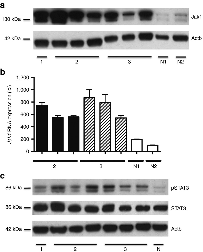
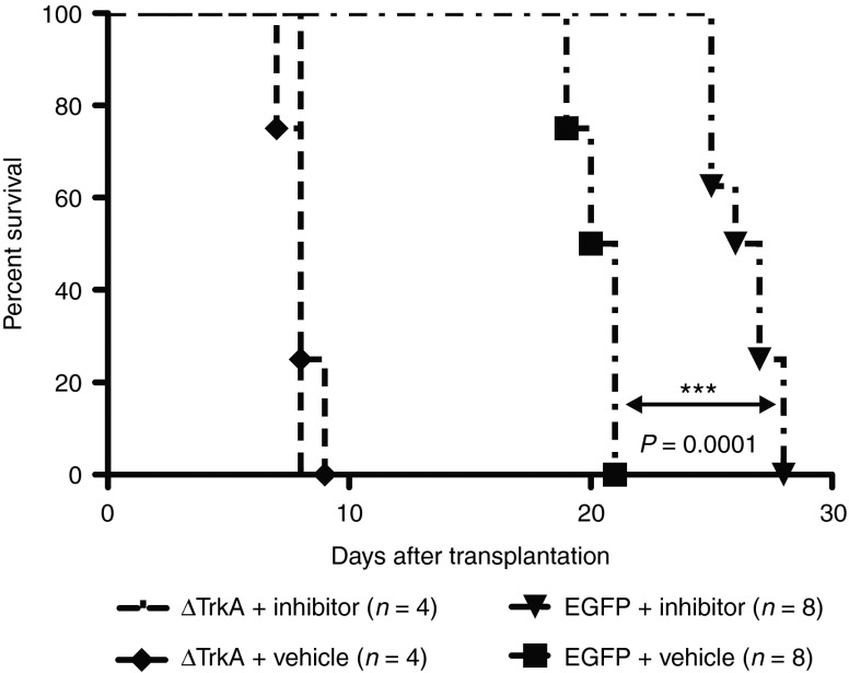
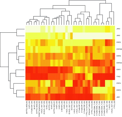
Retroviral vectors (RVs) are powerful tools in clinical gene therapy. However, stable genomic integration of RVs can be oncogenic, as reported in several animal models and in clinical trials. Previously, we observed that T-cell receptor (TCR) polyclonal mature T cells are resistant to transformation after gammaretroviral transfer of (proto-)oncogenes, whereas TCR-oligoclonal T cells were transformable in the same setting. Here, we describe the induction of a mature T-cell lymphoma (MTCL) in TCR-oligoclonal OT-I transgenic T cells, transduced with an enhanced green fluorescent protein (EGFP)-encoding gammaretroviral vector. The tumor cells were of a mature T-cell phenotype and serially transplantable. Integration site analysis revealed a proviral hit in Janus kinase 1 (Jak1), which resulted in Jak1 overexpression and Jak/STAT-pathway activation, particularly via signal transducer and activator of transcription 3 (STAT3). Specific inhibition of Jak1 markedly delayed tumor growth. A systematic meta-analysis of available gene expression data on human mature T-cell lymphomas/leukemias confirmed the relevance of Jak/STAT overexpression in sporadic human T-cell tumorigenesis. To our knowledge, this is the first study to describe RV-associated insertional mutagenesis in mature T cells.


Figures

Figure 1
Figure 1
T-cell lymphomagenesis induced by transduction of an EGFP-encoding gammaretroviral control vector. (a) The gammaretroviral vector used in this study comprised long terminal repeats (LTRs) from myeloproliferative sarcoma virus (MPSV) and a leader region from the murine embryonic stem cell virus (MESV leader 71) including a splice donor and a splice acceptor site. A posttranscriptional regulatory element of the Woodchuck hepatitis virus (WPRE) was located 3′ of the transgene enhanced green fluorescent protein (EGFP). (b) T lymphocytes from OT-I donor mice were isolated and transduced with gammaretroviral particles encoding for EGFP and transplanted into RAG1−/−–recipient mice. One year after transplantation, splenocytes and lymphocytes were reisolated and serially transplanted into secondary RAG1−/−–recipient mice. Four months after serial transplantation, mature T-cell lymphoma emerged. (c) Representative histological sections (HE stained) from lymph node, spleen, kidney, liver, and lung of a diseased animal (magnification 200× scale bar = 100 µm). (d) Cell surface characterization of control vector–induced malignancy. After single cell suspension of tumor samples, cells from massively enlarged spleens were stained for the α chain of the OT-I TCR (Vα2), CD3, CD8, and ICOS (inducible co-stimulator). Staining for CD4 was negative. Representative blots (gated on EGFP-positive cells) from the primary tumor are depicted. Cell surface marking was comparable in all secondary and tertiary recipients. Staining of lymph node samples showed the same results.
Figure 2
Figure 2
Serial transplantation of primary tumor cells reveals outgrowth of a T-cell clone with a retroviral insertion site in Jak1. (a) Tumor cells from the primary mature T-cell tumor (1st generation) were serially transplantable into secondary (2nd generation) and tertiary (3rd generation) recipients and established histological and immunophenotypical identical malignancies. 5 × 106 tumor cells from spleen and lymph node were injected intravenously into secondary and tertiary RAG1−/− recipients. (b) Retroviral integration site (RIS) analysis revealed eight unique proviral insertions in the primary tumor, whereof one RIS was detectable in all animals of the three generations. The RIS was amplified via integration site-specific PCR and verified by sequencing. Primary, secondary, and tertiary tumor samples are depicted by 1, 2, and 3, respectively. Negative control (N), untransduced OT-I cells. M, base pair marker. (c) Using ligation-mediated PCR (LM-PCR) and integration site-specific PCR, we identified the proviral insertion in Jak1, that was common to all animals, which succumbed to mature T-cell lymphoma. The EGFP-encoding provirus integrated between exon 1 and exon 2 of the Jak1 gene on chromosome 4. The start codon of the 25 exons spanning the Jak1 gene is depicted on exon 2. Proviral sequence was orientated in sense with Jak1.
Figure 3
Figure 3
Exclusion of an EGFP/Jak1 fusion product and detection of an aberrant transcript. (a) Detection of EGFP by western blot to exclude a fusion-protein of Jak1 and EGFP. EGFP was solely detectable at a molecular weight (MW) of 27 kDa in all diseased animals. This result was confirmed by PCR on cDNA. Primary, secondary, and tertiary tumor samples are depicted by 1, 2, and 3, respectively. Negative control (N), untransduced OT-I T cells. (b) Insertion site and orientation of EGFP-encoding provirus and schematic overview of different detectable Jak1 transcripts in tumor samples. Retroviral insertion site was located on chromosome 4 in the intron between exons 1 and 2 of Jak1 upstream of the start codon, which is depicted on exon 2. Black boxes represent the three 5′ exons of Jak1 (25 exons in total, whereof 24 are coding). Filled arrowheads schematically show the location of the proviral promoter in the 5′ LTR and 3′ LTR, the splice donor (SD), and the splice acceptor (SA) in the leader region (MESV-L), respectively. The presence of different transcripts was investigated by PCR on cDNA. In all recipients, the Jak1 nascent chain transcript (transcript 1) and the transgene transcript (transcript 3) could be detected. In tumor cells from the second generation, we found an aberrant transcript comprising of proviral LTR, the MESV leader region until the SD site and the 3′ located Jak1 exons (transcript 2). Secondary and tertiary tumor samples are depicted by 2 and 3, respectively. Negative control (N), untransduced OT-I T cells. Only vector-related transcripts are shown. Nascent chain transcript was detectable in all investigated samples including controls. M, base pair marker.
Figure 4
Figure 4
Methylation analysis of CpG islands within the proviral LTR in Jak1. DNA methylation analysis was carried out using sodium bisulfite sequencing to determine methylation status of CpG islands within the long terminal repeat (LTR) of the proviral integration in Jak1. The enhancer is located in the U3-region; promoter is spanning the U3-, R-, and U5-region. Filled boxes resemble methylated CpG islands; open boxes resemble non-methylated CpG islands.
Figure 5
Figure 5
Provirus-induced activation of the Jak/STAT-pathway. (a) Western blot analysis showing highly elevated levels of Jak1 in tumor tissue derived from spleen and lymph node carrying the insertion site in Jak1. Primary, secondary, and tertiary tumor samples are depicted by 1, 2, and 3, respectively. Untransduced OT-I cells isolated from RAG1−/− mice were used as negative control N1; ex vivo obtained EGFP-transduced OT-I T cells w/o RIS in Jak1 as negative control N2. (b) Elevated Jak1 RNA expression determined by real-time quantitative PCR. Measurements were performed in duplicates. Relative quantification was based on normalization on the housekeeping gene 18S rRNA. As negative controls, untransduced OT-I cells (N1) isolated from RAG1−/− mice and ex vivo obtained EGFP-transduced OT-I cells (N2) w/o RIS in Jak1 were used. RNA from the primary mature T-cell lymphoma was not isolated. (c) Western blot analysis showing the selective phosphorylation of STAT3 in tumor tissue obtained from spleen and lymph node carrying the RIS in Jak1. Phosphorylation of STAT5 could not be detected. Primary, secondary, and tertiary tumor samples are depicted by 1, 2, and 3, respectively. Untransduced OT-I T cells served as negative control (N).
Figure 6
Figure 6
Reduced tumor growth kinetics in vivo after specific Jak1 inhibition by INCB018424. Survival of animals transplanted with either EGFP control vector–transformed tumor cells, carrying the genetic lesion in Jak1 (n = 16), or ΔTrkA-induced tumor cells (n = 8) under treatment of Jak1/2 inhibitor INCB018424 or vehicle only. Treated as well as untreated animals transplanted with ΔTrkA-induced tumor cells developed hematopoietic malignancies within 8 days. Tumor development in recipients transplanted with T cells bearing the RIS in Jak1 was significantly delayed by the inhibitor INCB018424 (***P = 0.0001, log-rank).
Figure 7
Figure 7
Gene expression levels of Jak and STAT family members in human primary mature T-cell lymphomas. Array-based mRNA expression levels of the Jak and STAT family genes were retrieved from publically available primary in silico datasets of whole-genome profilings and summarized in a heatmap showing upregulation (red) and downregulation (yellow) across various human MTCL entities and subsets of normal T cells. On the X-axis, the first digit(s) indicate(s) the number of samples per entity per dataset; the second field abbreviates the entity followed by the primary data reference. Unsupervised clustering reveals the association of Jak1 with STAT3 by showing the closest resemblance of expression profiles across MTCL and normal T-cell isolates. The predominantly high expression of Jak1 and to a lesser degree of STAT3 in most MTCL except for some anaplastic large-cell lymphomas and normal T cells, emphasizes the relevance of these molecules in spontaneous mature T-cell tumorigenesis. Another Jak family member highly expressed in the analyzed MTCLs is Tyk2, which shows a closer resemblance with the STAT1 pattern. AB HSTL, α-β hepatosplenic T-cell lymphoma; AITL, Angioimmunoblastic T-cell lymphoma; ALCL-ALK, Anaplastic large-cell lymphoma ALK negative; ALCL-ALK+, Anaplastic large-cell lymphoma ALK positive; ATLL, Adult T-cell lymphoma; cALCL, primary cutaneous anaplastic large cell lymphoma; CTCL, Cutaneous T-cell lymphoma; EATL, Enteropathy-associated T-cell lymphoma; HSTL, hepatosplenic T-cell lymphoma; PTCL-NOS, Peripheral T-cell lymphoma not otherwise specified; T-cells DR+, T-cells DR, DR: HLA-DR, human leukocyte antigen receptor DR positive/negative; T-PLL, T-cell prolymphocytic leukemia; T-PLL no inv(14), T-cell prolymphocytic leukemia without inv(14)/t(14;14); T-PLL inv (14), T-cell prolymphocytic leukemia with inv(14)/t(14;14); T-cells CD8A, activated CD8 T cells; T-cells CD8R, resting CD8 T cells; T-cells CD4A, activated CD4 T cells; T-cells CD4R, resting CD4 T cells.

Comment in

  • T lymphocytes are not immune.
    Dotti G. Dotti G. Mol Ther. 2013 Jun;21(6):1114-5. doi: 10.1038/mt.2013.102. Mol Ther. 2013. PMID: 23728254 Free PMC article. No abstract available.

References

    1. Wu X, Li Y, Crise B, Burgess SM. Transcription start regions in the human genome are favored targets for MLV integration. Science. 2003;300:1749–1751. - PubMed
    1. Laufs S, Nagy KZ, Giordano FA, Hotz-Wagenblatt A, Zeller WJ, Fruehauf S. Insertion of retroviral vectors in NOD/SCID repopulating human peripheral blood progenitor cells occurs preferentially in the vicinity of transcription start regions and in introns. Mol Ther. 2004;10:874–881. - PubMed
    1. Bushman F, Lewinski M, Ciuffi A, Barr S, Leipzig J, Hannenhalli S, et al. Genome-wide analysis of retroviral DNA integration. Nat Rev Microbiol. 2005;3:848–858. - PubMed
    1. Staal FJ, Pike-Overzet K, Ng YY, van Dongen JJ. Sola dosis facit venenum. Leukemia in gene therapy trials: a question of vectors, inserts and dosage. Leukemia. 2008;22:1849–1852. - PubMed
    1. Li Z, Düllmann J, Schiedlmeier B, Schmidt M, von Kalle C, Meyer J, et al. Murine leukemia induced by retroviral gene marking. Science. 2002;296:497. - PubMed

Publication types

MeSH terms