Conjugated polyelectrolyte materials for promoting progenitor cell growth without serum
- PMID: 23609105
- PMCID: PMC3633151
- DOI: 10.1038/srep01702
Conjugated polyelectrolyte materials for promoting progenitor cell growth without serum
Abstract
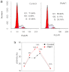
The discovery of new active biomaterials for promoting progenitor cell growth and differentiation in serum-free medium is still proving more challenging for the clinical treatments of degenerative diseases. In this work, a conjugated polyelectrolyte, polythiophene derivative (PMNT), was discovered to significantly drive the cell cycle progression from G₁ to S and G₂ phases and thus efficiently promote the cell growth without the need of serum. Furthermore, the fluorescent characteristic of PMNT makes it simultaneously able to trace its cellular uptake and localization by cell imaging. cDNA microarray study shows that PMNT can greatly regulate genes related to cell growth or differentiation. To the best of our knowledge, this is the first example of cell growth or differentiation promotion by polyelectrolyte material without the need of serum, thereby providing an important demonstration of degenerative biomaterial discovery through polymer design.
Figures


Similar articles
-
3D Bioprinting of Polythiophene Materials for Promoting Stem Cell Proliferation in a Nutritionally Deficient Environment.ACS Appl Mater Interfaces. 2021 Jun 9;13(22):25759-25770. doi: 10.1021/acsami.1c04967. Epub 2021 May 26. ACS Appl Mater Interfaces. 2021. PMID: 34036779
-
High-Throughput Assessment and Modeling of a Polymer Library Regulating Human Dental Pulp-Derived Stem Cell Behavior.ACS Appl Mater Interfaces. 2018 Nov 14;10(45):38739-38748. doi: 10.1021/acsami.8b12473. Epub 2018 Nov 5. ACS Appl Mater Interfaces. 2018. PMID: 30351898
-
The effect of topology of chitosan biomaterials on the differentiation and proliferation of neural stem cells.Acta Biomater. 2010 Sep;6(9):3630-9. doi: 10.1016/j.actbio.2010.03.039. Epub 2010 Apr 3. Acta Biomater. 2010. PMID: 20371303
-
Polymer microarray technology for stem cell engineering.Acta Biomater. 2016 Apr 1;34:60-72. doi: 10.1016/j.actbio.2015.10.030. Epub 2015 Oct 20. Acta Biomater. 2016. PMID: 26497624 Free PMC article. Review.
-
Development of Scaffolds for Vascular Tissue Engineering: Biomaterial Mediated Neovascularization.Curr Stem Cell Res Ther. 2017;12(2):155-164. doi: 10.2174/1574888X11666151203223658. Curr Stem Cell Res Ther. 2017. PMID: 26647912 Review.
Cited by
-
Artificial regulation of state transition for augmenting plant photosynthesis using synthetic light-harvesting polymer materials.Sci Adv. 2020 Aug 26;6(35):eabc5237. doi: 10.1126/sciadv.abc5237. eCollection 2020 Aug. Sci Adv. 2020. PMID: 32923652 Free PMC article.
References
-
- Flores I., Cayuela M. L. & Blasco M. A. Effects of telomerase and telomere length on epidermal stem cell behavior. Science 309, 1253–1256 (2005). - PubMed
-
- van der Valk J. et al. Optimization of chemically defined cell culture media – Replacing fetal bovine serum in mammalian in vitro methods. Toxicol. in Vitro 24, 1053–1063 (2010). - PubMed
-
- Toriniwa H. & Komiya T. Comparison of viral glycosylation using lectin blotting with Vero cell-derived and mouse brain-derived Japanese encephalitis vaccines. Vaccine 29, 1859–1862 (2011). - PubMed
Publication types
MeSH terms
Substances
LinkOut - more resources
Full Text Sources
Other Literature Sources
Medical

