. 2013 Jun 7;288(23):16855-16861.
doi: 10.1074/jbc.M113.473207. Epub 2013 Apr 22.

Sphingolipid domains in the plasma membranes of fibroblasts are not enriched with cholesterol

Affiliations

Sphingolipid domains in the plasma membranes of fibroblasts are not enriched with cholesterol

Jessica F Frisz et al. J Biol Chem. .

Abstract

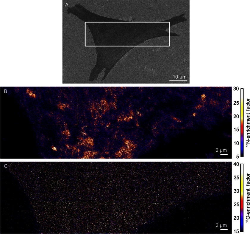
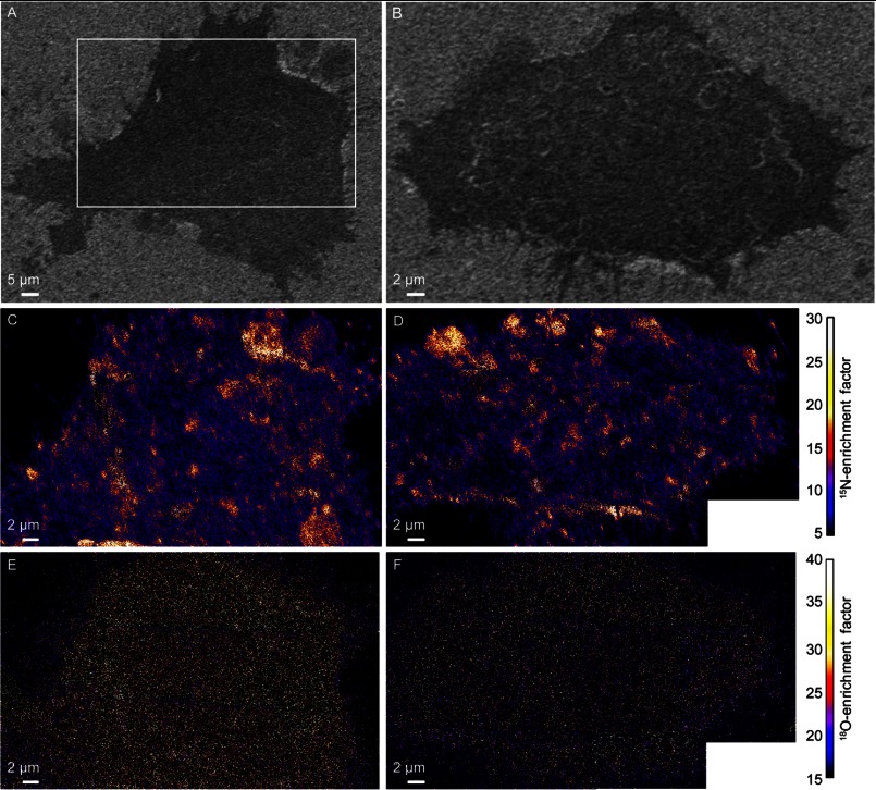
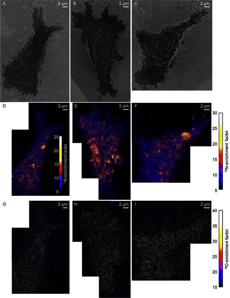
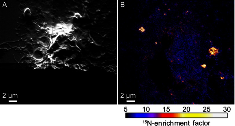
The plasma membranes of mammalian cells are widely expected to contain domains that are enriched with cholesterol and sphingolipids. In this work, we have used high-resolution secondary ion mass spectrometry to directly map the distributions of isotope-labeled cholesterol and sphingolipids in the plasma membranes of intact fibroblast cells. Although acute cholesterol depletion reduced sphingolipid domain abundance, cholesterol was evenly distributed throughout the plasma membrane and was not enriched within the sphingolipid domains. Thus, we rule out favorable cholesterol-sphingolipid interactions as dictating plasma membrane organization in fibroblast cells. Because the sphingolipid domains are disrupted by drugs that depolymerize the cells actin cytoskeleton, cholesterol must instead affect the sphingolipid organization via an indirect mechanism that involves the cytoskeleton.

Keywords: Cholesterol; Isotopic Tracers; Lipid Raft; Mass Spectrometry (MS); Membrane Structure; Plasma Membrane; Sphingolipid.


Figures

FIGURE 1.
FIGURE 1.
SEM and high-resolution SIMS images of a representative clone 15 fibroblast cell. A, SEM image of a clone 15 cell. The approximate location that was analyzed with high-resolution SIMS is outlined. B, the distribution of metabolically incorporated 15N-sphingolipids in the plasma membrane of the clone 15 cell was imaged by detecting the sphingolipid-specific 15N-enrichment with high-resolution SIMS. Orange and yellow regions represent plasma membrane domains that are enriched with 15N-sphingolipids. C, mosaic of 18O-enrichment images acquired with high-resolution SIMS shows the metabolically incorporated 18O-cholesterol is relatively uniformly distributed in the plasma membrane.
FIGURE 2.
FIGURE 2.
SEM and SIMS images of two NIH 3T3 mouse fibroblast cells that did not express the influenza membrane protein, hemagglutinin. A and B, SEM images show cell morphology. The region outlined in A shows the approximate location that was analyzed with high-resolution SIMS. C and D, mosaics of 15N-enrichment SIMS images were acquired on each metabolically labeled cell shown in A and B, respectively. Domains enriched with 15N-sphingolipids (orange, yellow, and white areas) are present in the plasma membrane. E and F, mosaics of 18O-enrichment SIMS images show the distribution of metabolically incorporated 18O-cholesterol in the plasma membrane.
FIGURE 3.
FIGURE 3.
SEM and SIMS images of cells that were treated with mβCD to reduce cellular cholesterol levels by 30%. A–C, SEM images show the morphology of three metabolically labeled clone 15 cells that were treated with mβCD to reduce their cellular cholesterol levels. D–F, mosaics of 15N-enrichment SIMS images show mβCD treatment reduced the number of 15N-sphingolipid domains (orange, yellow, and white areas) in the plasma membranes of the clone 15 cells. The false color scale that quantifies the 15N-enrichment at each pixel is shown in the inset in D, and at the right side of the figure for E and F. G–I, mosaics of 18O-enrichment SIMS images of the clone 15 cells show the remaining 18O-cholesterol in the plasma membrane of each cell appeared to be relatively evenly distributed. Images of B, C, E, and F were adapted from Ref. .
FIGURE 4.
FIGURE 4.
High-resolution SIMS images of a NIH 3T3 cell treated with latrunculin A to disrupt its cytoskeleton. A, montage of secondary electron images acquired with high-resolution SIMS shows latrunculin A treatment disrupted the cytoskeleton of the metabolically labeled NIH 3T3 cell. Secondary electrons were not detected near the bottom of the image due to the low primary ion beam current used for analysis. B, montage of 15N-enrichment SIMS images shows few 15N-sphingolipid domains were present in the plasma membrane of the NIH 3T3 cell after latrunculin A treatment. Latrunculin A treatment also disrupts the sphingolipid domains in the plasma membranes of clone 15 cells (11).

References

    1. Maxfield F. R., Menon A. K. (2006) Intracellular sterol transport and distribution. Curr. Opin. Cell Biol. 18, 379–385 - PubMed
    1. Leslie M. (2011) Mysteries of the cell: Do lipid rafts exist? Science 334, 1046–1047 - PubMed
    1. Lingwood D., Simons K. (2010) Lipid rafts as a membrane-organizing principle. Science 327, 46–50 - PubMed
    1. Simons K., Gerl M. J. (2010) Revitalizing membrane rafts: new tools and insights. Nat. Rev. Mol. Cell Biol. 11, 688–699 - PubMed
    1. Eggeling C., Ringemann C., Medda R., Schwarzmann G., Sandhoff K., Polyakova S., Belov V. N., Hein B., von Middendorff C., Schönle A., Hell S. W. (2009) Direct observation of the nanoscale dynamics of membrane lipids in a living cell. Nature 457, 1159–1162 - PubMed

Publication types

LinkOut - more resources