. 2013 Apr 29;201(3):439-48.
doi: 10.1083/jcb.201209023. Epub 2013 Apr 22.

Sanpodo controls sensory organ precursor fate by directing Notch trafficking and binding γ-secretase

Affiliations

Sanpodo controls sensory organ precursor fate by directing Notch trafficking and binding γ-secretase

Alok Upadhyay et al. J Cell Biol. .

Abstract

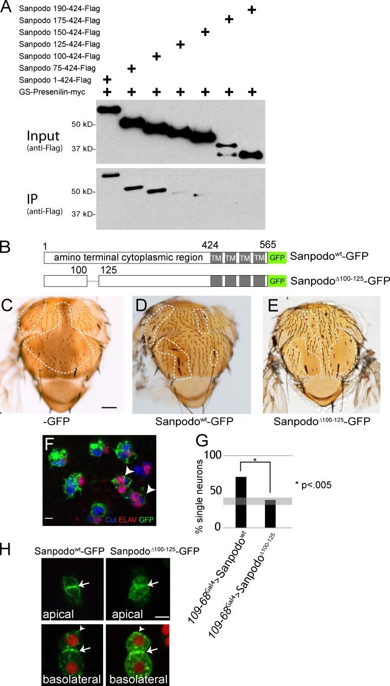
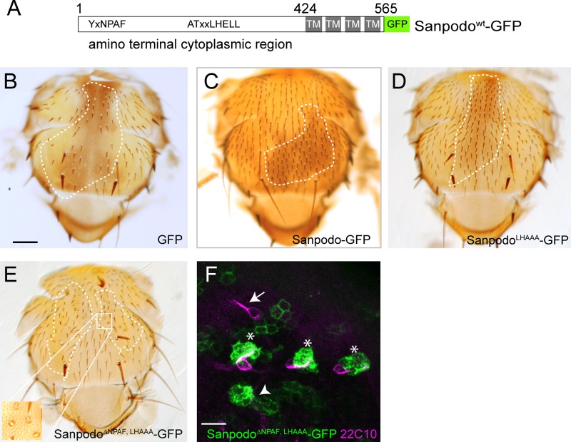
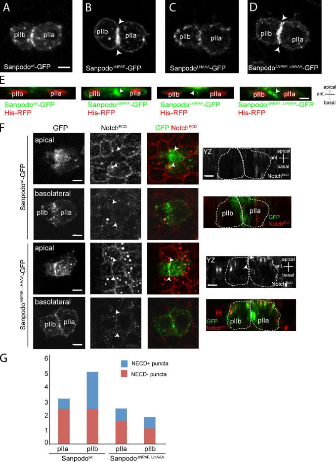
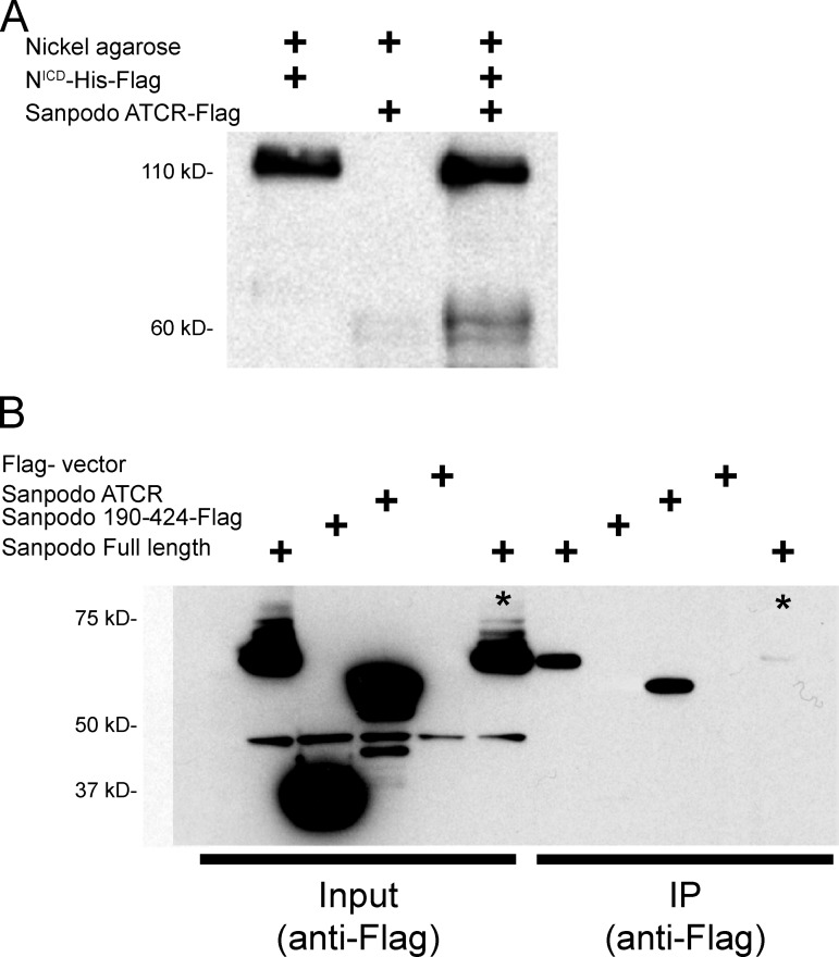
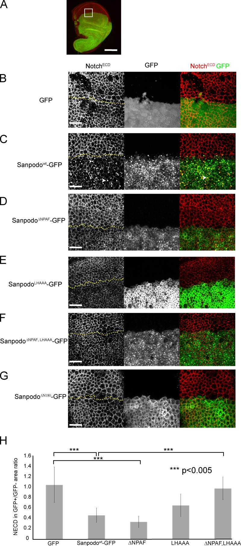
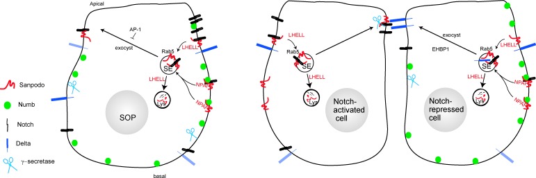
In Drosophila peripheral neurogenesis, Notch controls cell fates in sensory organ precursor (SOP) cells. SOPs undergo asymmetric cell division by segregating Numb, which inhibits Notch signaling, into the pIIb daughter cell after cytokinesis. In contrast, in the pIIa daughter cell, Notch is activated and requires Sanpodo, but its mechanism of action has not been elucidated. As Sanpodo is present in both pIIa and pIIb cells, a second role for Sanpodo in regulating Notch signaling in the low-Notch pIIb cell has been proposed. Here we demonstrate that Sanpodo regulates Notch signaling levels in both pIIa and pIIb cells via distinct mechanisms. The interaction of Sanpodo with Presenilin, a component of the γ-secretase complex, was required for Notch activation and pIIa cell fate. In contrast, Sanpodo suppresses Notch signaling in the pIIb cell by driving Notch receptor internalization. Together, these results demonstrate that a single protein can regulate Notch signaling through distinct mechanisms to either promote or suppress signaling depending on the local cellular context.


Figures

Figure 1.
Figure 1.
Sanpodo’s interaction with Presenilin is required for pIIa cell fate. Coexpression of the γ-secretase complex with myc-tagged Presenilin and Flag-tagged Sanpodo amino-terminal fragments in Drosophila S2 cells. (A) Binding of the amino-terminal cytoplasmic portion of Sanpodo (amino acids 1–424) to Presenilin-myc requires amino acids 100–125. (B) Schematic of SanpodoΔ100–125 mutant, deleting amino acids 100–125 within the amino terminus. (C–E) Thoraces of flies with sanpodoc55Sb e mutant clones (dotted line: approximate clone boundaries, as determined by e/e and Sb/Sb) with 109-68-Gal4–driven expression of GFP, Sanpodowt-GFP, or SanpodoΔ100–125–GFP using the MARCM system. Sanpodowt-GFP expression largely restores the wild-type bristle pattern to the thorax in sanpodo mutant clones (C; bar, 200 µm), whereas SanpodoΔ100–125–GFP expression fails to suppress the characteristic balding (D). Supernumerary neurons (marked with both Cut [blue] and ELAV [red]) in pupal sanpodoC55 mutant sensory organs expressing SanpodoΔ100–125–GFP (green) using the MARCM system (F, arrowhead). Bar, 5 µm. (G) Quantification of pupal sanpodo mutant sensory organs with only one ELAV+ neuron, indicating suppression of the sanpodo mutant phenotype; when expressing either Sanpodowt-GFP or SanpodoΔ100–125–GFP in MARCM clones using 109-68-Gal4, expression of SanpodoΔ100–125–GFP does not suppress the incompletely penetrant sanpodo mutant phenotype (gray bar indicates previously reported range of single neuron sensory organs in sanpodo null mutant clones; Jafar-Nejad et al., 2005; Roegiers et al., 2005). (H) SanpodoΔ100–125–GFP localizes to the apical and basolateral plasma membrane (white arrows) and intracellular puncta (white arrowheads) in pIIb/pIIa cells, similarly to wild-type Sanpodo-GFP (Histone RFP [red], Sanpodowt-GFP and SanpodoΔ100–125–GFP [green]). Bar, 2 µm.
Figure 2.
Figure 2.
Sanpodo NPAF and ELL motif are required for Notch inhibition in pIIb cells. (A) Schematic of Sanpodowt-GFP transgene with NPAF and LHELL motifs shown. (B–E) scabrous-Gal4–driven expression of wild-type and mutant Sanpodo transgenes in sanpodoc55Sb e mutant clones using the MARCM system (approximate clone borders are marked with dotted line). Bar, 200 µm. (B) GFP expression fails to restore the wild-type bristle pattern to the thorax in sanpodoc55Sb e mutant clones, whereas both Sanpodowt-GFP and SanpodoLHAAA-GFP expression (C and D) fully rescue the sanpodo mutant phenotype. (E) SanpodoΔNPAF-LHAAA–GFP expression suppresses the bristle loss phenotype associated with the Sanpodo mutant, but a number of rescued bristles exhibit supernumerary socket cells (inset). (F) Sensory organs from pupae Expressing SanpodoΔNPAF-LHAAA-GFP (green) in sanpodoc55Sb e mutant clones using the MARCM system, labeled with the neuronal marker 22C10 (purple). Bar, 10 µm. Single 22C10-positive neurons are visible in most GFP+ sanpodo mutant sensory organs (*), as well as outside the clone region (white arrow), but are absent in others (white arrowhead).
Figure 3.
Figure 3.
The NPAF and ELL motifs control Sanpodo trafficking in pIIa and pIIb cells. (A–D) Live-cell imaging of neuralized-Gal4–driven expression of GFP-tagged Sanpodo transgenes in pIIa/pIIb cell pairs; XY equatorial view. Bar, 2 µm. Sanpodowt-GFP (A) and SanpodoLHAAA-GFP (C) localize to the plasma membrane and in intracellular puncta, whereas SanpodoΔNPAF-GFP (B) and SanpodoΔNPAF, LHAAA–GFP (D) accumulate at the plasma membrane in both daughter cells, particularly at the membrane interface between pIIa/pIIb (B and D, white arrowheads). (E) Live-cell analysis of Sanpodo mutant transgenes (green) in pIIa and pIIb cells expressing His-RFP (red) 7 min after SOP mitotic exit; YZ view, apical at top, anterior to the left. Bar, 2 µm. SanpodoΔNPAF-GFP and SanpodoΔNPAF-LHAAA–GFP accumulate strongly at the basolateral interface (white arrowhead), whereas SanpodoLHAAA-GFP accumulates at the apical plasma membrane in pIIa and pIIb cells, compared with Sanpodowt-GFP. Notch (NotchECD, red) is depleted from apical (white arrowheads) pIIa/pIIb membrane interface in Sanpodo-GFP–expressing cells. (F) Apical membrane Notch (white arrowheads) is detected at low levels in cell rescued with SanpodoΔNPAF, LHAAA-GFP, whereas basolateral Notch levels are similar to wild type (YZ planes, apical is at the top, approximate cell outlines represented by dashed lines). Bar, 2 µm. (G) Quantification of colocalization of Sanpodo and NECD in intracellular vesicles in pIIa and pIIb cells. Wild-type Sanpodo-GFP accumulates in large intracellular (>0.5 µm) puncta, which partially colocalize with Notch, in higher numbers in pIIb cells. SanpodoΔNPAF, LHAAA-GFP puncta are less abundant and are equally distributed in pIIa and pIIb cells (standard deviation of 2.215–3.599 total Sanpodo + puncta per cell, n = 7 WT; n = 22 NPAF, LHAAA).
Figure 4.
Figure 4.
Sanpodo binds the Notch intracellular domain. (A) Binding of Flag-tagged Sanpodo amino-terminal cytoplasmic domain (424 amino acids, detected at ∼60 kD) to the nickel Agarose beads is strongly increased when preincubated with the Flag- and His-tagged NICD, which binds to nickel Agarose beads robustly and is detected at 110 kD with the anti-Flag antibody. (B) Full-length Notch coimmunoprecipitates with either transfected full-length Sanpodo (all Sanpodo transgenes are Flag tagged) or the Sanpodo cytoplasmic domain in the induced S2-MT-Notch Drosophila cell line, whereas the amino-terminal truncated Sanpodo cytoplasmic domain (190–424) does not (asterisk indicates Notch uninduced).
Figure 5.
Figure 5.
Sanpodo promotes Notch endocytosis. (A) apterous-Gal4–driven ectopic expression of GFP-tagged Sanpodo transgenes (green) in wing disc epithelial cells labeled with the NotchECD antibody (red). Bar, 100 µm. White box is the approximate region of disc shown in B–G. Merged apical XY confocal sections through wing disc epithelial cells (1.5-µm thickness, the border between GFP+ and GFP− regions are marked by a dashed yellow line). Expression of GFP alone has no effect on NECD localization to apical cell junctions in wing disc epithelial cells (B), whereas expression of Sanpodowt-GFP depletes Notch from the apical membrane in epithelial cells and causes Notch to accumulate in large apical vesicles, which in some cases colocalize with Sanpodo-GFP (C, white arrowheads). Both the ΔNPAF mutant (D) and the LHAAA mutant (E) retain the ability to deplete apical Notch, whereas the ΔNPAF, LHAAA mutant (F) or ΔN180 mutant of Sanpodo (G, ΔN180) abrogate Notch apical depletion. Quantification of apical NECD staining represented as a ratio of the area occupied by apical NECD staining in the GFP+/GFP− regions of merged apical XY planes from apterous-Gal4–driven ectopic expression of GFP-tagged Sanpodo transgenes and a GFP control 9H). Sanpodo-GFP and SanpodoΔNPAF-GFP significantly deplete membrane Notch levels, whereas Notch levels are decreased in the LHAAA; they do not attain statistical significance. In contrast, Notch levels in the SanpodoΔNPAF, LHAAA–GFP expression region were indistinguishable from GFP control expression, and significantly increased relative to Sanpodo-GFP. Bars, 25 µm.
Figure 6.
Figure 6.
A potential model for Sanpodo trafficking in SOP lineage cells. Sanpodo expression is induced in SOP cells and the bulk of Sanpodo (red) enters the endocytic system, along with Notch, from the basolateral membrane via an interaction of Numb (green) and the Sanpodo NPAF motif. An alternative route for Sanpodo and Notch endocytosis requires the ELL motif. Endocytosed Sanpodo is directed to the lysosome and shunted away from the recycling to the apical membrane by the ELL motif. Sanpodo targeting to the apical membrane is regulated by the exocyst complex and the AP-1 complex of endocytic adaptor proteins.

Similar articles

Cited by

References

    1. Babaoglan A.B., O’Connor-Giles K.M., Mistry H., Schickedanz A., Wilson B.A., Skeath J.B. 2009. Sanpodo: a context-dependent activator and inhibitor of Notch signaling during asymmetric divisions. Development. 136:4089–4098 10.1242/dev.040386 - DOI - PMC - PubMed
    1. Banks S.M.L., Cho B., Eun S.H., Lee J.-H., Windler S.L., Xie X., Bilder D., Fischer J.A. 2011. The functions of auxilin and Rab11 in Drosophila suggest that the fundamental role of ligand endocytosis in notch signaling cells is not recycling. PLoS ONE. 6:e18259 10.1371/journal.pone.0018259 - DOI - PMC - PubMed
    1. Ben-Yaacov S., Le Borgne R., Abramson I., Schweisguth F., Schejter E.D. 2001. Wasp, the Drosophila Wiskott-Aldrich syndrome gene homologue, is required for cell fate decisions mediated by Notch signaling. J. Cell Biol. 152:1–13 - PMC - PubMed
    1. Benhra N., Lallet S., Cotton M., Le Bras S., Dussert A., Le Borgne R. 2011. AP-1 controls the trafficking of Notch and Sanpodo toward E-cadherin junctions in sensory organ precursors. Curr. Biol. 21:87–95 10.1016/j.cub.2010.12.010 - DOI - PubMed
    1. Berdnik D., Török T., González-Gaitán M., Knoblich J.A. 2002. The endocytic protein alpha-Adaptin is required for numb-mediated asymmetric cell division in Drosophila. Dev. Cell. 3:221–231 10.1016/S1534-5807(02)00215-0 - DOI - PubMed

Publication types

MeSH terms