. 2013 Jun;41(11):5898-911.
doi: 10.1093/nar/gkt265. Epub 2013 Apr 22.

Exploring mRNA 3'-UTR G-quadruplexes: evidence of roles in both alternative polyadenylation and mRNA shortening

Affiliations

Exploring mRNA 3'-UTR G-quadruplexes: evidence of roles in both alternative polyadenylation and mRNA shortening

Jean-Denis Beaudoin et al. Nucleic Acids Res. 2013 Jun.

Abstract

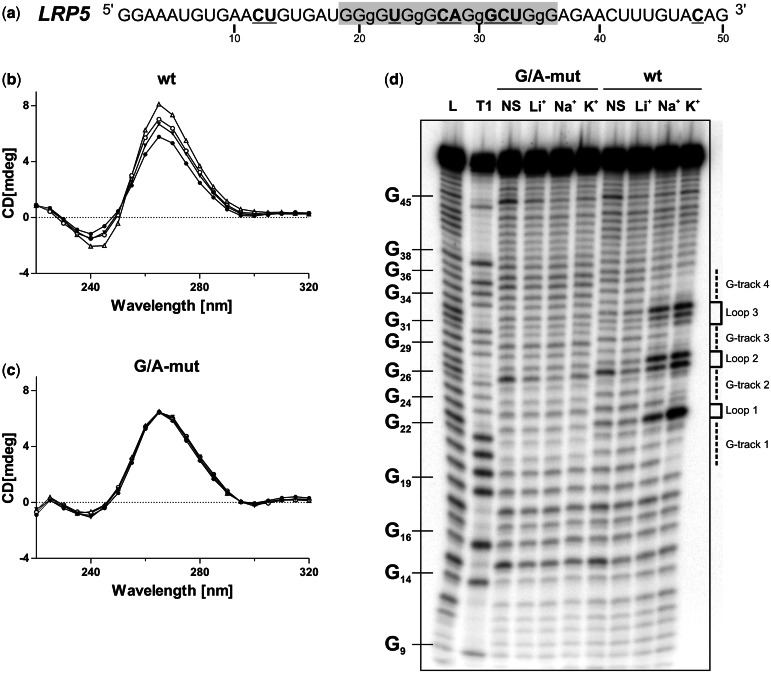
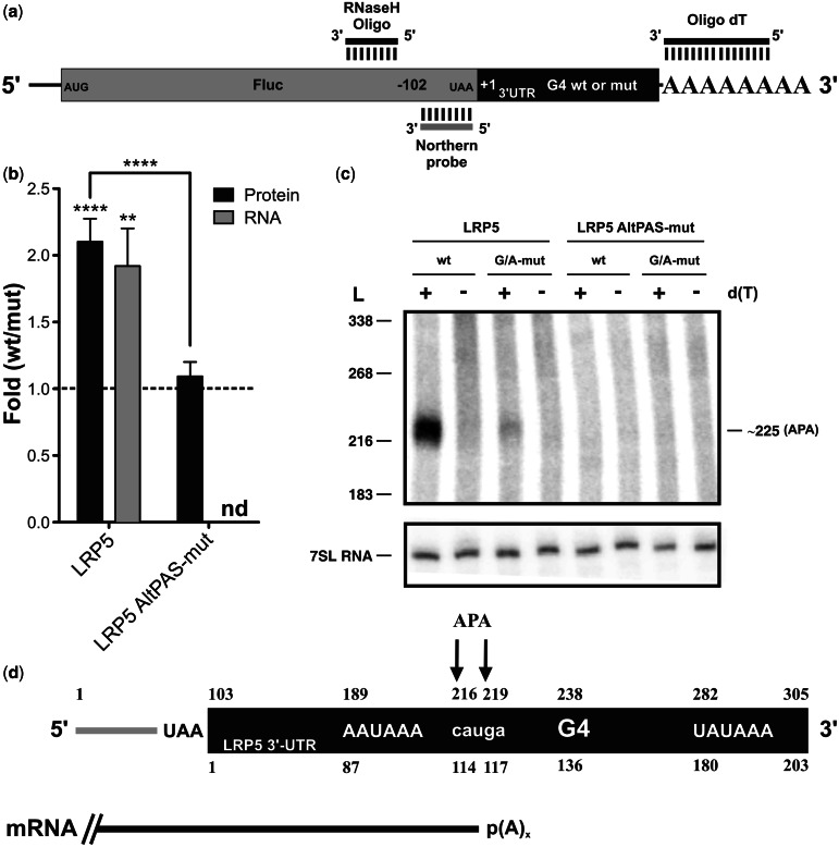
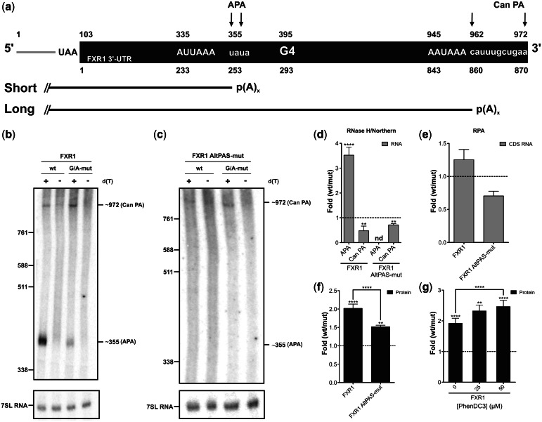
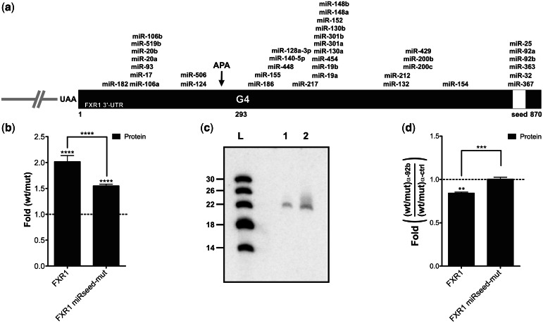
Guanine-rich RNA sequences can fold into non-canonical, four stranded helical structures called G-quadruplexes that have been shown to be widely distributed within the mammalian transcriptome, as well as being key regulatory elements in various biological mechanisms. That said, their role within the 3'-untranslated region (UTR) of mRNA remains to be elucidated and appreciated. A bioinformatic analysis of the 3'-UTRs of mRNAs revealed enrichment in G-quadruplexes. To shed light on the role(s) of these structures, those found in the LRP5 and FXR1 genes were characterized both in vitro and in cellulo. The 3'-UTR G-quadruplexes were found to increase the efficiencies of alternative polyadenylation sites, leading to the expression of shorter transcripts and to possess the ability to interfere with the miRNA regulatory network of a specific mRNA. Clearly, G-quadruplexes located in the 3'-UTRs of mRNAs are cis-regulatory elements that have a significant impact on gene expression.


Figures

Figure 1.
Figure 1.
LRP5 3′-UTR PG4 folds into a G4 structure in vitro. (a) Sequence and numbering of the wt LRP5 PG4 used in the in vitro experiments. The lowercase guanosines (g) correspond to those mutated to adenosines in the G/A-mutant version. Nucleotides that were hydrolyzed significantly more in the presence of KCl during the in-line probing are both in bold and underlined. (b, c) CD spectra for the LRP5 PG4 sequence using 4 µM of either the wt (b) or the G/A-mutant (c) versions performed either in the absence of salt (closed circle) or in the presence of 100 mM of either LiCl (inverted closed triangle), NaCl (open circle) or KCl (open triangle). (d) Autoradiogram of a 10% denaturing polyacrylamide gel of the in-line probing of the 5′-end-labeled LRP5 wt and G/A-mutant PG4 versions performed either in the absence of salt (NS), or in the presence of 100 mM of either LiCl, NaCl or KCl. Lanes L and T1 correspond to alkaline hydrolysis and RNase T1 mapping of the wt version, respectively. The positions of the guanosines are indicated on the left of the gel, whereas the domains of the G4 structure are indicated on the right.
Figure 2.
Figure 2.
The LRP5 3′-UTR G4 structure in cellulo. (a) Schematic representation of the Fluc–LRP5 construction. The Fluc-coding sequence is shown in gray, whereas the LRP5 3′-UTR is shown in black. The binding regions of the oligonucleotides used for the RNase H hydrolysis, as well as the luciferase-specific probe, are illustrated. (b) Gene expression levels of the different LRP5 constructs either at the protein level (black) or the mRNA level (gray). The x-axis identifies the constructions used and the y-axis the fold difference (i.e. wt result divided by G/A-mutated result) (for LRP5 protein n = 3, whereas for mRNA n = 5, for LRP5 AltPAS-mut protein n = 4, nd indicates not detectable). Error bars, mean ± SD, **P < 0.01 and ****P < 0.0001. (c) Northern blot hybridization of RNA samples subjected to an RNase H hydrolysis in the presence of a Fluc-specific DNA oligonucleotide and either in the absence (−) or the presence (+) of oligo-dT. The numbers on the left refer to the sizes of a molecular RNA ladder, whereas that on the right is the estimated size of the detected transcript. 7SL RNA was probed as internal control. (d) Schematic view of the RNA product resulting from the RNase H hydrolysis. The upper numbers correspond to the numbering from the 5′-end of the digestion product, whereas lower ones refer to the start of the LRP5 3′-UTR. The arrows map the different PA sites as determined by 3′-RACE, and the mRNA produced is depicted in black.
Figure 3.
Figure 3.
The FXR1 3′-UTR G4 structure in cellulo. (a) Schematic representation of the Fluc–FXR1 transcripts resulting from the RNase H hydrolysis. The upper numbers correspond to the numbering from the 5′-end of the hydrolyzed product, whereas lower ones refer to the start of the FXR1 3′-UTR (black part). The arrows map the different PA sites as determined by the 3′-RACE experiments [alternative (APA) and canonical (Can PA) sites]. The short and long mRNA isoforms produced are shown. (b, c) Northern blot hybridizations of the RNA samples previously subjected to RNase H hydrolysis in either the absence (−) or the presence (+) of oligo-dT. The numbers on the left refer to the sizes of a molecular RNA ladder, whereas those on the right are the estimated sizes of the two isoforms. 7SL RNA was probed as an internal control. (d–f) Gene expression levels of constructs either at the mRNA level as determined by northern blot hybridization (for FXR1 n = 5, whereas for FXR1 AltPAS-mut n = 3; nd indicates not detectable) (d), by RNase protection assay (FXR1 and FXR1 AltPAS-mut n = 3) (e) (gray) or at the protein level as determined by luciferase assay (FXR1 n = 7, FXR1 AltPAS-mut n = 3) (f) (black). The x-axis identifies the constructions used and the y-axis the fold difference (wt result divided by G/A-mutated result). (g) Luciferase assays in the presence of various concentrations of PhenDC3 (0–50 µM; n = 3). Error bars, mean ± SD, **P < 0.01 and ****P < 0.0001.
Figure 4.
Figure 4.
FXR1 3′-UTR shortening and the microRNAs regulatory network. (a) Schematic representation of the FXR1 3′-UTR. The numbering refers to the position from the start site of the FXR1 3′-UTR. All predicted microRNA target sites with a mirSVR score <−0.5 according to the miRanda algorithm are shown (40). The white region corresponds to the predicted shared microRNA seed region that was mutated in the FXR1 miRseed-mut constructions. (b) Gene expression levels of different FXR1 constructs at the protein level as determined by luciferase assays. The x-axis identifies the constructions used and the y-axis the fold difference (wt result divided by G/A-mutated result) (for both FXR1 and miRseed-mut n = 4). (c) Northern blot hybridization for the detection of miR-92b performed using either 5 µg (lane 1) of small RNAs (<200 nt) or 50 µg of total RNA (lane 2) extracted from untransfected HEK293T cells. The numbers on the left refer to the sizes of a molecular RNA ladder of 5′-end labeled in vitro transcripts (lane L). (d) Gene expression levels of different FXR1 constructs at the protein level as determined by luciferase assays in the presence of either 100 nM miR-92b inhibitor or of irrelevant control inhibitors. The x-axis identifies the constructions used and the y-axis the fold difference (ratio wt on G/A-mutated version obtained in the presence of the miR-92b inhibitor divided by that obtained in presence of the control inhibitor) (both FXR1 and miRseed-mut n = 3). **P < 0.01, ***P < 0.001 and ****P < 0.0001.

References

    1. Birney E, Stamatoyannopoulos JA, Dutta A, Guigó R, Gingeras TR, Margulies EH, Weng Z, Snyder M, Dermitzakis ET, Stamatoyannopoulos JA, et al. Identification and analysis of functional elements in 1% of the human genome by the ENCODE pilot project. Nature. 2007;447:799–816. - PMC - PubMed
    1. Huppert JL. Four-stranded nucleic acids: structure, function and targeting of G-quadruplexes. Chem. Soc. Rev. 2008;37:1375–1384. - PubMed
    1. Burge S, Parkinson GN, Hazel P, Todd AK, Neidle S. Quadruplex DNA: sequence, topology and structure. Nucleic Acids Res. 2006;34:5402–5415. - PMC - PubMed
    1. Neidle S, Balasubramanian S. Quadruplex Nucleic Acids. Cambridge: RSC Publishing; 2006. p. 301.
    1. Todd AK, Johnston M, Neidle S. Highly prevalent putative quadruplex sequence motifs in human DNA. Nucleic Acids Res. 2005;33:2901–2907. - PMC - PubMed

Publication types

MeSH terms

Substances