. 2013 Jun;41(11):5704-16.
doi: 10.1093/nar/gkt285. Epub 2013 Apr 22.

The Scaffold attachment factor b1 (Safb1) regulates myogenic differentiation by facilitating the transition of myogenic gene chromatin from a repressed to an activated state

Affiliations

The Scaffold attachment factor b1 (Safb1) regulates myogenic differentiation by facilitating the transition of myogenic gene chromatin from a repressed to an activated state

J Manuel Hernández-Hernández et al. Nucleic Acids Res. 2013 Jun.

Abstract

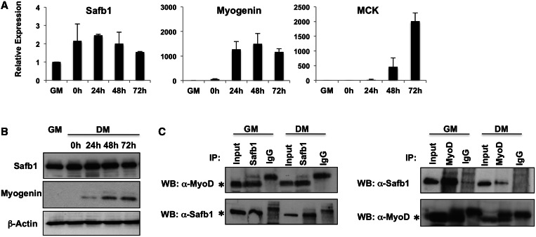
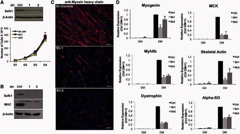
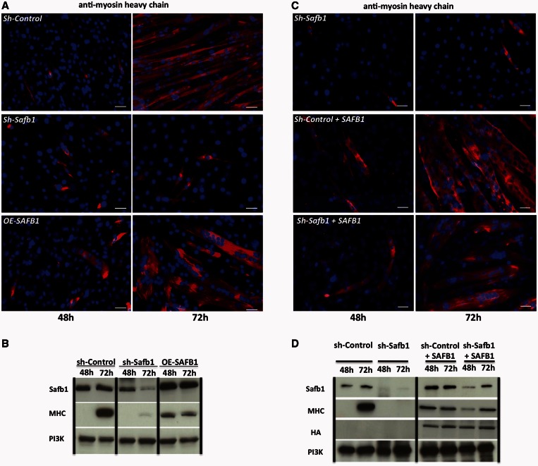
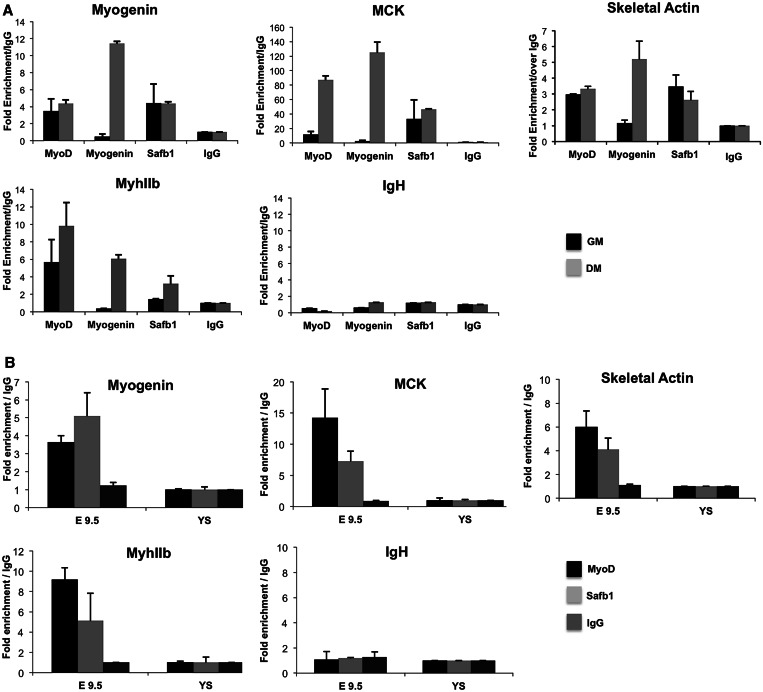
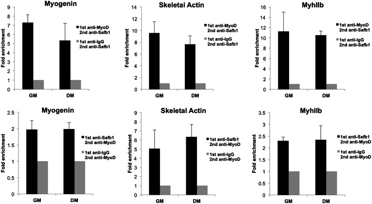
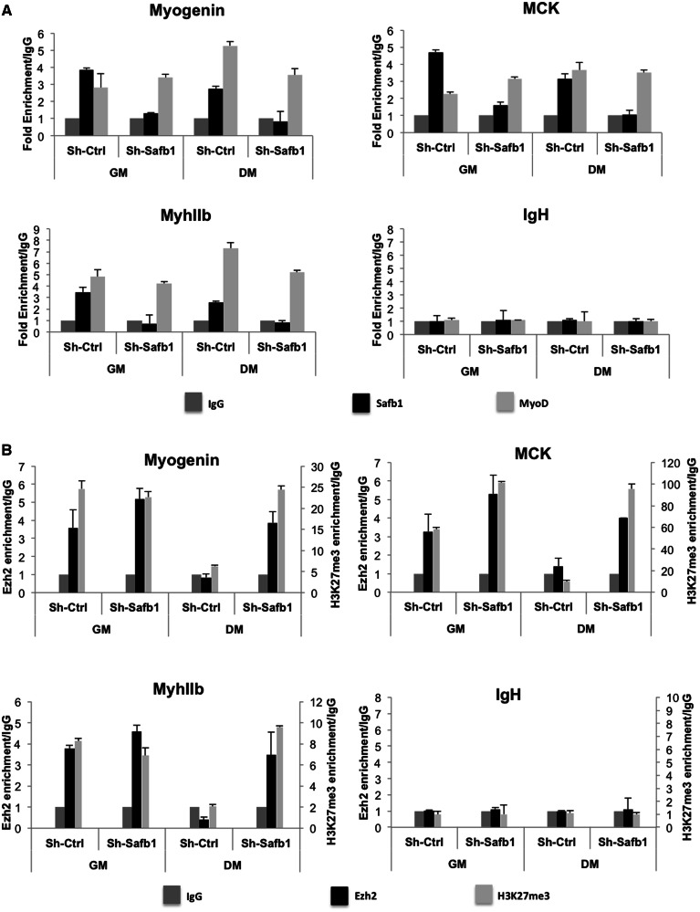
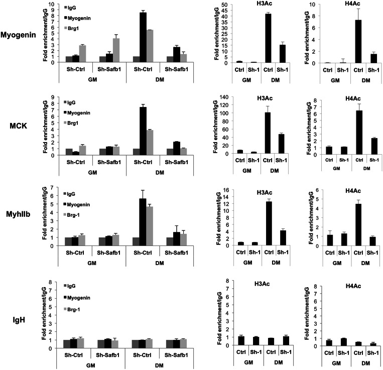
The regulation of skeletal muscle gene expression during myogenesis is mediated by lineage-specific transcription factors in combination with numerous cofactors, many of which modify chromatin structure. However, the involvement of scaffolding proteins that organize chromatin and chromatin-associated regulatory proteins has not extensively been explored in myogenic differentiation. Here, we report that Scaffold attachment factor b1 (Safb1), primarily associated with transcriptional repression, functions as a positive regulator of myogenic differentiation. Knockdown of Safb1 inhibited skeletal muscle marker gene expression and differentiation in cultured C2C12 myoblasts. In contrast, over-expression resulted in the premature expression of critical muscle structural proteins and formation of enlarged thickened myotubes. Safb1 co-immunoprecipitated with MyoD and was co-localized on myogenic promoters. Upon Safb1 knockdown, the repressive H3K27me3 histone mark and binding of the Polycomb histone methyltransferase Ezh2 persisted at differentiation-dependent gene promoters. In contrast, the appearance of histone marks and regulators associated with myogenic gene activation, such as myogenin and the SWI/SNF chromatin remodelling enzyme ATPase, Brg1, was blocked. These results indicate that the scaffold protein Safb1 contributes to the activation of skeletal muscle gene expression during myogenic differentiation by facilitating the transition of promoter sequences from a repressive chromatin structure to one that is transcriptionally permissive.


Figures

Figure 1.
Figure 1.
Safb1 co-immunoprecipitates with MyoD and Brg1 during C2C12 cell differentiation. (A and B) Total RNA and protein from proliferating myoblasts in growth media (GM), confluent myoblasts (time 0) and myoblasts exposed to differentiation media (DM) for up to 72 h were collected and analysed for Safb1 mRNA and protein levels. Myogenic markers of differentiation Myogenin and MCK were monitored as controls for C2C12 cell differentiation. β-actin was monitored as a loading control for the western blot. mRNA data are presented as the expression relative to EF-1α levels. The expression in growth media was normalized to 1. Data represent the average of three independent analyses ± SEM. t-test analysis of Safb1 mRNA levels indicated that only the 0 h timepoint gave a P value > 0.05 relative to the value obtained in proliferating myoblasts. ImageJ quantification of the western blot indicated that Safb1 levels varied by no more than 15% from the level present in growth media through 48 h post-differentiation and that Safb1 levels were reduced by 40% in differentiated cells (72 h post-differentiation). (C) Whole cell protein extracts from myoblast and myotubes were collected to perform co-immunoprecipitation assays for Safb1 and MyoD. Asterisks indicate the MyoD or Safb1 specific bands. GM, growth media; DM, differentiation media; IP, immunoprecipitation; WB, western blot.
Figure 2.
Figure 2.
Safb1 knockdown blocks C2C12 cell differentiation and inhibits skeletal muscle genes expression. (A) shRNA-mediated knockdown of Safb1 in proliferating myoblasts does not affect proliferation. Two different sequences against Safb1 mRNA (sh1 and sh2) were used. Knockdown of Safb1 in proliferating myoblasts was confirmed by western blot. PI3K levels were monitored as a loading control. Cell counts were taken at 24 h intervals after seeding of equal numbers of cells. Data represent the average of three independent analyses ± SEM. sh control cells (sh ctrl) express a scrambled sequence shRNA. (B) Confirmation of Safb1 knockdown in cells induced to differentiate for 72 h compared with cells expressing the control shRNA. MHC protein levels were also reduced in differentiated Safb1 knockdown cells. β-actin levels were monitored as a loading control. (C) Immunostaining indicates that the population of MHC-positive myotubes is markedly reduced in Safb1 knockdown C2C12 cells induced to differentiate. Nuclei were counterstained with DAPI. (D) Total RNA from C2C12 cells expressing shRNA against Safb1 was used to analyse expression levels of Myogenin, Muscle Creatine Kinase Myosin heavy chain IIb (Myh IIb), Skeletal Actin, Dystrophin and Alpha Sarcoglycan (alpha-SG) genes. Expression in differentiated sh-control (ctrl) cells was normalized to 1. Quantification represents the average of three independent experiments ± SEM t test analysis indicates that all values for mRNA levels in the differentiated knockdown cells are significantly reduced compared with the mRNA levels in control differentiated cells (P < 0.05 for Sh2 KD of myogenin, P < 0.01 for all other data points).
Figure 3.
Figure 3.
Over-expression of SAFB1 leads to formation of enlarged, thicker, MHC-positive myotubes. (A) C2C12 cells infected with the shRNA control vector, an shRNA against Safb1 or a vector encoding SAFB1 were immunostained for MHC after 48 and 72 h of differentiation. Nuclei were counterstained with DAPI. (B) Western blot of protein extracts obtained from duplicate plates of cells in (A) showing Safb1 and MHC levels. PI3K levels were monitored as a loading control. (C) Immunostaining indicates that exogenous expression of SAFB1 in Safb1 knockdown cells rescues MHC expression and results in myotube formation similar to that observed in sh-control cells over-expressing SAFB1. Nuclei were counterstained with DAPI. (D) Rescue of Safb1 expression after knockdown of endogenous Safb1 (shSafb1 + SAFB1), reestablishes MHC expression at 48 h of differentiation. HA signal indicates the presence of exogenous SAFB1, which is HA-tagged. PI3K levels were monitored as a loading control.
Figure 4.
Figure 4.
Safb1 is recruited to regulatory sequences of skeletal muscle-specific genes before and during C2C12 cell differentiation and in somite-enriched tissue isolated from mouse embryos at E9.5. (A) ChIP analysis of skeletal muscle promoters Myogenin, Muscle Creatine Kinase, Myosin heavy chain IIb (Myh IIb) and IgH (as a negative control) in C2C12 myoblasts (GM) and myotubes differentiated for 72 h (DM). (B) ChIP analysis of skeletal muscle promoters in somite-enriched tissue obtained from mouse embryos at E9.5 dpc. Yolk sac (YS) from E9.5 embryos was used as negative control. Quantification represents the mean of three independent experiments ± SEM. t test analyses indicate that all binding events except for myogenin binding to myogenic gene promoters in growth media conditions are statistically significant (P < 0.05) compared with the IgG controls (panel A) and that all MyoD and Safb1 binding in E9.5 somite-enriched tissue is statistically significant (P < 0.01) compared with binding in yolk sac (panel B).
Figure 5.
Figure 5.
Safb1 and MyoD are co-localized at regulatory sequences of myogenic promoters in myoblasts prior to the activation of transcription. ReChIPs were performed by immunoprecipitating chromatin with an antibody against MyoD followed by a second immunoprecipitation using antibody against Safb1 (upper panel) or using Safb1 as the first immunoprecipitating antibody (lower panel) in undifferentiated myoblasts (GM) and 72 h differentiated myotubes (DM). ChIP values were normalized against the input and expressed as relative enrichment of the material precipitated by the indicated antibody. Data represent the mean of three independent experiments ± SEM. t test analyses indicate there is no statistical difference when comparing MyoD/Safb1 ReChIP signals in growth media with the signals obtained in differentiation media. MyoD/Safb1 ReChIP signals relative to IgG controls were statistically significant (P < 0.05).
Figure 6.
Figure 6.
shRNA-mediated knockdown of Safb1 does not alter MyoD recruitment to myogenic sequences but increases Polycomb repressive marks. Proliferating (GM) or 72 h differentiated (DM) C2C12 cells expressing control shRNA or shRNA against Safb1 were fixed and processed for ChIP assays using (A) antibodies against MyoD and Safb1 (top panel) or (B) antibodies against Ezh2 and H3K27me3 at the indicated gene regulatory sequences. H3K27me3 enrichment is presented on the right-hand y-axis in the bottom panel. Data represent the mean of three independent experiments ± SEM. t test analyses indicate that when comparing binding in sh-Safb1-treated differentiated cells with sh-Control differentiated cells, the binding of Safb1 was significantly reduced (P < 0.01), the binding of MyoD was not significantly reduced, except for the binding to the MyhIIb promoter (P = 0.045), and the binding of Ezh2 and H3K27me3 were significantly reduced (P < 0.01 and P < 0.005, respectively).
Figure 7.
Figure 7.
Recruitment of transcriptional activators and associated chromatin marks is inhibited in Safb1 knockdown cells. Proliferating (GM) or 72 h differentiated (DM) C2C12 cells expressing control shRNA or shRNA against Safb1 were fixed and processed for ChIP assays using antibodies against Myogenin, Brg1, acetylated H3 or acetylated H4 at the indicated gene regulatory sequences. Data represent the mean of three independent experiments ± SEM. t test analyses indicate that binding of myogenin and Brg1 and incorporation of acetylated H3 and H4 was significantly reduced in differentiated sh-Safb1-treated cells compared with binding/incorporation in differentiated sh-Control cells (myogenin P < 0.05, Brg1 P < 0.002, acetylated H3 P < 0.005, acetylated H4 P < 0.002).

Similar articles

Cited by

References

    1. Albini S, Puri PL. SWI/SNF complexes, chromatin remodeling and skeletal myogenesis: it's time to exchange! Exp. Cell Res. 2010;316:3073–3080. - PMC - PubMed
    1. Perdiguero E, Sousa-Victor P, Ballestar E, Munoz-Canoves P. Epigenetic regulation of myogenesis. Epigenetics. 2009;4:541–550. - PubMed
    1. Caretti G, Palacios D, Sartorelli V, Puri PL. Phosphoryl-EZH-ion. Cell Stem Cell. 2011;8:262–265. - PMC - PubMed
    1. Harada A, Okada S, Konno D, Odawara J, Yoshimi T, Yoshimura S, Kumamaru H, Saiwai H, Tsubota T, Kurumizaka H, et al. Chd2 interacts with H3.3 to determine myogenic cell fate. EMBO J. 2012;31:2994–3007. - PMC - PubMed
    1. Lee H, Quinn JC, Prasanth KV, Swiss VA, Economides KD, Camacho MM, Spector DL, Abate-Shen C. PIAS1 confers DNA-binding specificity on the Msx1 homeoprotein. Genes Dev. 2006;20:784–794. - PMC - PubMed

Publication types