. 2013 Aug;62(8):2717-27.
doi: 10.2337/db13-0062. Epub 2013 Apr 22.

Resistance to aerobic exercise training causes metabolic dysfunction and reveals novel exercise-regulated signaling networks

Affiliations

Resistance to aerobic exercise training causes metabolic dysfunction and reveals novel exercise-regulated signaling networks

Sarah J Lessard et al. Diabetes. 2013 Aug.

Abstract

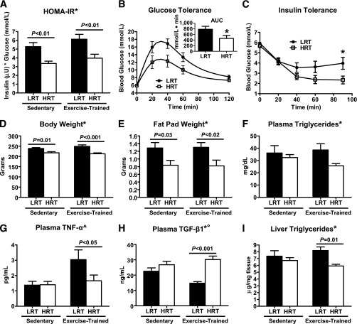
Low aerobic exercise capacity is a risk factor for diabetes and a strong predictor of mortality, yet some individuals are "exercise-resistant" and unable to improve exercise capacity through exercise training. To test the hypothesis that resistance to aerobic exercise training underlies metabolic disease risk, we used selective breeding for 15 generations to develop rat models of low and high aerobic response to training. Before exercise training, rats selected as low and high responders had similar exercise capacities. However, after 8 weeks of treadmill training, low responders failed to improve their exercise capacity, whereas high responders improved by 54%. Remarkably, low responders to aerobic training exhibited pronounced metabolic dysfunction characterized by insulin resistance and increased adiposity, demonstrating that the exercise-resistant phenotype segregates with disease risk. Low responders had impaired exercise-induced angiogenesis in muscle; however, mitochondrial capacity was intact and increased normally with exercise training, demonstrating that mitochondria are not limiting for aerobic adaptation or responsible for metabolic dysfunction in low responders. Low responders had increased stress/inflammatory signaling and altered transforming growth factor-β signaling, characterized by hyperphosphorylation of a novel exercise-regulated phosphorylation site on SMAD2. Using this powerful biological model system, we have discovered key pathways for low exercise training response that may represent novel targets for the treatment of metabolic disease.


Figures

FIG. 1.
FIG. 1.
Exercise capacity in rats bred for LRT and HRT. Exercise capacity (m) was measured using an incremental treadmill running test to exhaustion in (A) sedentary rats at 10 and 20 weeks of age and in (B) a separate group of age-matched trained rats before (pretraining) and after (posttraining) 8 weeks (3 days/week) of treadmill running exercise. C: Exercise response was calculated as the difference in exercise capacity before and after exercise training (exercise-trained) for each rat. The change in exercise capacity between 10 and 20 weeks of age is shown for animals in the control group (sedentary). *P < 0.05 for phenotype main effect; ^P < 0.05 for exercise main effect; °P < 0.05 for phenotype–exercise interaction by two-way ANOVA. P values obtained by Tukey post hoc testing are shown. n = 10–12/group.
FIG. 2.
FIG. 2.
Whole-body metabolic dysfunction in LRT. Fasting blood samples were collected from sedentary and exercise-trained LRT/HRT. A: Plasma glucose and insulin values were used to calculate the homeostasis model of insulin resistance (HOMA-IR). B: Glucose tolerance was assessed in sedentary rats after an intraperitoneal (IP) injection of 2 g/kg glucose and the area under curve (AUC) was calculated. C: Insulin tolerance was assessed after IP injection of 0.75 units/kg insulin. Body weight (D) and gonadal fat pad weight (E) were measured in sedentary and exercise-trained LRT and HRT. Plasma triglycerides (F), TNF-α (G), and TGF-β1 (H) concentrations were analyzed by ELISA. I: Liver triglycerides were estimated from total liver glycerol content. *P < 0.05 for phenotype main effect; ^P < 0.05 for exercise main effect; °P < 0.05 for phenotype–exercise interaction by two-way ANOVA. P values obtained by Tukey post hoc testing are displayed. n = 10–12/group.
FIG. 3.
FIG. 3.
HRT have fewer oxidative muscle fibers and impaired exercise-induced angiogenesis. Plantaris muscles from sedentary (SED) and exercise-trained (EXT) rats were frozen in N2-cooled isopentane and cut into 6-µm cross-sections. A: Sections were stained with antibodies against laminin (white) and myosin heavy chain I (green) and visualized using fluorescent secondary antibodies under 100× magnification. Type I fiber content was expressed as % of total muscle fibers counted. B: Capillary density (capillaries/mm2) was calculated in sections stained with an antibody against the endothelial marker CD31 (red). Nuclei were visualized with DAPI stain (blue). n = 4–5/group. *P < 0.05 for phenotype main effect by two-way ANOVA. P values obtained by Tukey post hoc testing are displayed.
FIG. 4.
FIG. 4.
Analysis of gene transcription in response to an acute bout of exercise identifies dysregulation of SMAD, CREB, and HDAC activity in LRT. RNA was extracted from the soleus muscles of rats under resting conditions or 3 h after an acute bout of treadmill running exercise. Genes that were significantly upregulated (red) or downregulated (blue) in response to exercise in LRT/HRT were identified using Affymetrix Rat ST 1.0 chips and analyzed using ingenuity pathway analysis (IPA; false discovery rate [FDR] = 5%, no fold-change filter). Transcription factor analysis in IPA clearly identified activation of SMAD3 (Z score = 2.3; P = 1.1 × 10⁻⁷) and CREB1 (Z score = 2.1; P = 6.7 × 10⁻⁶) target genes, whereas HDAC-regulated genes were inhibited (Z score = −2.8; P = 1.1 × 10⁻⁷) in response to exercise in LRT. Direct transcription factor/target gene relationships are indicated by solid arrows, and indirect relationships are indicated by broken arrows. No transcription factor enrichment was found in the HRT-regulated gene list, which was a set of entirely upregulated genes (n = 156, FDR = 5%, no fold-change filter) that bore no ontological or pathway overlap with the LRT dataset in IPA. n = 5–6 chips/group.
FIG. 5.
FIG. 5.
Hyperphosphorylation of CaMKII, SMAD2, and MAPK in LRT. A–E: Phosphorylation of proteins involved in calcium, MAPK, and TGF-β MAPK signaling were measured by Western blotting in lysates from gastrocnemius muscle under resting conditions (basal) or immediately after an acute bout of exercise (exercise). ERK, extracellular signal–regulated kinase; P38, p38 mitogen-activated protein kinase; SMAD, mothers against decapentaplegic homolog. n = 6–7/group. *P < 0.05 for phenotype main effect; ^P < 0.05 for exercise main effect; °P < 0.05 for phenotype–exercise interaction by 2-way ANOVA. P values obtained by Tukey post hoc testing are displayed. AU, arbitrary units.
FIG. 6.
FIG. 6.
Muscle signaling in LRT. A proposed sequence of signaling and transcriptional regulatory events that occurred in LRT was generated based on bioinformatic analysis of exercise-stimulated transcription and Western blotting analysis of skeletal muscle samples. In response to acute exercise, LRT have hyperactivation of JNK and P38 MAPK, leading to elevated phosphorylation of SMAD2 in its linker region at Ser245/250/255. Increased exercise-induced SMAD2 linker region phosphorylation results in altered gene expression by its binding partners SMAD3 and CREB1. Constitutive activation of CaMKII by phosphorylation of its autoregulatory site Thr286 also may contribute to altered transcription in LRT via its regulatory effects on HDAC and CREB1. Altered signal transduction and gene transcription likely lead to impaired remodeling of skeletal muscle in LRT, which, in turn, may contribute to decreased exercise capacity and whole-body metabolic dysfunction. ERK, extracellular signal–regulated kinase; P38, p38 mitogen-activated protein kinase; PM, plasma membrane; SMAD, mothers against decapentaplegic homolog.

References

    1. Hansen D, Dendale P, van Loon LJ, Meeusen R. The impact of training modalities on the clinical benefits of exercise intervention in patients with cardiovascular disease risk or type 2 diabetes mellitus. Sports Med 2010;40:921–940 - PubMed
    1. Sanz C, Gautier JF, Hanaire H. Physical exercise for the prevention and treatment of type 2 diabetes. Diabetes Metab 2010;36:346–351 - PubMed
    1. Slentz CA, Houmard JA, Kraus WE. Exercise, abdominal obesity, skeletal muscle, and metabolic risk: evidence for a dose response. Obesity (Silver Spring) 2009;17(Suppl. 3):S27–S33 - PMC - PubMed
    1. Blair SN, Kohl HW, 3rd, Paffenbarger RS, Jr, Clark DG, Cooper KH, Gibbons LW. Physical fitness and all-cause mortality. A prospective study of healthy men and women. JAMA 1989;262:2395–2401 - PubMed
    1. Blair SN, Kampert JB, Kohl HW, 3rd, et al. Influences of cardiorespiratory fitness and other precursors on cardiovascular disease and all-cause mortality in men and women. JAMA 1996;276:205–210 - PubMed

Publication types