Microphthalmia transcription factor regulates pancreatic β-cell function
- PMID: 23610061
- PMCID: PMC3717881
- DOI: 10.2337/db12-1464
Microphthalmia transcription factor regulates pancreatic β-cell function
Abstract
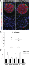
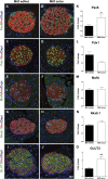
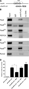
Precise regulation of β-cell function is crucial for maintaining blood glucose homeostasis. Pax6 is an essential regulator of β-cell-specific factors like insulin and Glut2. Studies in the developing eye suggest that Pax6 interacts with Mitf to regulate pigment cell differentiation. Here, we show that Mitf, like Pax6, is expressed in all pancreatic endocrine cells during mouse postnatal development and in the adult islet. A Mitf loss-of-function mutation results in improved glucose tolerance and enhanced insulin secretion but no increase in β-cell mass in adult mice. Mutant β-cells secrete more insulin in response to glucose than wild-type cells, suggesting that Mitf is involved in regulating β-cell function. In fact, the transcription of genes critical for maintaining glucose homeostasis (insulin and Glut2) and β-cell formation and function (Pax4 and Pax6) is significantly upregulated in Mitf mutant islets. The increased Pax6 expression may cause the improved β-cell function observed in Mitf mutant animals, as it activates insulin and Glut2 transcription. Chromatin immunoprecipitation analysis shows that Mitf binds to Pax4 and Pax6 regulatory regions, suggesting that Mitf represses their transcription in wild-type β-cells. We demonstrate that Mitf directly regulates Pax6 transcription and controls β-cell function.
Figures


Similar articles
-
Pax6 is crucial for β-cell function, insulin biosynthesis, and glucose-induced insulin secretion.Mol Endocrinol. 2012 Apr;26(4):696-709. doi: 10.1210/me.2011-1256. Epub 2012 Mar 8. Mol Endocrinol. 2012. PMID: 22403172 Free PMC article.
-
A regulatory loop involving PAX6, MITF, and WNT signaling controls retinal pigment epithelium development.PLoS Genet. 2012 Jul;8(7):e1002757. doi: 10.1371/journal.pgen.1002757. Epub 2012 Jul 5. PLoS Genet. 2012. PMID: 22792072 Free PMC article.
-
High-fat diet induces early-onset diabetes in heterozygous Pax6 mutant mice.Diabetes Metab Res Rev. 2014 Sep;30(6):467-75. doi: 10.1002/dmrr.2572. Diabetes Metab Res Rev. 2014. PMID: 24925705
-
Regulation of insulin synthesis and secretion and pancreatic Beta-cell dysfunction in diabetes.Curr Diabetes Rev. 2013 Jan 1;9(1):25-53. Curr Diabetes Rev. 2013. PMID: 22974359 Free PMC article. Review.
-
Glucagon gene expression in the endocrine pancreas: the role of the transcription factor Pax6 in α-cell differentiation, glucagon biosynthesis and secretion.Diabetes Obes Metab. 2011 Oct;13 Suppl 1:31-8. doi: 10.1111/j.1463-1326.2011.01445.x. Diabetes Obes Metab. 2011. PMID: 21824254 Review.
Cited by
-
Transcriptomic signatures of subcutaneous adipose tissue in patients with diabetes and coronary artery disease: a pilot study.Front Cardiovasc Med. 2025 Feb 5;12:1524605. doi: 10.3389/fcvm.2025.1524605. eCollection 2025. Front Cardiovasc Med. 2025. PMID: 39974596 Free PMC article.
-
Identifying potential pathogenesis and immune infiltration in diabetic foot ulcers using bioinformatics and in vitro analyses.BMC Med Genomics. 2023 Dec 1;16(1):313. doi: 10.1186/s12920-023-01741-2. BMC Med Genomics. 2023. PMID: 38041124 Free PMC article.
-
Pax6 influences expression patterns of genes involved in neuro- degeneration.Ann Neurosci. 2015 Oct;22(4):226-31. doi: 10.5214/ans.0972.7531.220407. Ann Neurosci. 2015. PMID: 26525840 Free PMC article.
-
MafB-dependent neurotransmitter signaling promotes β cell migration in the developing pancreas.Development. 2023 Mar 15;150(6):dev201009. doi: 10.1242/dev.201009. Epub 2023 Mar 27. Development. 2023. PMID: 36897571 Free PMC article.
-
MiRNA-144-3p inhibits high glucose induced cell proliferation through suppressing FGF16.Biosci Rep. 2019 Jul 25;39(7):BSR20181788. doi: 10.1042/BSR20181788. Print 2019 Jul 31. Biosci Rep. 2019. PMID: 31292167 Free PMC article.
References
-
- Jonsson J, Carlsson L, Edlund T, Edlund H. Insulin-promoter-factor 1 is required for pancreas development in mice. Nature 1994;371:606–609 - PubMed
-
- Ashery-Padan R, Zhou X, Marquardt T, et al. Conditional inactivation of Pax6 in the pancreas causes early onset of diabetes. Dev Biol 2004;269:479–488 - PubMed
-
- Stoffers DA, Ferrer J, Clarke WL, Habener JF. Early-onset type-II diabetes mellitus (MODY4) linked to IPF1. Nat Genet 1997;17:138–139 - PubMed
Publication types
MeSH terms
Substances
LinkOut - more resources
Full Text Sources
Other Literature Sources
Medical
Molecular Biology Databases

