. 2013 May 7;110(19):7814-9.
doi: 10.1073/pnas.1220466110. Epub 2013 Apr 22.

Rhomboid domain-containing protein 3 is a negative regulator of TLR3-triggered natural killer cell activation

Affiliations

Rhomboid domain-containing protein 3 is a negative regulator of TLR3-triggered natural killer cell activation

Juan Liu et al. Proc Natl Acad Sci U S A. .

Abstract

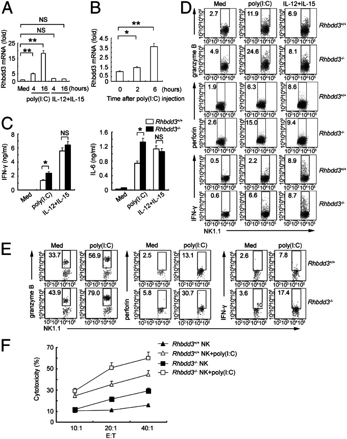
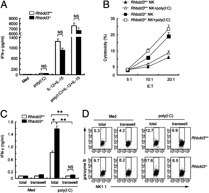
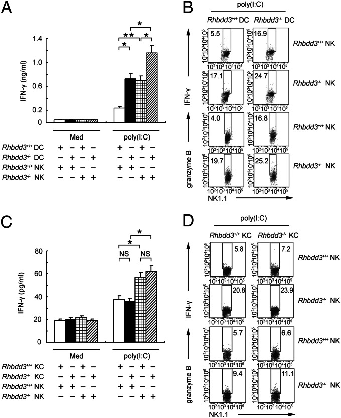
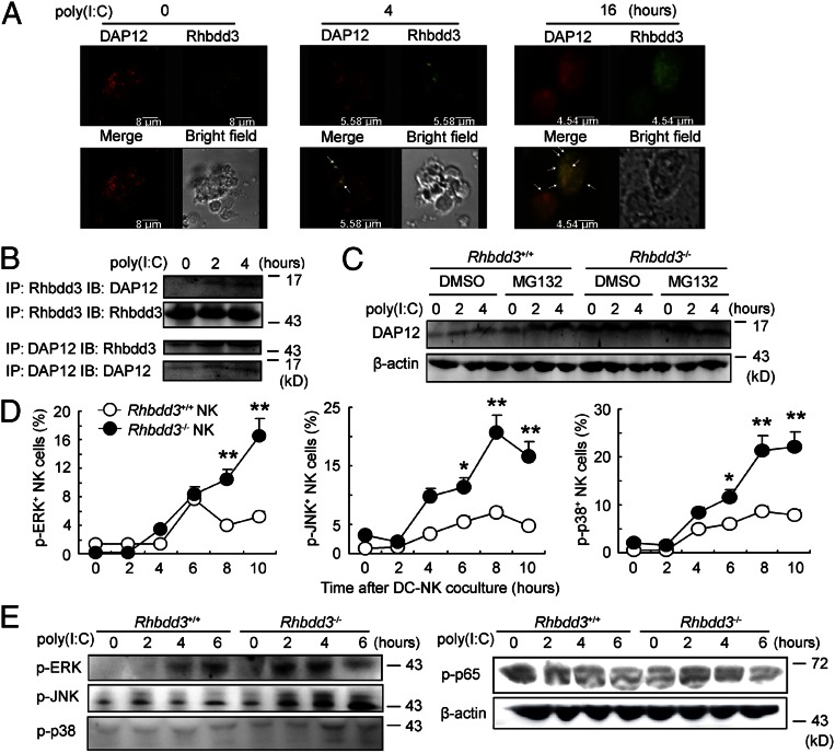
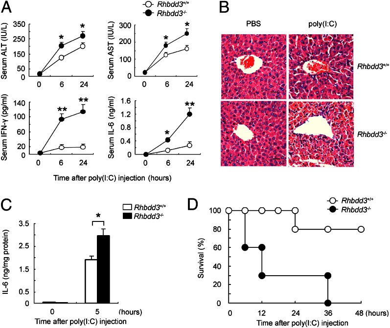
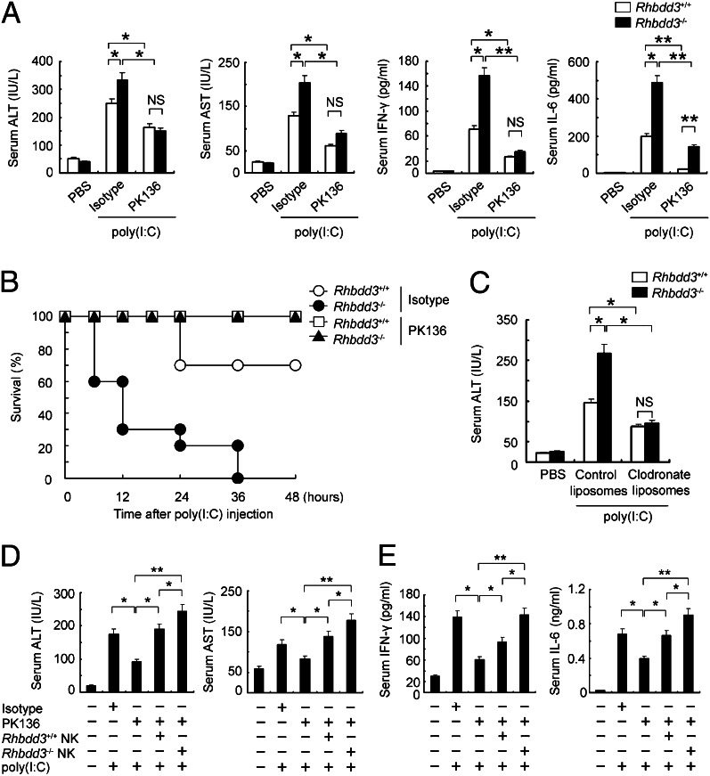
Rhomboid domain-containing protein 3 (Rhbdd3), which belongs to a family of proteins with rhomboid domain, is widely expressed in immune cells; however, the roles of the Rhbdd members, including Rhbdd3, in immunity remain unknown. Natural killer (NK) cells are critical for host immune defense and also can mediate inflammatory diseases such as hepatitis. Although much is known about how NK cells are activated, the detailed mechanisms for negative regulation of NK cell activation remain to be fully understood. Using Rhbdd3-deficient mice, we reveal that Rhbdd3, selectively up-regulated in NK cells upon Toll-like receptor 3 (TLR3) stimulation, negatively regulates TLR3-mediated NK cell activation in a feedback manner. Rhbdd3 inhibits TLR3-triggered IFN-γ and granzyme B expression of NK cells in cell-cell contact dependence of accessory cells such as dendritic cells and Kupffer cells. Rhbdd3 interacts with DNAX activation protein of 12 kDa and promotes its degradation, inhibiting MAPK activation in TLR3-triggered NK cells. Furthermore, Rhbdd3 plays a critical role in attenuating TLR3-triggered acute inflammation by controlling NK cell activation and accumulation in liver and disrupting NK cell-Kupffer cell interaction. Therefore, Rhbdd3 is a feedback inhibitor of TLR3-triggered NK cell activation. Our study outlines a mechanism for the negative regulation of NK cell activation and also provides clues for the function of the rhomboid proteins in immunity.

Keywords: immune regulation; innate immunity; poly(I:C).


Conflict of interest statement

The authors declare no conflict of interest.

Figures

Fig. 1.
Fig. 1.
Rhbdd3 negatively regulates TLR3-induced NK cell activation in vitro and in vivo. (A) Splenocytes were stimulated with poly(I:C) or IL-12/15. NK cells were purified then and assessed for Rhbdd3 mRNA expression using quantitative PCR. (B) Splenic NK cells collected from mice i.p. injected with poly(I:C) and D-GalN were assessed for Rhbdd3 mRNA expression using quantitative PCR. (C and D) Rhbdd3+/+ and Rhbdd3−/− splenocytes were stimulated with medium (Med), poly(I:C), or IL-12/15. The levels of IFN-γ and IL-6 in supernatants (C) and intracellular expression of granzyme B, perforin, and IFN-γ in NK1.1+ cells (D) were, respectively, measured by cytometric bead array (CBA) and FACS. (E and F) Rhbdd3+/+ and Rhbdd3−/− mice (six mice per group) were i.p. injected with poly(I:C) and D-GalN. After 6 h, the liver mononuclear cells were collected. The intracellular granzyme B, perforin, and IFN-γ in liver NK1.1+ cells were determined by FACS (E). Liver NK cells were further isolated and assessed for their cytotoxicity against YAC-1 cells. E:T, effector versus target (F). The data shown are the means ± SD from three independent experiments (AC and F). *P < 0.05; **P < 0.01; NS, not significant.
Fig. 2.
Fig. 2.
Accessory cells are required for the suppression of TLR3-triggered NK cell activation by Rhbdd3 in a cell–cell contact-dependent manner. (A) Rhbdd3+/+ and Rhbdd3−/− splenic NK cells were stimulated with poly(I:C), IL-12/15, or poly(I:C) plus IL-12/15, and IFN-γ levels in supernatants were assessed by CBA. (B) Rhbdd3+/+ and Rhbdd3−/− splenic NK cells were stimulated with poly(I:C) and then cultured with 5,6-carboxyfluorescein diacetate succinimidyl ester-labeled Yac-1 cells at indicated E:T ratios. The cytotoxicity of NK cells against YAC-1 cells were analyzed by FACS. (C and D) Splenic DX5+ NK cells and the left DX5 splenocytes, which were, respectively, seeded in the upper and lower compartments of the Transwell system (transwell), or total splenocytes (total) were stimulated with poly(I:C). IFN-γ levels in supernatants (C) and intracellular granzyme B in NK1.1+ cells (D) were respectively assessed by CBA and FACS. The data shown are the means ± SD (AC) from three independent experiments. *P < 0.05; **P < 0.01; NS, not significant.
Fig. 3.
Fig. 3.
DCs and KCs are required for the suppression of NK cell activation by Rhbdd3. Rhbdd3+/+ or Rhbdd3−/− splenic NK cells were cocultured with Rhbdd3+/+ or Rhbdd3−/− bone marrow-derived (BM) DCs (A and B) or liver KCs (C and D) at a ratio of 5:1 and stimulated with poly(I:C). IFN-γ levels in supernatants (A and C) and intracellular production of IFN-γ and granzyme B in NK1.1+ cells (B and D) were, respectively, assessed using CBA or FACS. The data shown are the means ± SD (A and C) from three independent experiments. *P < 0.05; **P < 0.01; NS, not significant.
Fig. 4.
Fig. 4.
Inhibition of TLR3-triggered NK cell DAP12 expression and MAPK activation by Rhbdd3. (A) Splenocytes were stimulated with poly(I:C) for the indicated time, and then NK cells were isolated. The intracellular localization of Rhbdd3 (red) and DAP12 (green) were determined by confocal analysis. (Objective, 40×; numerical aperture, 1.4.) Bar lengths are as indicated. (B) Cell lysates from poly(I:C)-activated splenic NK cells were immunoprecipitated and immunoblotted by anti-Rhbdd3 antibody or anti-DAP12 antibody as indicated. (C) Rhbdd3+/+ or Rhbdd3−/− splenic NK cells were treated with MG132 (20 μM) or DMSO (vehicle control) for 2 h and then stimulated by poly(I:C) for the indicated time. The expression of DAP12 was analyzed by Western blotting. (D) Rhbdd3+/+ or Rhbdd3−/− splenic NK cells were stimulated with poly(I:C) in the presence of BMDCs. The levels of p-ERK, p-JNK, and p-p38 in NK1.1+ NK cells were determined using a Phosflow method (BD Biosciences). (E) Splenic NK cells were, respectively, purified from Rhbdd3+/+ and Rhbdd3−/− mice i.p. treated with poly(I:C) and D-GalN. The expression of p-ERK, p-JNK, p-p38, and p-p65 were evaluated by Western blotting. The data shown are the means ± SD (D) from three independent experiments. *P < 0.05; **P < 0.01; NS, not significant.
Fig. 5.
Fig. 5.
Rhbdd3 attenuates poly(I:C)-induced acute liver inflammation. (AD) Rhbdd3+/+ and Rhbdd3−/− mice (six mice per group) were i.p. injected with poly(I:C) and D-GalN. (A) The levels of ALT, AST, IFN-γ, and IL-6 in serum were, respectively, assayed using ELISA (ALT and AST) and CBA (IFN-γ and IL-6). (B and C) After 6 h, liver tissues were collected. The pathology of liver was analyzed by microscopy. [Original magnification, 400×; bars, 10 μm (B).] IL-6 levels in liver homogenates were determined by CBA and then normalized against milligrams of total protein determined by BCA assay (C). (D) The survival rate of Rhbdd3+/+ and Rhbdd3−/− mice (10 mice per group) was monitored for 48 h. P < 0.01 by Wilcoxon test. The data shown are the means ± SD (A and C) from three independent experiments. *P < 0.05; **P < 0.01.
Fig. 6.
Fig. 6.
Inhibition of NK cell function by Rhbdd3 contributes to attenuation of poly(I:C)-mediated acute liver inflammation. (A and B) Rhbdd3+/+ and Rhbdd3−/− mice were pretreated i.p. with anti-NK1.1 mAb (PK136) or isotype Ig (Isotype), followed by poly(I:C) and D-GalN injection 36 h later. After 24 h, serum ALT, AST, IFN-γ, and IL-6 levels were determined (six mice per group) (A). The survival rates were monitored for 48 h (10 mice per group). P < 0.01 by Wilcoxon test (B). (C) Rhbdd3+/+ and Rhbdd3−/− mice (six mice per group) were pretreated i.p. with clodronate liposomes or control liposomes, followed by poly(I:C) and D-GalN injection 36 h later. After 24 h, serum ALT levels were determined by ELISA. (D and E) Splenic NK cells from Rhbdd3+/+ or Rhbdd3−/− mice were i.v. transferred into WT mice, which were predepleted with NK cells by anti-NK1.1 mAb (4 × 10⁶ cells per mouse) 36 h before. Mice were then i.p. injected with poly(I:C) and D-GalN 24 h later. After 24 h, serum ALT, AST, IFN-γ, and IL-6 levels were determined. The data shown are the means ± SD (A and CE) from three independent experiments. *P < 0.05; **P < 0.01; NS, not significant.

Similar articles

Cited by

  • Comparative Analysis of MicroRNA and mRNA Profiles of Sperm with Different Freeze Tolerance Capacities in Boar (Susscrofa) and Giant Panda (Ailuropodamelanoleuca).
    Ran MX, Zhou YM, Liang K, Wang WC, Zhang Y, Zhang M, Yang JD, Zhou GB, Wu K, Wang CD, Huang Y, Luo B, Qazi IH, Zhang HM, Zeng CJ. Ran MX, et al. Biomolecules. 2019 Sep 1;9(9):432. doi: 10.3390/biom9090432. Biomolecules. 2019. PMID: 31480517 Free PMC article.
  • Genome-wide association and transcriptome studies identify target genes and risk loci for breast cancer.
    Ferreira MA, Gamazon ER, Al-Ejeh F, Aittomäki K, Andrulis IL, Anton-Culver H, Arason A, Arndt V, Aronson KJ, Arun BK, Asseryanis E, Azzollini J, Balmaña J, Barnes DR, Barrowdale D, Beckmann MW, Behrens S, Benitez J, Bermisheva M, Białkowska K, Blomqvist C, Bogdanova NV, Bojesen SE, Bolla MK, Borg A, Brauch H, Brenner H, Broeks A, Burwinkel B, Caldés T, Caligo MA, Campa D, Campbell I, Canzian F, Carter J, Carter BD, Castelao JE, Chang-Claude J, Chanock SJ, Christiansen H, Chung WK, Claes KBM, Clarke CL; EMBRACE Collaborators; GC-HBOC Study Collaborators; GEMO Study Collaborators; Couch FJ, Cox A, Cross SS, Czene K, Daly MB, de la Hoya M, Dennis J, Devilee P, Diez O, Dörk T, Dunning AM, Dwek M, Eccles DM, Ejlertsen B, Ellberg C, Engel C, Eriksson M, Fasching PA, Fletcher O, Flyger H, Friedman E, Frost D, Gabrielson M, Gago-Dominguez M, Ganz PA, Gapstur SM, Garber J, García-Closas M, García-Sáenz JA, Gaudet MM, Giles GG, Glendon G, Godwin AK, Goldberg MS, Goldgar DE, González-Neira A, Greene MH, Gronwald J, Guénel P, Haiman CA, Hall P, Hamann U, He W, Heyworth J, Hogervorst FBL, Hollestelle A, Hoover RN, Hopper JL, Hulick PJ, Humphreys K, Imyanitov EN; ABCTB Investigators; HEBON Inve… See abstract for full author list ➔ Ferreira MA, et al. Nat Commun. 2019 Apr 15;10(1):1741. doi: 10.1038/s41467-018-08053-5. Nat Commun. 2019. PMID: 30988301 Free PMC article.
  • Cellular and molecular regulation of innate inflammatory responses.
    Liu J, Cao X. Liu J, et al. Cell Mol Immunol. 2016 Nov;13(6):711-721. doi: 10.1038/cmi.2016.58. Epub 2016 Oct 31. Cell Mol Immunol. 2016. PMID: 27818489 Free PMC article. Review.
  • Substrate binding and specificity of rhomboid intramembrane protease revealed by substrate-peptide complex structures.
    Zoll S, Stanchev S, Began J, Skerle J, Lepšík M, Peclinovská L, Majer P, Strisovsky K. Zoll S, et al. EMBO J. 2014 Oct 16;33(20):2408-21. doi: 10.15252/embj.201489367. Epub 2014 Sep 12. EMBO J. 2014. PMID: 25216680 Free PMC article.
  • Rhomboids, signalling and cell biology.
    Freeman M. Freeman M. Biochem Soc Trans. 2016 Jun 15;44(3):945-50. doi: 10.1042/BST20160035. Biochem Soc Trans. 2016. PMID: 27284064 Free PMC article. Review.

References

    1. Yokoyama WM. Mistaken notions about natural killer cells. Nat Immunol. 2008;9(5):481–485. - PubMed
    1. Waggoner SN, Cornberg M, Selin LK, Welsh RM. Natural killer cells act as rheostats modulating antiviral T cells. Nature. 2012;481(7381):394–398. - PMC - PubMed
    1. Lang PA, et al. Natural killer cell activation enhances immune pathology and promotes chronic infection by limiting CD8+ T-cell immunity. Proc Natl Acad Sci USA. 2012;109(4):1210–1215. - PMC - PubMed
    1. Baxter AG, Smyth MJ. The role of NK cells in autoimmune disease. Autoimmunity. 2002;35(1):1–14. - PubMed
    1. Mondelli MU, Varchetta S, Oliviero B. Natural killer cells in viral hepatitis: Facts and controversies. Eur J Clin Invest. 2010;40(9):851–863. - PubMed

Publication types

MeSH terms