. 2013 May 7;110(19):E1734-42.
doi: 10.1073/pnas.1300725110. Epub 2013 Apr 22.

Castor is required for Hedgehog-dependent cell-fate specification and follicle stem cell maintenance in Drosophila oogenesis

Affiliations

Castor is required for Hedgehog-dependent cell-fate specification and follicle stem cell maintenance in Drosophila oogenesis

Yu-Chiuan Chang et al. Proc Natl Acad Sci U S A. .

Abstract

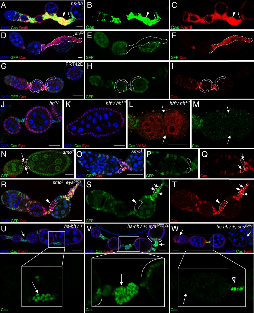
Asymmetric division of stem cells results in both self-renewal and differentiation of daughters. Understanding the molecules and mechanisms that govern differentiation of specific cell types from adult tissue stem cells is a major challenge in developmental biology and regenerative medicine. Drosophila follicle stem cells (FSCs) represent an excellent model system to study adult stem cell behavior; however, the earliest stages of follicle cell differentiation remain largely mysterious. Here we identify Castor (Cas) as a nuclear protein that is expressed in FSCs and early follicle cell precursors and then is restricted to differentiated polar and stalk cells once egg chambers form. Cas is required for FSC maintenance and polar and stalk cell fate specification. Eyes absent (Eya) is excluded from polar and stalk cells and represses their fate by inhibiting Cas expression. Hedgehog signaling is essential to repress Eya to allow Cas expression in polar and stalk cells. Finally, we show that the complementary patterns of Cas and Eya reveal the gradual differentiation of polar and stalk precursor cells at the earliest stages of their development. Our studies provide a marker for cell fates in this model and insight into the molecular and cellular mechanisms by which FSC progeny diverge into distinct fates.

Keywords: germarium; polar cell; polar stalk precursor.


Conflict of interest statement

The authors declare no conflict of interest.

Figures

Fig. 1.
Fig. 1.
Castor expression in polar, stalk, and stem cells and its requirement in FSC maintenance. (A) A schematic drawing of a Drosophila ovariole and a magnified germarium showing regions 1–3. (BK) Confocal micrographs of Cas expression in wild-type ovarioles. (B and C) 3D reconstructions show Cas (green) expressed in stalk cells (open arrows) and polar cells (arrows), which are marked by β-galactosidase (red), expressed with the polar-cell–specific driver updGAL4. (D and E) Cas (green) is expressed in the polar cells (arrow, marked by enriched FasIII staining in red), stalk cells (open arrow), and follicle precursor cells in the germarium. (FH) FSCs (one of which is indicated by an arrowhead) express Cas and are located at the border of regions 2a and 2b, anterior to the FasIII expression domain (red). (IK) Cas (red) is expressed in the most anterior cells of FSC clones, which are marked by GFP (green). In B and I, DAPI (blue) labels nuclei. In D, F, and G, VASA (blue) marks the germline cells. (L) The percentage of germaria containing follicle clones for the control (blue), casΔ1 (red), and rescue of casΔ1 (green) as a function of time after clone induction. In the control, the percentage decreases over time because of natural stem cell turnover. A greater percentage of mutant stem cells are lost. The total number of germaria analyzed is shown above each bar. Data are shown as mean ± SD. (Scale bars: 20 μm in B and C; 10 μm in DK.)
Fig. 2.
Fig. 2.
Complementary expression patterns and functions of Cas and Eya. (A and B) Confocal micrographs of ovarioles stained for Cas (green) and Eya (red). DAPI (blue) labels nuclei. Eya is expressed in main-body follicle cells of egg chambers but not in polar or stalk cells marked by Cas. (CH) Loss of Cas causes fused egg chambers. (CE) In control follicle cell clones marked by GFP, Cas (red) labels polar and stalk cells. FasIII (white) stains polar cells and immature follicle cells. (FH) Ovarioles containing casΔ1 follicle cell clones, marked by GFP, contain fused egg chambers. Polar and stalk cells are absent between three germline clusters (F), as is FasIII (G) and Cas staining (H). D, E, G, and H show higher magnifications of the boxed regions shown in C and F, respectively. (IL) Loss of Eya leads to ectopic Cas expression. Homozygous mutant clones are GFP and are outlined by dashed lines. Ovarioles are stained for Cas (red) and DAPI (blue). eya54C2-mutant clones (JL) ectopically expressed Cas in many follicle cells (arrows) and caused long, abnormal stalks (arrowheads) in 24 of 24 ovarioles that contained clones between egg chambers when analyzed 7 d ACI. Clones confined to the main body epithelium (n = 78) transformed into ectopic polar cells as previously described (24), and all such cells expressed Cas (Fig. S2 MP). Control clones appear normal (J). (MO) Normal Eya expression following overexpression of Cas with c306GAL4. (PR) Loss of polar and stalk cells and egg-chamber fusions (arrowheads) caused by ectopic expression of Eya in polar and stalk cells using c306GAL4. Germline cysts are outlined in white. (SU) Coexpression of Cas partially suppresses the fused egg chamber phenotype caused by ectopic Eya expression (see also Table 1). DAPI (blue), Cas (green), and Eya (red) staining are shown in MU. (VX) Follicle cells expressing Cas from two UAS-cas transgenes using the FLP-OUT system (GFP+, green) exhibit normal Eya expression (red). DAPI labels all nuclei in blue. Flies carrying genotype P[hsp70-flp]; UAS-cas/AyGAL4, UAS-gfp; UAS-cas, were heat-shocked at 37 °C for 1 h and then were incubated at 25 °C for 2 d before dissection. (Scale bars: 20 μm.)
Fig. 3.
Fig. 3.
Hh-mediated somatic cell proliferation and polar/stalk cell fate specification require Cas. (AF) Hyperactivity of Hh signaling ectopically induces Cas. (AC) Heat shock of Hh induced enlarged stalks that express Cas (green) and FasIII (red) (arrowhead). (DF) ptcS2-mutant clones that are GFP (marked by dashed lines) also cause massive stalks and extra Cas+ cells (red). (GI) No such phenotype was observed in control clones. (JM) Loss of Hh activity causes fused egg chambers. (J) hhts/+ control ovarioles are stained for Cas (green) and Eya (red). Each egg chamber has 16 germ cells [large DAPI-stained nuclei (blue)], six of which appear in this focal plane. (K) An hhts/hhAC fused egg chamber contains 32 germ cell nuclei, 14 of which appear in this focal plane. (L and M) Two germline clusters marked by VASA (red) are encapsulated in one hhts/hhAC fused egg chamber lacking polar or stalk cells labeled by Cas (green). (NQ) smo3-mutant (GFP) intercyst cells (outlined in white) show reduced Cas (red). (RT) smo3, eya54C2 double-mutant clones (GFP; white outline) exhibit ectopic expression of Cas (red) in the main-body follicle cells (arrowhead) and enlarged stalks (arrows). (UW) Ovarioles stained for Cas (green) to show stalk cells (arrows) and FasIII (red). (V) Enhancement of the hs-hh phenotype by mutation of one eya allele. Stalks were enlarged further compared with hs-hh alone (arrows), and extra Cas-expressing cells were present near the poles (white arcs). (W) Expression of a Cas RNAi line using hs-GAL4 suppressed the hs-hh phenotype. Stalks reverted to normal (open arrowhead) or to even smaller-than-normal (arrows) size. Boxed regions are shown at higher magnification below. (Scale bars: 20 μm.)
Fig. 4.
Fig. 4.
Follicle cell fate specification by Hh, Eya, and Cas. (AJ) The combination of Cas (green) and EYA (red) expression. FSCs and the early progeny (arrowheads in CE) express both proteins. Cas expression increases in the region between two cysts (arrows) but gradually is down-regulated in presumptive main-body follicle cells in region 3 (arc) where Eya expression (red) is up-regulated. (FJ) High magnification of germaria showing successive stages of follicle cell fate specification. VASA staining of the germline is shown in blue. Presumptive polar/stalk precursor cells at the border between two cysts are indicated by brackets, and the presumptive main-body follicle precursors are indicated by arcs. CJ are confocal micrographs. A and B are 3D reconstructions of micrographs taken from the ovariole shown in Fig. 2 A and B. (Scale bars: 20 μm.) (KM) Schematic diagram illustrating the progression of follicle precursor differentiation. Green and red represent cells expressing high levels of Cas and Eya, respectively. Yellow indicates cells expressing similar levels of Cas and Eya. Orange represents high Eya expression with a residual low level of Cas. (K) The FSC (dark green) gives rise to progeny that continue to divide and gradually differentiate into polar/stalk precursors (bracket) at the border between regions 2b and 3. Meanwhile, the presumptive main-body follicles (arcs) gradually lose Cas protein. (L) The polar/stalk precursors (bracket) separate the cysts. (M) Some of the polar/stalk precursors (bracket) differentiate into polar cells (asterisks). Polar cells secrete Unpaired to induce the neighboring precursor cells to form the stalk. Finally, after egg-chamber budding, only polar and stalk cells (green) express Cas, and Eya expression is restricted to the main-body follicle cells (red). (N) Positive and negative signals controlling the indicated stages of normal follicle cell development.

Similar articles

Cited by

References

    1. Spradling AC. Developmental genetics of oogenesis. In: Martinez-Arias B, editor. The Development of Drosophila melanogaster. Cold Spring Harbor, NY: Cold Spring Harbor Laboratory; 1993. pp. 1–70.
    1. Grammont M, Irvine KD. Fringe and Notch specify polar cell fate during Drosophila oogenesis. Development. 2001;128(12):2243–2253. - PubMed
    1. Grammont M, Irvine KD. Organizer activity of the polar cells during Drosophila oogenesis. Development. 2002;129(22):5131–5140. - PubMed
    1. Vachias C, Couderc JL, Grammont M. A two-step Notch-dependant mechanism controls the selection of the polar cell pair in Drosophila oogenesis. Development. 2010;137(16):2703–2711. - PubMed
    1. Xi R, McGregor JR, Harrison DA. A gradient of JAK pathway activity patterns the anterior-posterior axis of the follicular epithelium. Dev Cell. 2003;4(2):167–177. - PubMed

Publication types