. 2013 May 7;110(19):7586-91.
doi: 10.1073/pnas.1217796110. Epub 2013 Apr 22.

Geometric control of vascular networks to enhance engineered tissue integration and function

Affiliations

Geometric control of vascular networks to enhance engineered tissue integration and function

Jan D Baranski et al. Proc Natl Acad Sci U S A. .

Abstract

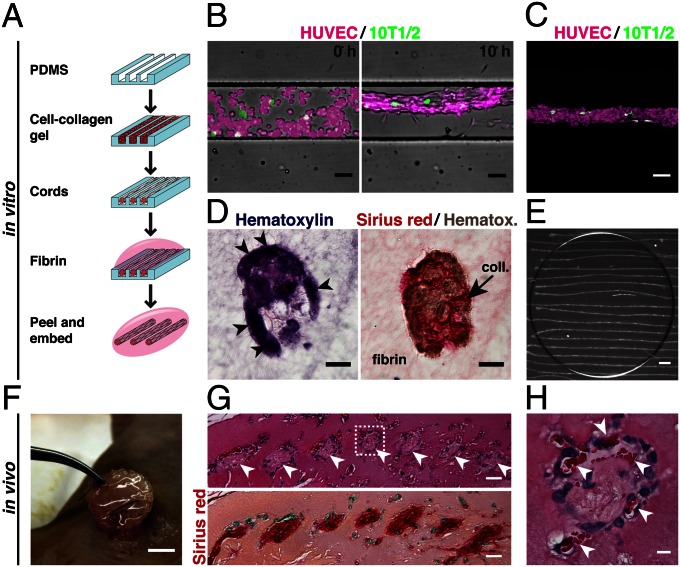
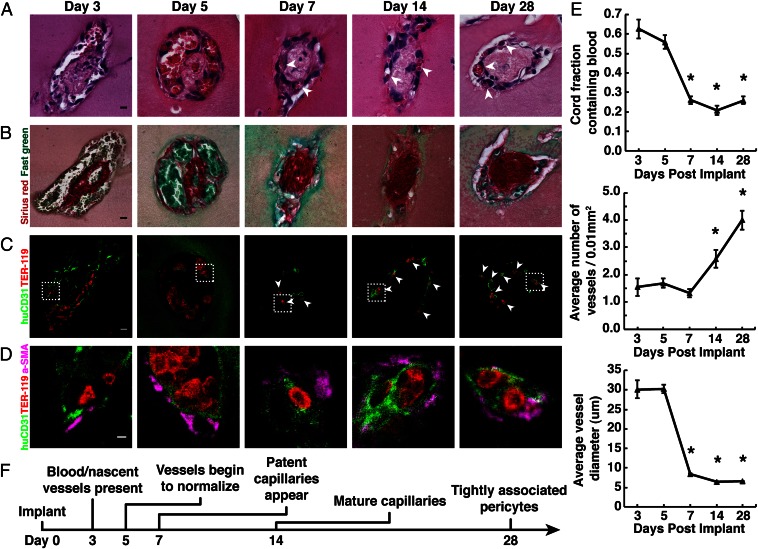
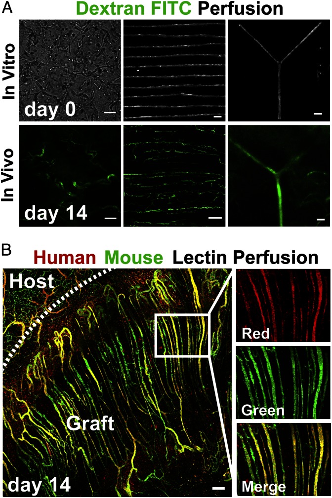
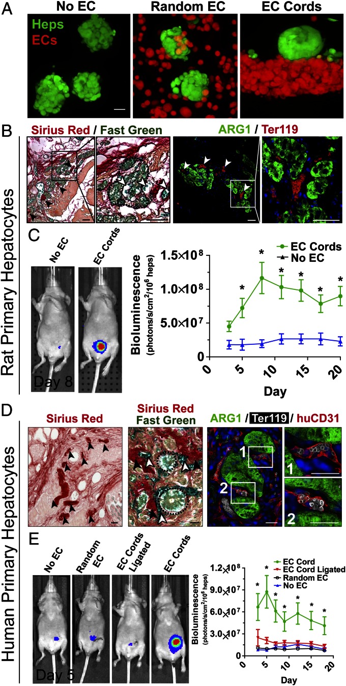
Tissue vascularization and integration with host circulation remains a key barrier to the translation of engineered tissues into clinically relevant therapies. Here, we used a microtissue molding approach to demonstrate that constructs containing highly aligned "cords" of endothelial cells triggered the formation of new capillaries along the length of the patterned cords. These vessels became perfused with host blood as early as 3 d post implantation and became progressively more mature through 28 d. Immunohistochemical analysis showed that the neovessels were composed of human and mouse endothelial cells and exhibited a mature phenotype, as indicated by the presence of alpha-smooth muscle actin-positive pericytes. Implantation of cords with a prescribed geometry demonstrated that they provided a template that defined the neovascular architecture in vivo. To explore the utility of this geometric control, we implanted primary rat and human hepatocyte constructs containing randomly organized endothelial networks vs. ordered cords. We found substantially enhanced hepatic survival and function in the constructs containing ordered cords following transplantation in mice. These findings demonstrate the importance of multicellular architecture in tissue integration and function, and our approach provides a unique strategy to engineer vascular architecture.

Keywords: angiogenesis; liver; regenerative medicine; tissue engineering; vascular biology.


Conflict of interest statement

The authors declare no conflict of interest.

Figures

Fig. 1.
Fig. 1.
Generation of EC cords. (A) Schematic representation of the process used to generate cords. Collagen, red; fibrin, pink. (B) Merged phase and fluorescence images of cord formation at 0 and 10 h within PDMS microchannels (HUVECs, calcein AM red-orange; 10T1/2s, calcein AM green; bar, 50 μm). (C) Maximum-intensity z-projection of fluorescently labeled HUVECs (magenta) and 10T1/2s (green) (bar, 50 μm). (D) H&E and Sirius red/hematoxylin staining of paraffin-embedded cord constructs showing distribution of cells and collagen in cross-section (bar, 20 μm). (E) Bright field micrograph of EC cord constructs after removal from PDMS substrate and embedding within fibrin gel. Dark circle indicates the portion of the gel that was cut with a biopsy punch in preparation for implantation (bar, 500 μm). (F) Tissue construct containing cords sutured in place adjacent to the parametrial fat pad of an athymic mouse. (G) H&E and Sirius red staining of cord-containing tissue constructs resected after 7 d in vivo. Arrowheads indicate the location of cords (bar, 25 μm). (H) Inset showing higher magnification H&E staining of single cord. Arrowheads indicate areas of blood around the periphery of the cord (bar, 5 μm).
Fig. 2.
Fig. 2.
Implanted EC cords drive formation of stable capillaries. (A) H&E staining of EC cords resected at days 3, 5, 7, 14, and 28 PI suggests the presence of blood within vessels that organize into small capillaries by day 7 (arrowheads) (bar, 10 μm). (B) Sirius red/fast-green staining of collagen within the cords after harvest (bar, 10 μm). (C) Ter-119 (red) and human-specific CD31 (green) staining positively identify RBCs and ECs and suggest that vessels were of human origin (bar, 10 μm). (D) α-SMA–positive (magenta) cells with a perivascular localization are seen at higher magnification (bar, 20 μm). (E) Quantification of blood area, vessel diameter, and vessel numbers over 28 d. *P < 0.05 for comparison of days 7, 14, and 28 vs. 3 and 5 in blood area and vessel diameter measurements, and days 14 and 28 vs. 3, 5, and 7 in vessel number measurements. Error bars: SEM, n ≥ 20, one-way ANOVA followed by Tukey’s post hoc test. (F) Timeline representation of vessel maturation.
Fig. 3.
Fig. 3.
Patterned EC cords integrate with host vasculature. (A) Bright field images of fibrin gels in vitro before implantation, which contain self-organized networks of HUVECs and 10T1/2s, patterned EC cords, or EC cords patterned into a branched topology (Upper row; bars: 250 μm, 500 μm, and 500 μm, respectively). (Upper row) These samples were implanted in the intraperitoneum of nude mice, and after 14 d FITC-dextran was perfused via tail vein injection. (Lower row) Representative FITC-dextran images (bars, 100 μm). (B) To further distinguish human from mouse endothelium, gels containing parallel arrays of cords were implanted, and mice were perfused with human-specific lectin (UEA-1–TRITC) and mouse-specific lectin (HPA–Alexa 488) via tail vein injection at 14 d PI. Representative images demonstrate that the resultant perfused microvascular network of the graft is composed of a parallel array of patent capillaries that are chimeric in composition (red, human; green, mouse; bar, 150 μm).
Fig. 4.
Fig. 4.
EC cords within engineered hepatic tissue improve function. (A) Three types of tissue constructs were generated: (i) hepatocyte aggregates only (No EC), (ii) randomly seeded HUVECs and 10T1/2s with hepatocyte aggregates (Random EC), and (iii) hepatocyte aggregates adjacent to EC cords (EC Cord; bar, 20 μm) using labeled hepatocytes (green, calcein AM) and HUVECs (calcein, red-orange). (B) Sirius red/fast-green staining (Left) revealed cellular aggregates (dotted line) close to spatially patterned collagen structures (black arrows) indicative of cords as well as to vessels that appeared to carry fast-green–stained blood (white arrows; bar, 50 μm). Immunostaining for Ter-119 (red; erythrocytes) and ARG-1 (green; hepatocytes) confirmed the presence of RBCs directly adjacent to viable hepatocyte aggregates at 20 d PI (Right bars, 25 μm). (C) Luciferase activity showed significantly increased albumin promoter activity in the tissue constructs containing patterned rat hepatocyte aggregates and EC cords at least 20 d PI. Error bars: SEM, n = 13 and 5 for EC Cords and No EC groups, respectively. *P < 0.05, one-way ANOVA followed by Tukey’s post hoc test. (D) Similar to histochemistry for rat hepatic tissues, Sirius red staining demonstrated the presence of patterned collagen remnants of cords (Left). Further addition of fast green identified capillaries containing blood (white arrows) as well as cellular aggregates (dotted line) near these collagen cores (Center). Triple immunostaining for ARG-1 (green; hepatocytes), Ter-119 (white; erythrocytes), and huCD31 (red; human endothelial cells) demonstrated localization of RBCs within capillaries that were lined with human endothelium and immediately adjacent to viable hepatocyte aggregates (Right; bars, 25 μm). (E) Representative images of luciferase activity under the control of the albumin promoter show increased primary human hepatocyte function in constructs containing patterned EC cords. Constructs containing human hepatocytes patterned with EC cords performed significantly better than all control groups for at least 18 d PI. Albumin promoter activity was similar among control groups, which contained EC cords but were ligated upon implantation (EC Cord Ligated), randomly seeded HUVECs and 10T1/2s (Random EC), or no cells (No EC). Error bars: SEM, n = 11, 7, 5, and 6 for EC Cord, No EC, EC Cord Ligated, and Random EC groups, respectively. *P < 0.05, one-way ANOVA followed by Tukey’s post hoc test.

References

    1. Vacanti JP, Langer R. Tissue engineering: The design and fabrication of living replacement devices for surgical reconstruction and transplantation. Lancet. 1999;354(Suppl 1):SI32–SI34. - PubMed
    1. Lovett M, Lee K, Edwards A, Kaplan DL. Vascularization strategies for tissue engineering. Tissue Eng Part B Rev. 2009;15(3):353–370. - PMC - PubMed
    1. Jain RK. Transport of molecules, particles, and cells in solid tumors. Annu Rev Biomed Eng. 1999;1:241–263. - PubMed
    1. Folkman J. Looking for a good endothelial address. Cancer Cell. 2002;1(2):113–115. - PubMed
    1. Radisic M, et al. High-density seeding of myocyte cells for cardiac tissue engineering. Biotechnol Bioeng. 2003;82(4):403–414. - PubMed

Publication types