. 2013 May 7;110(19):E1761-70.
doi: 10.1073/pnas.1222303110. Epub 2013 Apr 22.

Soluble IL7Rα potentiates IL-7 bioactivity and promotes autoimmunity

Affiliations

Soluble IL7Rα potentiates IL-7 bioactivity and promotes autoimmunity

Wangko Lundström et al. Proc Natl Acad Sci U S A. .

Abstract

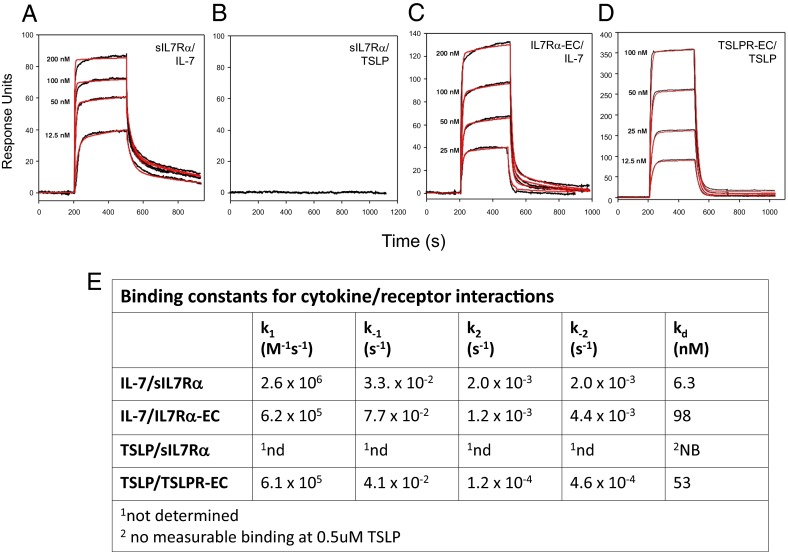
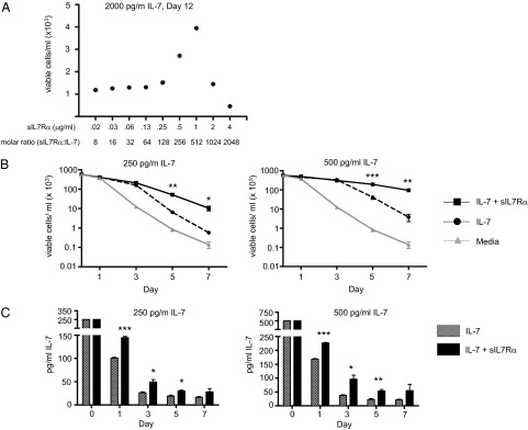
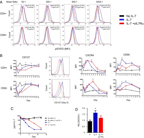
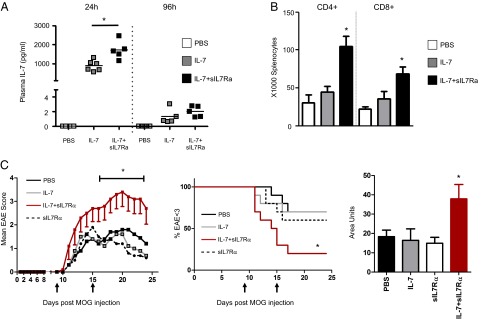
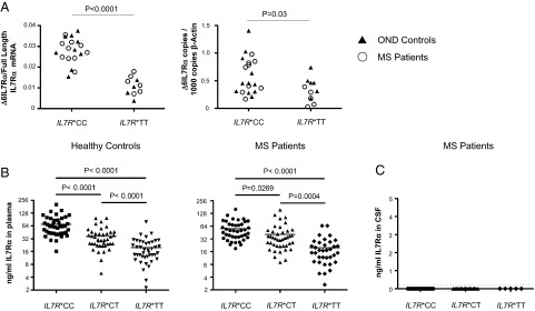
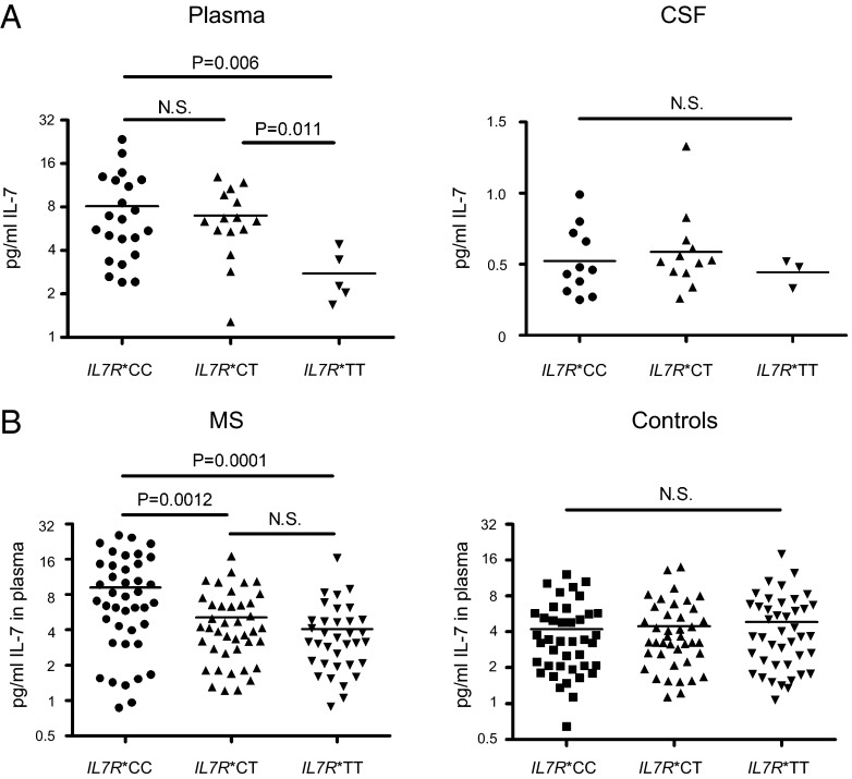
Human soluble interleukin-7 receptor (sIL7R)α circulates in high molar excess compared with IL-7, but its biology remains unclear. We demonstrate that sIL7Rα has moderate affinity for IL-7 but does not bind thymic stromal lymphopoietin. Functionally, sIL7Rα competes with cell-associated IL-7 receptor to diminish excessive IL-7 consumption and, thus, enhances the bioactivity of IL-7 when the cytokine is limited, as it is presumed to be in vivo. IL-7 signaling in the presence of sIL7Rα also diminishes expression of CD95 and suppressor of cytokine signaling 1, both regulatory molecules. Murine models confirm diminished consumption of IL-7 in the presence of sIL7Rα and also demonstrate a potentiating effect of sIL7Rα on IL-7-mediated homeostatic expansion and experimental autoimmune encephalomyelitis exacerbation. In multiple sclerosis and several other autoimmune diseases, IL7R genotype influences susceptibility. We measured increased sIL7Rα levels, as well as increased IL-7 levels, in multiple sclerosis patients with the predisposing IL7R genotype, consistent with diminished IL-7 consumption in vivo. This work demonstrates that sIL7Rα potentiates IL-7 bioactivity and provides a basis to explain the increased risk of autoimmunity observed in individuals with genotype-induced elevations of sIL7Rα.

Keywords: immunology; soluble receptors; tolerance.


Conflict of interest statement

The authors declare no conflict of interest.

Figures

Fig. 1.
Fig. 1.
sIL7Rα binds IL-7 but not TSLP. The binding kinetics for all of the cytokine/receptor interactions fit best to a two-step binding reaction model using two on-rate (k1 and k2) and off-rate (k-1 and k-2) constants. (A–D) Surface plasmon resonance sensorgrams are shown for each designated protein pair. Black lines represent raw data and red lines represent the global fitting analysis of the sensorgrams to a two-step binding reaction model using ClampXP. (E) Summary of binding affinities measured in A–D. Experiments were performed in triplicate with errors on the order of 5–10% for the rate constants.
Fig. 2.
Fig. 2.
sIL7RΑ potentiates IL-7–mediated survival and diminishes consumption of IL-7. (A) Survival of the IL-7–dependent cell line 2E8 was measured in the presence of rhIL-7 (2,000 pg/mL) plus varying concentrations of sIL7Rα. No effects were seen at any time point using molar ratios of 128:1 and below. However, on day 12, increased survival was observed at sIL7Rα:IL-7 molar ratios of 256–512:1, which was diminished at higher molar ratios. (B) Using lower concentrations of rhIL-7 [250 pg/mL (Left) and 500 pg/mL (Right)], survival of 2E8 from days 0–7 is shown with no cytokine, rhIL-7 alone, or sIL7Rα:rhIL7 molar ratio of 500:1. sIL7RΑ significantly increased survival on days 5 and 7 (*P < 0.05; **P < 0.01; ***P < 0.001). Error bars represent SEM of triplicate experiments. (C) IL-7 levels measured in the cultures described in B. Significantly increased IL-7 levels were measured in the presence of sIL7Rα on days 1, 3, and 5 of culture (*P < 0.05; **P < 0.01; ***P < 0.001). Error bars represent SEM of triplicate experiments. This experiment was performed three times with similar results.
Fig. 3.
Fig. 3.
sIL7Rα-mediated modulation of IL-7 signaling on human T cells leads to diminished IL-7 consumption and diminished SOCS1 and CD95 Induction. Human PBMCs were coincubated with rhIL-7 with or without sIL7Rα and then analyzed at the times shown. Molar ratios were as noted in A, whereas 500:1 sIL7Rα:rhIL7 molar ratio was used in BD. (A) IL-7–induced STAT-5 phosphorylation was measured in gated CD4+ and CD8+ T cells after 15 min. Cells were serum-starved before IL-7 was added. No effect was seen at 50:1 molar ratio, incomplete inhibition was seen at 500:1 ratio (similar effects at 1 and 10 ng/mL rhIL-7), and complete inhibition was seen at 5,000:1 ratio. Controls using human serum albumin and rat IgG2a at similar concentrations showed no significant impact on STAT-5 signaling. (B) Modulation of IL-7–induced changes in CD127, CXCR4, and CD95 expression by sIL7Rα. Representative flow-cytometric histograms of CD127 expression at day 5 on CD4+ and CD8+ T cells are shown on the right. (C) IL-7 consumption by human PBMCs is diminished in the presence of sIL7Rα, as measured by ELISA. (D) SOCS1 levels are reduced in the presence of sIL7Rα after 24 h of incubation with IL-7. Relative quantity (RQ) of SOCS1/GAPDH mRNA expression compared with untreated cells was determined. Error bars represent 95% CI of triplicate experiments. A total of three independent experiments were performed using PBMCs from different donors with comparable results. Statistical significance shown (*P < 0.05) reflects comparisons between rhIL-7 alone and rhIL-7 plus sIL7Rα using a two-tailed t test.
Fig. 4.
Fig. 4.
sIL7Rα diminishes IL-7 clearance in vivo and increases IL-7–mediated homeostatic peripheral expansion. (A) On day 0, IL-7−/− mice received one dose of rhIL-7 (5 μg) with or without sIL7Rα (100 μg) coinjected in the same syringe. A 10:1 molar ratio was chosen because of limited availability of recombinant sIL7Rα. Plasma levels were measured at 24 and 96 h. Mice injected with rhIL-7 plus sIL7Rα have higher plasma IL-7 levels after 24 h than those receiving rhIL-7 alone. (B) On day 0, mice received IL-7 with or without sIL7Rα, as in A above, plus 2 × 10⁶ congenic lymph node cells. On day 8, mice injected with rhIL-7 plus sIL7Rα showed higher numbers of adoptively transferred CD4+ and CD8+ splenocytes compared with mice receiving rhIL-7 alone. Asterisks denote significant differences. These experiments were repeated once for two independent experiments with similar results (n = 5 per group). (C) C57BL/6 mice (n = 10/group) were immunized with MOG and then scored for EAE symptoms. PBS, rhIL-7, sIL7Rα, or rhIL-7 plus sIL7Rα was administered i.p. on days 9 and 15 postimmunization (arrows). Mice injected with rhIL-7 plus sIL7Rα showed increased EAE scores (Left), more rapid progression to EAE score 3 (Center), and higher overall disability as measured by area under the curve of EAE score over time per individual mouse (Right). *P < 0.05 as determined by Mann–Whitney test. No significant differences were seen when injecting IL-7 with a control protein (αhIL6Rα) compared with IL-7 alone, using the models shown in A, B, or C. This experiment was carried out three times with similar results.
Fig. 5.
Fig. 5.
Individuals with the autoimmunity-associated IL7R*CC genotype have increased Δ6IL7Rα mRNA and increased levels of circulating sIL7Rα. (A) Quantitative, isoform-specific real-time PCR measured Δ6IL7Rα mRNA and full-length IL7Rα mRNA in PBMCs obtained from IL7R*CC and IL7R*TT MS patients and controls with ONDs. Each shape represents one patient’s sample. IL7R*CC individuals had increased Δ6IL7Rα/full-length mRNA ratios (Left) and increased Δ6IL7Rα mRNA levels (Right) compared with IL7R*TT MS patients and OND controls. (B) IL7R genotype modulates plasma protein levels of sIL7Rα in healthy controls (n = 41 of each genotype) and MS patients (n = 35–41 of each genotype) as measured using ELISA. The patient and control cohorts analyzed in B are distinct from those analyzed in A. (C) sIL7Rα was not detected in the CSF of MS patients, regardless of genotype. The dotted line indicates detection limit of the assay. P values were calculated using an unpaired two-tailed t test.
Fig. 6.
Fig. 6.
IL7R*CC MS patients have increased plasma IL-7 levels. (A) IL-7 levels were measured in plasma and CSF from patients with MS according to genotype. IL7R*CC patients had significantly higher mean plasma IL-7 levels than IL7R*TT patients, whereas no difference was observed between IL7R*CC and IL7R*CT heterozygotes. No correlation between CSF IL-7 levels and IL7R genotype was observed. The dotted line denotes limit of detection. (B) Plasma IL-7 levels were measured in plasma from a second independent cohort of patients with MS, as well as a separate cohort of healthy controls. IL7R*CC MS patients have significantly higher mean IL-7 levels than IL7R*TT MS patients, whereas no difference was observed according to genotype in healthy controls. Each shape represents a plasma or CSF sample analyzed from one patient or healthy control.

Similar articles

Cited by

References

    1. Tan JT, et al. IL-7 is critical for homeostatic proliferation and survival of naive T cells. Proc Natl Acad Sci USA. 2001;98(15):8732–8737. - PMC - PubMed
    1. Schluns KS, Kieper WC, Jameson SC, Lefrançois L. Interleukin-7 mediates the homeostasis of naïve and memory CD8 T cells in vivo. Nat Immunol. 2000;1(5):426–432. - PubMed
    1. Park JH, et al. Suppression of IL7Ralpha transcription by IL-7 and other prosurvival cytokines: A novel mechanism for maximizing IL-7-dependent T cell survival. Immunity. 2004;21(2):289–302. - PubMed
    1. Goldrath AW, Bevan MJ. Low-affinity ligands for the TCR drive proliferation of mature CD8+ T cells in lymphopenic hosts. Immunity. 1999;11(2):183–190. - PMC - PubMed
    1. Ernst B, Lee DS, Chang JM, Sprent J, Surh CD. The peptide ligands mediating positive selection in the thymus control T cell survival and homeostatic proliferation in the periphery. Immunity. 1999;11(2):173–181. - PubMed

Publication types

MeSH terms