. 2013 May 1;73(9):2718-36.
doi: 10.1158/0008-5472.CAN-12-4213. Epub 2013 Apr 22.

Animal models of human prostate cancer: the consensus report of the New York meeting of the Mouse Models of Human Cancers Consortium Prostate Pathology Committee

Affiliations

Animal models of human prostate cancer: the consensus report of the New York meeting of the Mouse Models of Human Cancers Consortium Prostate Pathology Committee

Michael Ittmann et al. Cancer Res. .

Abstract

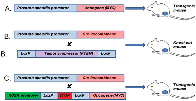
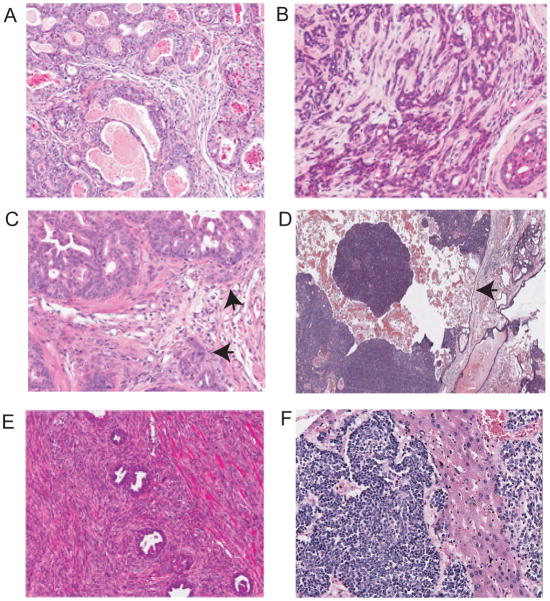
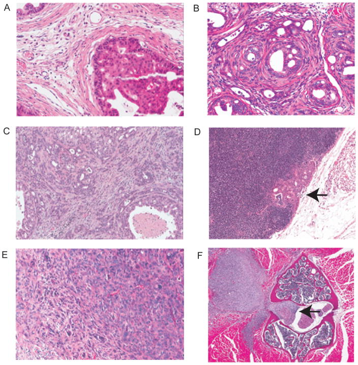
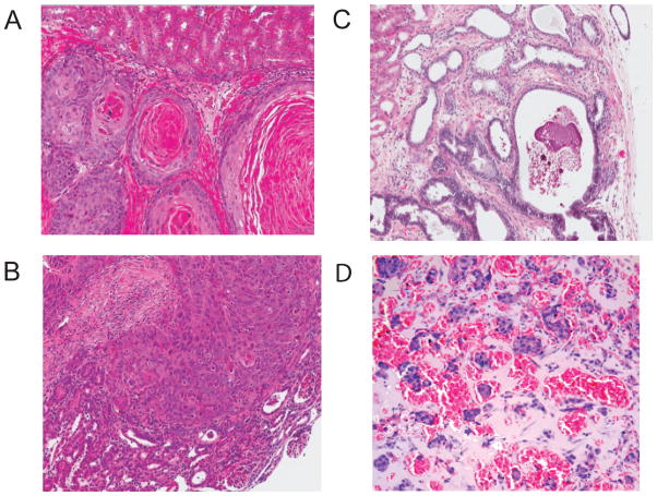
Animal models, particularly mouse models, play a central role in the study of the etiology, prevention, and treatment of human prostate cancer. While tissue culture models are extremely useful in understanding the biology of prostate cancer, they cannot recapitulate the complex cellular interactions within the tumor microenvironment that play a key role in cancer initiation and progression. The National Cancer Institute (NCI) Mouse Models of Human Cancers Consortium convened a group of human and veterinary pathologists to review the current animal models of prostate cancer and make recommendations about the pathologic analysis of these models. More than 40 different models with 439 samples were reviewed, including genetically engineered mouse models, xenograft, rat, and canine models. Numerous relevant models have been developed over the past 15 years, and each approach has strengths and weaknesses. Analysis of multiple genetically engineered models has shown that reactive stroma formation is present in all the models developing invasive carcinomas. In addition, numerous models with multiple genetic alterations display aggressive phenotypes characterized by sarcomatoid carcinomas and metastases, which is presumably a histologic manifestation of epithelial-mesenchymal transition. The significant progress in development of improved models of prostate cancer has already accelerated our understanding of the complex biology of prostate cancer and promises to enhance development of new approaches to prevention, detection, and treatment of this common malignancy.


Figures

Figure 1
Figure 1. Methods for generation of genetically engineered mouse models of prostate cancer
A. A prostate specific promoter (such as the enhanced ARR2Pb probasin promoter) is used to drive prostate specific expression of a gene of interest, typically an oncogene. The oncogene is expressed in the prostate epithelial cells at the onset of sexual maturity. B. Mice are generated with loxP sites flanking critical exons in a gene of interest, typically a tumor suppressor gene. These mice are then crossed with mice expressing the Cre recombinase under control of the probasin or other prostate specific promoter resulting in excision of key exons and inactivation of the targeted gene in prostatic epithelial cells bearing the targeted sequence in one or both alleles of the gene of interest. C. Mice are generated with the gene of interest downstream of a strong constitutive promoter such as ROSA26. Transcription/translation is inhibited by a lox-stop-lox cassette upstream of the gene of interest. These mice can be crossed to probasin Cre mice leading to excision of the stop sequence and expression of the gene of interest. This method has the advantage that expression of the gene of interest is no longer dependent on androgen receptor and other prostate specific factors whose activity may be altered by tumor progression or treatment.
Figure 2
Figure 2. Prostatic intraepithelial neoplasia (PIN) in GEM mouse models
A. Low grade mPIN in 73 week old wild type mouse. Arrow shows focal epithelial proliferation with mild atypia. B. Low grade mPIN in Pten null model (K5 promoter) 12 week old mouse C. High grade mPIN in Pten null model (K14 promoter). Note central necrosis and proliferation of atypical cells filling the duct but with well demarcated border (medium power). Chronic inflammation is noted (arrow). D. High grade mPIN in Pten null model. Arrow shows nucleus with striking pleomorphic atypia.
Figure 3
Figure 3. Prostate malignancies in GEM of prostate cancer
A. Invasive adenocarcinoma, Hi-myc model. B. Invasive adenocarcinoma, Pten null X Sox9 overexpression model. C. Microinvasive adenocarcinoma. Pten null Smad4 null model 10 weeks. Focal microinvasion (arrows). D. Intracystic carcinoma; APC model. Masses of poorly differentiated adenocarcinoma within a cystic space (arrow indicates wall of cystic space). E. Sarcomatoid carcinoma, Pten null p53 null model. Masses of atypical proliferating spindle cells entrapping residual glands with PIN. F. Neuroendocrine carcinoma, TRAMP model, metastatic to liver
Figure 4
Figure 4. Pathology in Pten deletion models
A. HG mPIN in Pten/p53 null model. Elsewhere in this same tumor there were areas of invasive adenocarcinoma and sarcomatoid carcinoma. B. Invasive adenocarcinoma Pten/p53 null; C. Invasive adenocarcinoma Pten/Smad4 null model. The invasive adenocarcinomas both show obvious reactive stroma. D. Metastatic adenocarcinoma in lumbosacral lymph node. Pten/p53/Smad4 null model. E. Pten/p53 null telomerase reactivation model. Sarcomatoid carcinoma in the prostate. Area on left shows very poorly differentiated carcinoma with a pleomorphic spindle cell pattern on the right. F. Sarcomatoid carcinoma invading vertebral bone (Pten/p53 null telomerase model)
Figure 5
Figure 5. Pathology of genetically engineered mouse models
A. Metastasis to femur in LADY X Hepsin transgenic mouse. Sheets of epithelial cells within bone are shown. B. Sheets of poorly differentiated adenocarcimoma with focal squamous areas. AR Osr1 model. C. Adenocarcinoma in ubi-CAT mouse D, HG mPIN in mouse with loss of stromal TGF-β signaling.
Figure 6
Figure 6. Representative microscopic images of patient-derived prostate carcinoma xenografts
A. Tissue sections of a poorly differentiated PCa xenograft stained with hematoxylin and eosin (H&E) or immunostained for AR or PSA. Upper panels show invasive growth into the host kidney parenchyma. Lower panels show intravascular tumor emboli in the host lungs. B. Sub-renal capsule xenograft derived from a poorly differentiated neuroendocrine carcinoma of the prostate. Tissue sections stained with H&E or immunostained for AR, PSA, or synaptophysin (SYN) are shown.
Figure 7
Figure 7. Tissue recombination models of prostate cancer
A–C. Representative microscopic images of H&E stained sections of tissue recombinants generated using BPH-1 cells with rUGM (A), or CAFs (B). C. Representative microscopic image of H&E stained sections of tissue recombinants generated using NHPrE1 cells and rUGM. D. DRS model. Nests of tumors cells surrounded by blood vessels and stroma with eosinophilic material derived from Matrigel.
Figure 8
Figure 8. Non-murine models of prostate cancer
A. Transgenic rat expressing SV40 large-T antigen. Adenocarcinoma with associated chronic inflammation. B. Transgenic rat expressing SV40 large-T antigen. Neuroendocrine carcinoma infiltrating around focal area of hyperplasia (arrow). C. Poorly differentiated adenocarcinoma of canine prostate with chronic inflammatory infiltrate.

References

    1. Taylor BS, Schultz N, Hieronymus H, Gopalan A, Xiao Y, Carver BS, et al. Integrative genomic profiling of human prostate cancer. Cancer Cell. 2010;18:11–22. - PMC - PubMed
    1. Bavik C, Coleman I, Dean JP, Knudsen B, Plymate S, Nelson PS. The gene expression program of prostate fibroblast senescence modulates neoplastic epithelial cell 1proliferation through paracrine mechanisms. Cancer Res. 2006;66:794–802. - PubMed
    1. Bianchi-Frias D, Vakar-Lopez F, Coleman IM, Plymate SR, Reed MJ, Nelson PS. The effects of aging on the molecular and cellular composition of the prostate microenvironment. PLoS One. 2010;5 - PMC - PubMed
    1. Zong Y, Xin L, Goldstein AS, Lawson DA, Teitell MA, Witte ON. ETS family transcription factors collaborate with alternative signaling pathways to induce carcinoma from adult murine prostate cells. Proc Natl Acad Sci U S A. 2009;106:12465–12470. - PMC - PubMed
    1. Memarzadeh S, Xin L, Mulholland DJ, Mansukhani A, Wu H, Teitell MA, et al. Enhanced paracrine FGF10 expression promotes formation of multifocal prostate adenocarcinoma and an increase in epithelial androgen receptor. Cancer Cell. 2007;12:572–585. - PMC - PubMed

Publication types