. 2013 Jan 11;3(1):12-29.
doi: 10.1159/000346180. Print 2013 Jan.

Development of a chronic kidney disease model in C57BL/6 mice with relevance to human pathology

Affiliations

Development of a chronic kidney disease model in C57BL/6 mice with relevance to human pathology

Linghong Huang et al. Nephron Extra. .

Abstract

Background: Genetically modified mice are used to investigate disease and assess potential interventions. However, research into kidney fibrosis is hampered by a lack of models of chronic kidney disease (CKD) in mice. Recently, aristolochic acid nephropathy (AAN), characterised by severe tubulointerstitial fibrosis, has been identified as a cause of end stage kidney disease and proposed as a model of CKD. Published studies have used various dosing regimens, species and strains, with variable outcomes. Therefore, we aimed to develop a standardised protocol to develop tubulointerstitial fibrosis using pure aristolochic acid I (AAI) in C57BL/6 mice.

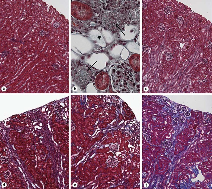
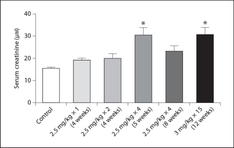
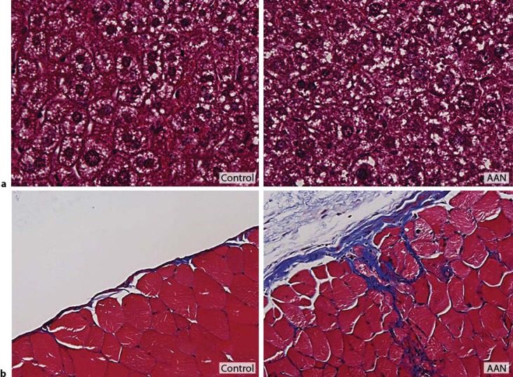
Methods: AAI dose optimisation was performed by intraperitoneal injection of AAI at varying dose, frequency and duration. Kidney function was assessed by serum creatinine. Fibrosis was quantified by hydroxyproline levels and Masson's Trichrome staining. Specific collagens were measured by immunofluorescent staining.

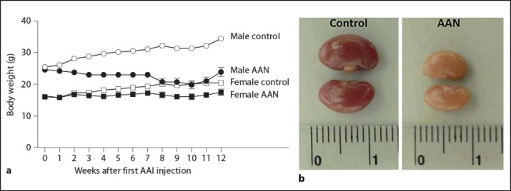
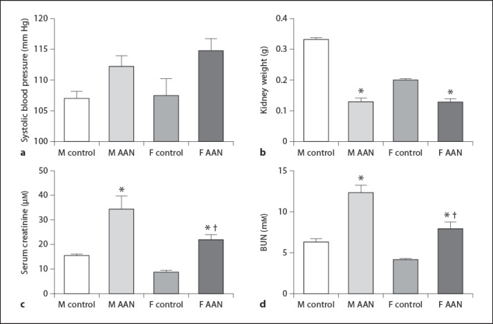
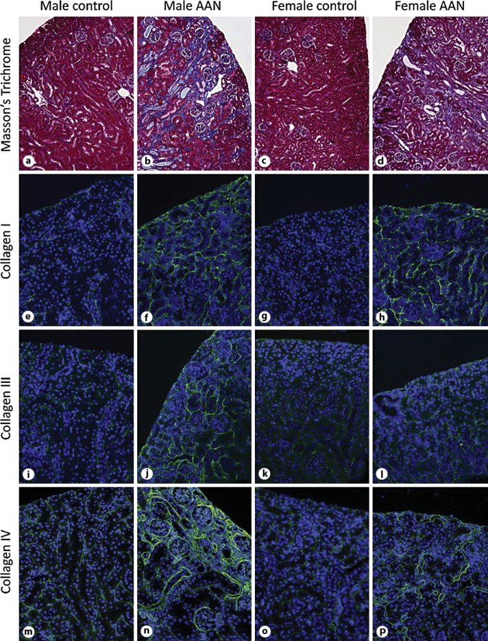
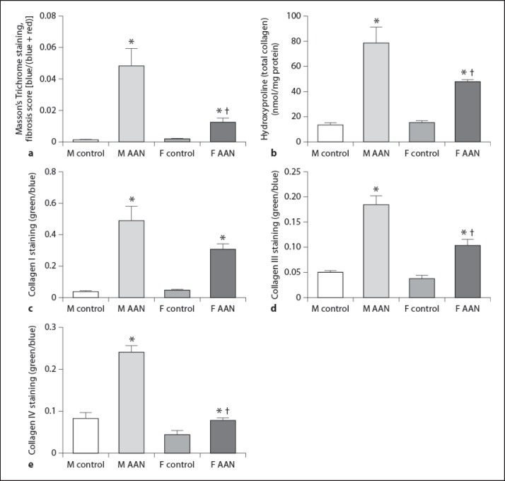
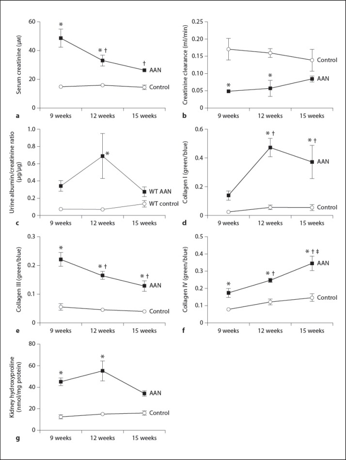
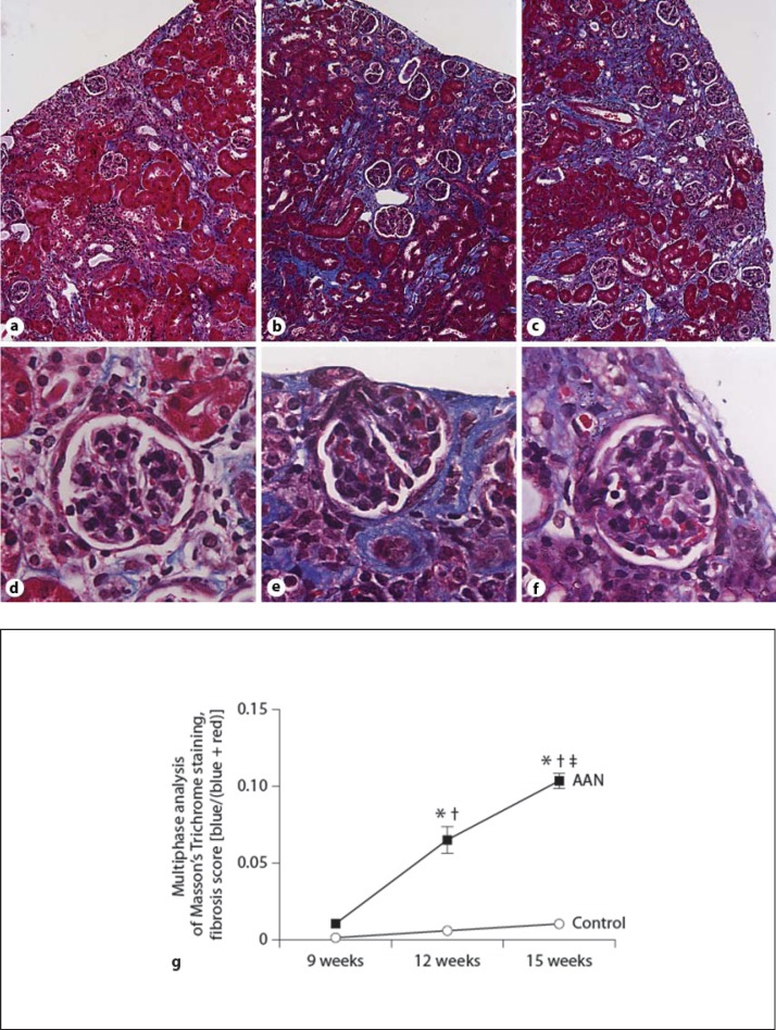
Results: Single doses of AAI of >10 mg/kg caused acute kidney failure and death. Lower doses of 2.5 mg/kg needed to be administrated more than weekly to cause significant fibrosis. 3 mg/kg once every 3 days for 6 weeks followed by a disease development time of 6 weeks after AAI led to reduced kidney weight and function. Substantial tubulointerstitial fibrosis occurred, with males more severely affected. Increased deposition of collagen I, III and IV contributed to fibrosis, with collagen III and IV higher in males.

Conclusions: AAN can be induced in C57BL/6 mice. The regimen of 3 mg/kg every 3 days for 6 weeks followed by 6 weeks of disease development time gives substantial tubulointerstitial fibrosis with lesions similar to those in humans.

Keywords: Aristolochic acid nephropathy; C57BL/6 mice; Chronic kidney disease; Extracellular matrix; Fibrosis.


Figures

Fig. 1
Fig. 1
Masson's Trichrome staining of kidneys from male C57BL/6 mice treated with AAI at varying dose and frequency. a Control. b 10 mg/kg single dose (×400 magnification). c 2.5 mg/kg once every 2 weeks for 4 weeks. d 2.5 mg/kg/week for 4 weeks with 1 week of disease development time. e 2.5 mg/kg/week for 4 weeks with 4 weeks of disease development time. f 3 mg/kg once every 3 days for 6 weeks with 6 weeks of disease development time. Magnification is ×100 unless stated.
Fig. 2
Fig. 2
Kidney function in male C57BL/6 mice treated with AAI at varying dose and frequency. Kidney function was assessed by measuring serum creatinine concentrations (μM) in mice treated with different doses of AAI (the x-axis labels show AAI concentration used per injection with number of injections given and overall duration in brackets). Data represent mean ± SEM; n = 3–5 per group. * Indicates statistical significance compared to the control group.
Fig. 3
Fig. 3
Body weight and kidney size in control and AAN mice. Mice were treated with 3 mg/kg of AAI for 6 weeks and then maintained for further 6 weeks. Mouse body weight was measured at least once a week throughout the 12 weeks (a). Kidneys were removed at termination and directly compared. Male kidneys are shown (b). Data represent mean ± SEM; n = 5 per group.
Fig. 4
Fig. 4
Systolic blood pressure, kidney weight and function differences in male and female mice. Male (M) and female (F) mice were treated with 3 mg/kg of AAI for 6 weeks and then maintained for a further 6 weeks. Systolic blood pressure measurement was performed using a BP-2000 blood pressure analysis system (a). Kidneys were removed at termination and weighed (b). Kidney function was measured by serum creatinine (c) and BUN (d). Data represent mean ± SEM; n = 5 per group. * Indicates statistical significance compared to the control group. Indicates statistical significance compared to male AAN mice.
Fig. 5
Fig. 5
Masson's Trichrome (×100) and immunofluorescent staining of collagens (×200). Male (left hand columns) and female (right hand columns) mice were treated with 3 mg/kg of AAI for 6 weeks and then maintained for a further 6 weeks. Paraffin sections were stained with Masson's Trichrome (a–d), collagen I (e–h), collagen III (i–l) and collagen IV (m–p). Nuclei were stained with DAPI.
Fig. 6
Fig. 6
Comparison of fibrosis levels and collagen I, III and IV immunostaining between male and female C57BL/6 mice. Male (M) and female (F) mice were treated with 3 mg/kg of AAI for 6 weeks and then maintained for a further 6 weeks. Masson's Trichrome staining (a) and immunofluorescence for collagen I (c), collagen III (d) and collagen IV (e) shown in figure 5 were quantified by multiphase image analysis of 10 fields per section. Kidney hydroxyproline in the same mice was expressed as nmol per mg protein (b). Data represent mean ± SEM; n = 5 per group. * Indicates statistical significance compared to the control group. Indicates statistical significance compared to male AAN mice.
Fig. 7
Fig. 7
Immunofluorescent staining of infiltrating inflammatory cells and fibroblasts in the AAN kidney. 4 μm thick sections from 12-week-old male AAN mice were stained with CD45 (A), S100A4 in red and α-SMA in green (B), and F4/80 (C). Nuclei were stained with DAPI. Boxed area on ×200 image is shown at ×400 magnification in the right column.
Fig. 8
Fig. 8
Comparison of kidney function, levels of collagen I, III and IV immunostaining and hydroxyproline in male mice with progression of AAN. Kidney function in the mice described in figure 7 was assessed using serum creatinine (a), creatinine clearance (b) and urine albumin/creatinine ratio (c). Quantification of immunofluorescence for collagen I (d), collagen III (e) and collagen IV (f) in the kidney were assessed by multiphase image analysis of 10 fields per section. Levels of hydroxyproline were expressed as nmol per mg protein (g). Data represent mean ± SEM; n = 5–10 per group. * Indicates statistical significance compared to the control group. and indicate changes from 9 and 12 weeks AAN groups, respectively.
Fig. 9
Fig. 9
Masson's Trichrome staining in male mice with the progression of AAN. Male mice were treated with either AAI for 3 weeks followed by 6 weeks remodelling time (9 weeks), AAI for 6 weeks followed by 6 weeks remodelling time (12 weeks) or AAI for 6 weeks followed by 9 weeks remodelling time (15 weeks). The development of kidney fibrosis is shown by Masson's Trichrome-stained slides (a–f). Quantification of Masson's Trichrome staining (g) was assessed by multiphase image analysis of 10 fields per section. Data represent mean ± SEM; n = 5–10 per group. * Indicates statistical significance compared to the control group. and indicate changes from 9 and 12 weeks AAN groups, respectively.
Fig. 10
Fig. 10
Masson's Trichrome staining of liver (a) and peritoneum (b). 4 μm thick sections from 12-week-old male mice treated with AA1 were stained with Masson's Trichrome (×400 magnification).

References

    1. Cole J, Quach DL, Sundaram K, Corvol P, Capecchi MR, Bernstein KE. Mice lacking endothelial angiotensin-converting enzyme have a normal blood pressure. Circ Res. 2002;90:87–92. - PubMed
    1. Pacholczyk G, Suhag R, Mazurek M, Dederscheck SM, Koni PA. Generation of C57BL/6 knockout mice using C3H × BALB/c blastocysts. Biotechniques. 2008;44:413–416. - PubMed
    1. Seong E, Saunders TL, Stewart CL, Burmeister M. To knockout in 129 or in C57BL/6: that is the question. Trends Genet. 2004;20:59–62. - PubMed
    1. Eddy AA, Lopez-Guisa JM, Okamura DM, Yamaguchi I. Investigating mechanisms of chronic kidney disease in mouse models. Pediatr Nephrol. 2012;27:1233–1247. - PMC - PubMed
    1. Vielhauer V, Anders HJ, Mack M, Cihak J, Strutz F, Stangassinger M, Luckow B, Grone HJ, Schlondorff D. Obstructive nephropathy in the mouse: progressive fibrosis correlates with tubulointerstitial chemokine expression and accumulation of CC chemokine receptor 2- and 5-positive leukocytes. J Am Soc Nephrol. 2001;12:1173–1187. - PubMed

LinkOut - more resources