

Time-dependent gene expression and immunohistochemical analysis of the injured anterior cruciate ligament
- PMID: 23610654
- PMCID: PMC3626253
- DOI: 10.1302/2046-3758.110.2000118
Time-dependent gene expression and immunohistochemical analysis of the injured anterior cruciate ligament
Abstract
Objectives: This study aimed to investigate time-dependent gene expression of injured human anterior cruciate ligament (ACL), and to evaluate the histological changes of the ACL remnant in terms of cellular characterisation.
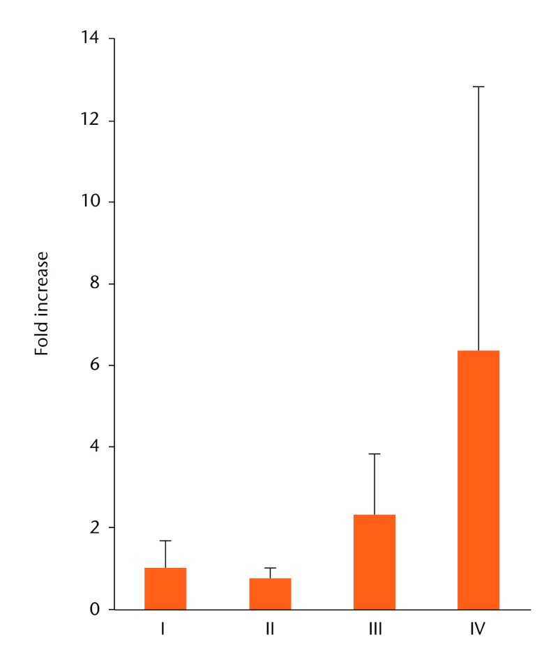
Methods: Injured human ACL tissues were harvested from 105 patients undergoing primary ACL reconstruction and divided into four phases based on the period from injury to surgery. Phase I was < three weeks, phase II was three to eight weeks, phase III was eight to 20 weeks, and phase IV was ≥ 21 weeks. Gene expressions of these tissues were analysed in each phase by quantitative real-time polymerase chain reaction using selected markers (collagen types 1 and 3, biglycan, decorin, α-smooth muscle actin, IL-6, TGF-β1, MMP-1, MMP-2 and TIMP-1). Immunohistochemical staining was also performed using primary antibodies against CD68, CD55, Stat3 and phosphorylated-Stat3 (P-Stat3).
Results: Expression of IL-6 was mainly seen in phases I, II and III, collagen type 1 in phase II, MMP-1, 2 in phase III, and decorin, TGF-β1 and α-smooth muscle actin in phase IV. Histologically, degradation and scar formation were seen in the ACL remnant after phase III. The numbers of CD55 and P-Stat3 positive cells were elevated from phase II to phase III.
Conclusions: Elevated cell numbers including P-Stat3 positive cells were not related to collagens but to MMPs' expressions.
Keywords: ACL; Anterior cruciate ligament; Gene expression; Immunohistochemical analysis; Matrix metalloproteinases (MMPs); Stat3; Synovial fibroblast-like cell.
Conflict of interest statement
Figures


Similar articles
-
Histological changes in the human anterior cruciate ligament after rupture.J Bone Joint Surg Am. 2000 Oct;82(10):1387-97. doi: 10.2106/00004623-200010000-00004. J Bone Joint Surg Am. 2000. PMID: 11057466
-
The effect of platelet-rich plasma on patterns of gene expression in a dog model of anterior cruciate ligament reconstruction.J Surg Res. 2013 Mar;180(1):80-8. doi: 10.1016/j.jss.2012.10.036. Epub 2012 Nov 8. J Surg Res. 2013. PMID: 23174707
-
Patterns of gene expression in a rabbit partial anterior cruciate ligament transection model: the potential role of mechanical forces.Am J Sports Med. 2010 Feb;38(2):348-56. doi: 10.1177/0363546509348052. Epub 2009 Dec 4. Am J Sports Med. 2010. PMID: 19966107
-
TGF-beta1 induces the different expressions of lysyl oxidases and matrix metalloproteinases in anterior cruciate ligament and medial collateral ligament fibroblasts after mechanical injury.J Biomech. 2013 Mar 15;46(5):890-8. doi: 10.1016/j.jbiomech.2012.12.019. Epub 2013 Jan 26. J Biomech. 2013. PMID: 23357697 Clinical Trial.
-
Passive anterior tibia translation in anterior cruciate ligament-injured, anterior cruciate ligament-reconstructed and healthy knees: a systematic review.Musculoskelet Surg. 2019 Aug;103(2):121-130. doi: 10.1007/s12306-018-0572-6. Epub 2018 Oct 16. Musculoskelet Surg. 2019. PMID: 30328030 Free PMC article.
Cited by
-
Platelet-rich plasma pretreatment protects anterior cruciate ligament fibroblasts correlated with PI3K-Akt-mTOR pathway under hypoxia condition.J Orthop Translat. 2022 Mar 10;34:102-112. doi: 10.1016/j.jot.2022.02.002. eCollection 2022 May. J Orthop Translat. 2022. PMID: 35891713 Free PMC article.
-
Therapeutic potential of vascular stem cells for anterior cruciate ligament reconstruction.Ann Transl Med. 2019 Dec;7(Suppl 8):S286. doi: 10.21037/atm.2019.11.79. Ann Transl Med. 2019. PMID: 32016005 Free PMC article. No abstract available.
-
Bio-enhanced repair of the anterior cruciate ligament.Arthroscopy. 2015 May;31(5):990-7. doi: 10.1016/j.arthro.2014.11.016. Epub 2015 Jan 14. Arthroscopy. 2015. PMID: 25595694 Free PMC article.
-
Migrating Myofibroblastic Iliotibial Band-Derived Fibroblasts Represent a Promising Cell Source for Ligament Reconstruction.Int J Mol Sci. 2019 Apr 22;20(8):1972. doi: 10.3390/ijms20081972. Int J Mol Sci. 2019. PMID: 31013670 Free PMC article.
-
Comparison of cellular activity and gene expression in repaired versus reconstructed anterior cruciate ligaments: A case-control study.J Exp Orthop. 2025 Jul 21;12(3):e70364. doi: 10.1002/jeo2.70364. eCollection 2025 Jul. J Exp Orthop. 2025. PMID: 40693219 Free PMC article.
References
-
- O’Donoghue DH, Frank GR, Jeter GL, et al. Repair and reconstruction of the anterior cruciate ligament in dogs: factors influencing long-term results. J Bone Joint Surg [Am] 1971;53-A:710–718 - PubMed
-
- Cabaud HE, Rodkey WG, Feagin JA. Experimental studies of acute anterior cruciate ligament injury and repair. Am J Sports Med 1979;7:18–22 - PubMed
-
- Woo SL, Vogrin TM, Abramowitch SD. Healing and repair of ligament injuries in the knee. J Am Acad Orthop Surg 2000;8:364–372 - PubMed
-
- Murray MM, Martin SD, Martin TL, Spector M. Histological changes in the human anterior cruciate ligament after rupture. J Bone Joint Surg [Am] 2000;82-A:1387–1397 - PubMed
-
- Murray MM, Spindler KP, Devin C, et al. Use of a collagen-platelet rich plasma scaffold to stimulate healing of a central defect in the canine ACL. J Orthop Res 2006;24:820–830 - PubMed
LinkOut - more resources
Full Text Sources
Research Materials
Miscellaneous
