

Crystal structure of the capsular polysaccharide synthesizing protein CapE of Staphylococcus aureus
- PMID: 23611437
- PMCID: PMC3699295
- DOI: 10.1042/BSR20130017
Crystal structure of the capsular polysaccharide synthesizing protein CapE of Staphylococcus aureus
Abstract
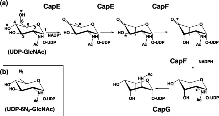
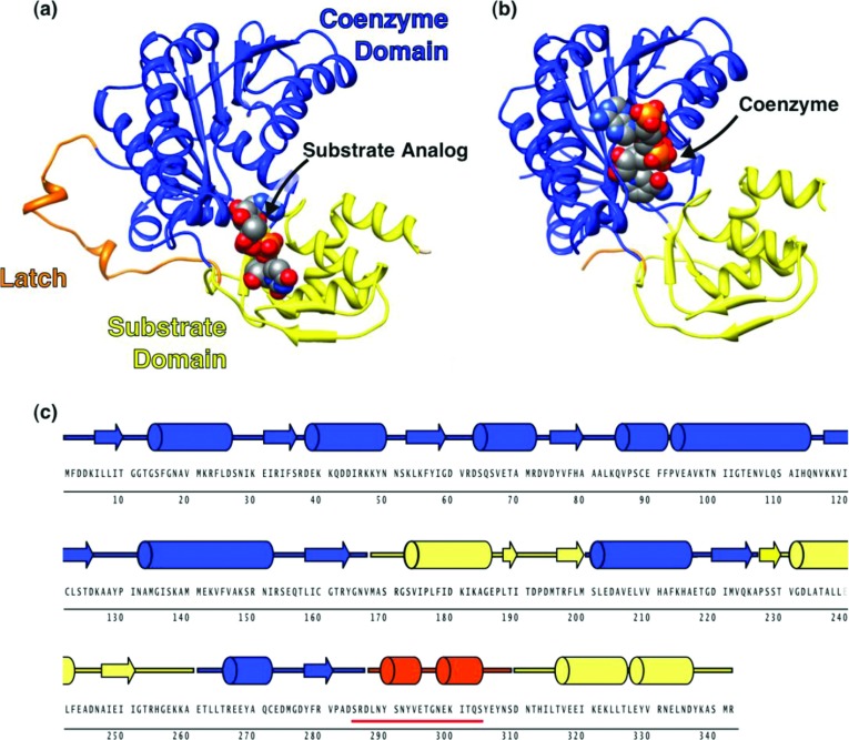
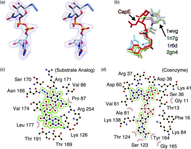
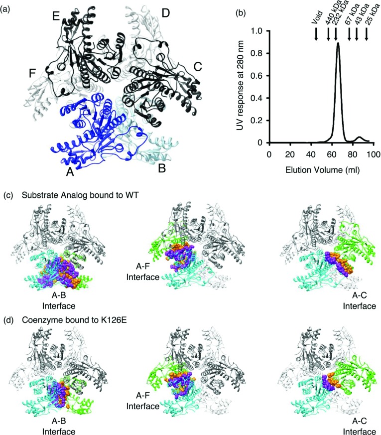
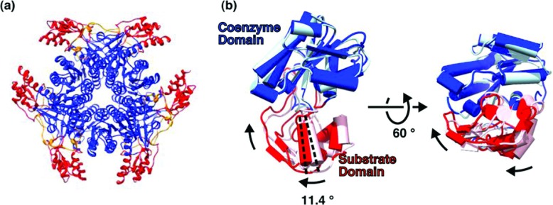
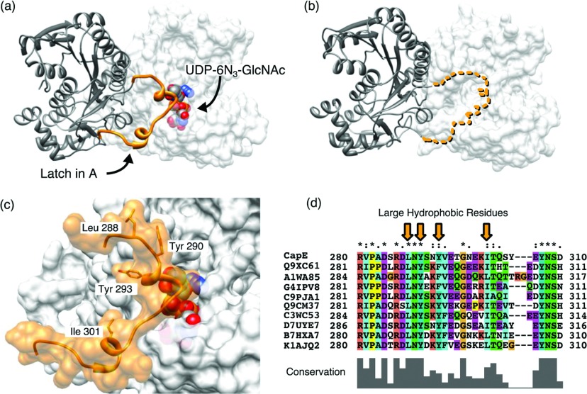
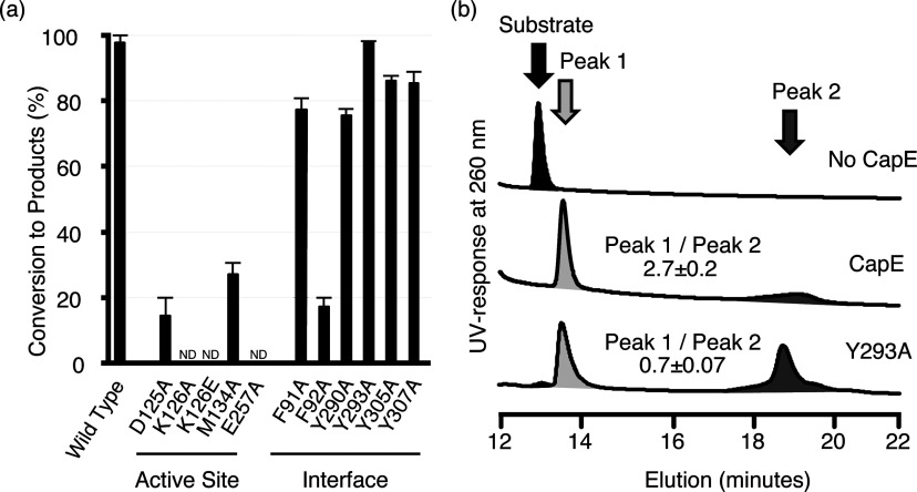
Enzymes synthesizing the bacterial CP (capsular polysaccharide) are attractive antimicrobial targets. However, we lack critical information about the structure and mechanism of many of them. In an effort to reduce that gap, we have determined three different crystal structures of the enzyme CapE of the human pathogen Staphylococcus aureus. The structure reveals that CapE is a member of the SDR (short-chain dehydrogenase/reductase) super-family of proteins. CapE assembles in a hexameric complex stabilized by three major contact surfaces between protein subunits. Turnover of substrate and/or coenzyme induces major conformational changes at the contact interface between protein subunits, and a displacement of the substrate-binding domain with respect to the Rossmann domain. A novel dynamic element that we called the latch is essential for remodelling of the protein-protein interface. Structural and primary sequence alignment identifies a group of SDR proteins involved in polysaccharide synthesis that share the two salient features of CapE: the mobile loop (latch) and a distinctive catalytic site (MxxxK). The relevance of these structural elements was evaluated by site-directed mutagenesis.
Figures
Similar articles
-
Dynamic elements govern the catalytic activity of CapE, a capsular polysaccharide-synthesizing enzyme from Staphylococcus aureus.FEBS Lett. 2013 Nov 29;587(23):3824-30. doi: 10.1016/j.febslet.2013.10.009. Epub 2013 Oct 21. FEBS Lett. 2013. PMID: 24157361
-
Structure-function analysis of heterodimer formation, oligomerization, and receptor binding of the Staphylococcus aureus bi-component toxin LukGH.J Biol Chem. 2015 Jan 2;290(1):142-56. doi: 10.1074/jbc.M114.598110. Epub 2014 Nov 3. J Biol Chem. 2015. PMID: 25371205 Free PMC article.
-
The structure of SAV1646 from Staphylococcus aureus belonging to a new `ribosome-associated' subfamily of bacterial proteins.Acta Crystallogr D Biol Crystallogr. 2015 Feb;71(Pt 2):332-7. doi: 10.1107/S1399004714025619. Epub 2015 Jan 23. Acta Crystallogr D Biol Crystallogr. 2015. PMID: 25664743
-
Function and regulation of Staphylococcus aureus wall teichoic acids and capsular polysaccharides.Int J Med Microbiol. 2019 Sep;309(6):151333. doi: 10.1016/j.ijmm.2019.151333. Epub 2019 Jul 18. Int J Med Microbiol. 2019. PMID: 31362856 Review.
-
The SarA protein family of Staphylococcus aureus.Int J Biochem Cell Biol. 2008;40(3):355-61. doi: 10.1016/j.biocel.2007.10.032. Epub 2007 Nov 13. Int J Biochem Cell Biol. 2008. PMID: 18083623 Free PMC article. Review.
Cited by
-
The Biosynthesis of UDP-D-QuiNAc in Bacillus cereus ATCC 14579.PLoS One. 2015 Jul 24;10(7):e0133790. doi: 10.1371/journal.pone.0133790. eCollection 2015. PLoS One. 2015. PMID: 26207987 Free PMC article.
-
The sps Genes Encode an Original Legionaminic Acid Pathway Required for Crust Assembly in Bacillus subtilis.mBio. 2020 Aug 18;11(4):e01153-20. doi: 10.1128/mBio.01153-20. mBio. 2020. PMID: 32817102 Free PMC article.
-
Coordination of capsule assembly and cell wall biosynthesis in Staphylococcus aureus.Nat Commun. 2019 Mar 29;10(1):1404. doi: 10.1038/s41467-019-09356-x. Nat Commun. 2019. PMID: 30926919 Free PMC article.
-
Discovery and characterization of natural tropolones as inhibitors of the antibacterial target CapF from Staphylococcus aureus.Sci Rep. 2015 Oct 16;5:15337. doi: 10.1038/srep15337. Sci Rep. 2015. PMID: 26471247 Free PMC article.
-
Biochemical analysis of a sugar 4,6-dehydratase from Acanthamoeba polyphaga Mimivirus.Protein Sci. 2020 May;29(5):1148-1159. doi: 10.1002/pro.3843. Epub 2020 Mar 4. Protein Sci. 2020. PMID: 32083779 Free PMC article.
References
-
- Lowy F. D. Medical progress: Staphylococcus aureus infections. N. Engl. J. Med. 1998;339:520–532. - PubMed
-
- Lowy F. D. How Staphylococcus aureus adapts to its host. N. Engl. J. Med. 2011;364:1987–1990. - PubMed
-
- Smith T. L., Pearson M. L., Wilcox K. R., Cruz C., Lancaster M. V., Robinson-Dunn B., Tenover F. C., Zervos M. J., Band J. D., et al. Emergence of vancomycin resistance in Staphylococcus aureus. N. Engl. J. Med. 1999;340:493–501. - PubMed
-
- Rybak M. J., Akins R. L. Emergence of methicillin-resistant Staphylococcus aureus with intermediate glycopeptide resistance–clinical significance and treatment options. Drugs. 2001;61:1–7. - PubMed
-
- Payne D. J., Gwynn M. N., Holmes D. J., Pompliano D. L. Drugs for bad bugs: confronting the challenges of antibacterial discovery. Nat. Rev. Drug Discovery. 2007;6:29–40. - PubMed
Publication types
MeSH terms
Substances
LinkOut - more resources
Full Text Sources
Other Literature Sources
Miscellaneous