Comparative Study
. 2013 Jun;19(6):789-802.
doi: 10.1261/rna.037523.112. Epub 2013 Apr 23.

Escherichia coli rimM and yjeQ null strains accumulate immature 30S subunits of similar structure and protein complement

Affiliations
Comparative Study

Escherichia coli rimM and yjeQ null strains accumulate immature 30S subunits of similar structure and protein complement

Vivian Leong et al. RNA. 2013 Jun.

Abstract

Assembly of the Escherichia coli 30S ribosomal subunits proceeds through multiple parallel pathways. The protein factors RimM, YjeQ, RbfA, and Era work in conjunction to assist at the late stages of the maturation process of the small subunit. However, it is unclear how the functional interplay between these factors occurs in the context of multiple parallel pathways. To understand how these factors work together, we have characterized the immature 30S subunits that accumulate in ΔrimM cells and compared them with immature 30S subunits from a ΔyjeQ strain. The cryo-EM maps obtained from these particles showed that the densities representing helices 44 and 45 in the rRNA were partially missing, suggesting mobility of these motifs. These 30S subunits were also partially depleted in all tertiary ribosomal proteins, particularly those binding in the head domain. Using image classification, we identified four subpopulations of ΔrimM immature 30S subunits differing in the amount of missing density for helices 44 and 45, as well as the amount of density existing in these maps for the underrepresented proteins. The structural defects found in these immature subunits resembled those of the 30S subunits that accumulate in the ΔyjeQ strain. These findings are consistent with an "early convergency model" in which multiple parallel assembly pathways of the 30S subunit converge into a late assembly intermediate, as opposed to the mature state. Functionally related factors will bind to this intermediate to catalyze the last steps of maturation leading to the mature 30S subunit.

Keywords: 30S subunit; RimM protein; YjeQ protein; cryo-electron microscopy; ribosome assembly.


Figures
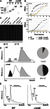
FIGURE 1.
FIGURE 1.
Cold-sensitive phenotype and ribosome profiling of the ΔrimM E. coli strain. (A) PCR screening of the ΔrimM strain. The image shows the obtained PCR products loaded in a 1% agarose gel. The wild-type parental E. coli BW25113 strain shows a defined 400-bp product amplified by RimM primers that anneal in a sequence internal to the intact rimM gene (lane 2). Using the set of primers annealing sequences upstream and downstream, the rimM gene produced a product of 600 bp corresponding to the complete 549-bp rimM gene plus 50 bp from the flanking sequences (lane 3). In contrast, the ΔrimM strain has a well-defined band of ∼1.2 kb amplified by primers annealing in sequences P1 and P2 flanking the kanamycin resistance cassette introduced during gene deletion (lane 4). This set of primers only produced various unspecific bands of different sizes with the parental strain (lane 1). There was no amplification with RimM primers internal to the rimM gene (lane 5); however, using the set of primers annealing rimM flanking sequences showed a large band of slightly >1.2 kb corresponding to the successfully inserted kanamycin resistance cassette flanked by these sequences (lane 6). (B) Growth profiles of the parental and ΔrimM strain in LB liquid media. The growth profile of the ΔrimM strain is shown for the untreated strain as well as after being transformed with an empty high-copy plasmid pCA24N (labeled as p-empty) or having reintroduced the rimM gene in this vector (labeled as p-rimM). (C) Dilution plating experiment of saturated cultures of ΔrimM and parental strains. The ΔrimM strain complemented with the pCA24N empty vector or encoding the rimM gene are also shown. Cultures were diluted in 10-fold increments, spotted on LB agar plates, and incubated at the indicated temperature. (D) Ribosomes from the parental (top panel) and ΔrimM strain were fractionated on 10%–30% sucrose gradients, providing the sedimentation profiles shown in the figure. Peaks corresponding to the ribosomal subunits and 70S ribosomes are labeled. Peak area for the 30S subunit (shaded in black) was measured with respect to the area under the 70S peak (shaded in gray) to calculate the percentage of free 30S subunit in both strains (pie charts in the right panels). (E) The effects of the rimM deletion are shown in the polysome profiles. Ribosome particles are labeled above the corresponding peaks.
FIGURE 2.
FIGURE 2.
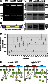
Ribosomal RNA and protein complement of immature 30S subunits accumulated in the ΔrimM strain. (A) Total rRNA purified from ΔrimM and ΔyjeQ cells resolved in a polyacrylamide gel and stained with ethidium bromide. The 23S, 16S, and 17S rRNA bands are indicated. (B) Diagram of the 17S rRNA molecule showing the 5′ and 3′ precursor sequences. The stars indicate the annealing sites for the oligonucleotide probes used in the Northern blot analysis. PS stands for precursor sequence. (C) The identity of small ribosomal rRNA species in the wild-type, ΔrimM, and ΔyjeQ strains from the Keio collection was revealed by Northern blot hybridization using 3′ end DIG-dUTP-labeled oligonucleotide probes visualized using a chemiluminescent reaction. The 16S and 17S rRNA bands are labeled. The asterisk in A and C indicates a lower molecular band corresponding to a degradation product or incorrectly processed 16S rRNA (Jomaa et al. 2011a). (D) iTRAQ analysis of the 30S subunits purified from the ΔrimM strain under associating conditions. The relative level for each r-protein with respect to wild-type parental cells is expressed as the ratio ΔrimM/WT. The ratio for each protein is calculated as the average of the ratios obtained for all the peptides assigned to each r-protein. Error bars represent the standard deviations of the average for each ratio. (E) The ΔrimM/WT average ratios were plotted in the Nomura assembly maps (lower panels) and shown along with a similar analysis performed for the free 30S subunits purified from the ΔyjeQ strain under identical conditions (data taken from Jomaa et al. 2011a). The proportion of the box colored in yellow is proportional to the degree of underrepresentation of each r-protein in the 30S subunits purified from ΔrimM and ΔyjeQ cells. The groups of primary (1°), secondary (2°), and tertiary (3°) proteins are indicated.
FIGURE 3.
FIGURE 3.
Conformational subpopulations of immature 30S subunits purified from ΔrimM cells. Front (top panel) and back (bottom panel) views of the cryo-EM maps representing four conformational subpopulations (labeled CL 1 to CL 4) of immature 30S subunits purified from ΔrimM cells. Particle classification was obtained using a maximum likelihood-based classification approach. Main domains of the 30S subunit are labeled in the structure for subpopulation class 4. The Fourier shell correlation plots estimating the resolution of these maps are shown in Supplemental Figure S4.
FIGURE 4.
FIGURE 4.
Structural defects in the 3′ minor domain in the immature 30S subunits purified from ΔrimM cells. (A) Cryo-EM maps of the mature 30S subunit and immature 30S subunit purified from ΔyjeQ cells. The X-ray structure of the 16S rRNA (PDB ID 2AVY) was fitted into the cryo-EM maps, and helices 44 and 45 are shown as a ribbon representation. The remaining of the 16S rRNA and r-proteins from the X-ray structure are not shown for clarity. Main landmarks of the 30S subunit are indicated in the left panel. (B) Front and platform view of the cryo-EM map of subpopulation four (CL 4) of the immature 30S subunit purified from ΔrimM cells. The X-ray structure of the mature 30S subunit was docked into the map, and helices 44 and 45 are shown.
FIGURE 5.
FIGURE 5.
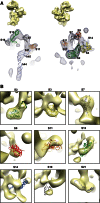
Structural defects in the head domain in the immature 30S subunits purified from ΔrimM cells. (A) Front and back views of the difference map obtained by subtracting the cryo-EM map of the immature 30S subunits (CL 4) from ΔrimM cells from that of the mature 30S subunit (EMDB ID 1775). Densities in the different maps are represented as a mesh, and the X-ray structure of the mature 30S subunit (PDB ID 2AVY) is shown docked into the difference map. The cryo-EM maps above each view are an aid for orientation, and they represent the cryo-EM map of the immature 30S subunit in the same view as the difference map below. The difference map showed densities overlapping with helices 44 and 45 of the 16 rRNA from the docked X-ray structure of the 30S subunit, indicating that these structural motifs do not have a correspondent density in the cryo-EM maps of the immature 30S subunits. The densities in the head domain of the difference map colocalized with the r-proteins from the structure of the mature 30S subunit that iTRAQ analysis identified as underrepresented in the immature 30S subunits from ΔrimM cells. (B) Images in this panel provide a detailed view of the docking of underrepresented r-proteins from the X-ray structure of the mature 30S subunit into the densities of the difference map. A higher proportion of a r-protein being enclosed in the density of the difference map indicates a larger amount of the corresponding density for that r-protein missing in the cryo-EM map of the immature 30S subunit. The densities in the difference map are shown as a semitransparent surface. The cryo-EM map for subpopulation 4 of immature 30S subunits was used to produce the difference map displayed in this panel. A similar difference map analysis performed with the cryo-EM maps for the other three subpopulations is shown in Supplemental Figures S5, S6, and S7.
FIGURE 6.
FIGURE 6.
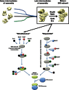
Early convergence model for the assembly of the 30S subunit. The “early convergency model” presented in this diagram (top panel) suggests that multiple parallel assembly pathways converge into a late assembly intermediate. A group of functionally related assembly factors (RimM, RbfA, YjeQ, and Era) will target this intermediate and catalyze the latest steps of maturation. Binding of these factors to the assembly intermediate will occur following a defined hierarchy. The bottom panel illustrates the two alternative models for the mechanism of action of RimM, RbfA, YjeQ, and Era. In the “simultaneous” model, all these assembly factors bind to the same intermediate to catalyze together the last maturation steps. In an alternative model, each factor performs its function independently and is released before the next factor binds to the maturing 30S subunit. A “hybrid” model is also possible (not displayed) where some factors may function sequentially, and others may act simultaneously. The question mark beside the name of each factor indicates that the order of binding is unknown.

Similar articles

Cited by

References

    1. Adilakshmi T, Bellur DL, Woodson SA 2008. Concurrent nucleation of 16S folding and induced fit in 30S ribosome assembly. Nature 455: 1268–1272 - PMC - PubMed
    1. Anantharaman V, Aravind L 2002. The PRC-barrel: A widespread, conserved domain shared by photosynthetic reaction center subunits and proteins of RNA metabolism. Genome Biol 3: RESEARCH0061. - PMC - PubMed
    1. Baba T, Ara T, Hasegawa M, Takai Y, Okumura Y, Baba M, Datsenko KA, Tomita M, Wanner BL, Mori H 2006. Construction of Escherichia coli K-12 in-frame, single-gene knockout mutants: The Keio collection. Mol Syst Biol 2: 2006.0008 10.1038/msb4100050 - DOI - PMC - PubMed
    1. Britton RA 2009. Role of GTPases in bacterial ribosome assembly. Annu Rev Microbiol 63: 155–176 - PubMed
    1. Brow DA, Noller HF 1983. Protection of ribosomal RNA from kethoxal in polyribosomes. Implication of specific sites in ribosome function. J Mol Biol 163: 27–46 - PubMed

Publication types

MeSH terms

LinkOut - more resources