

Generation of CD34+ cells from human embryonic stem cells using a clinically applicable methodology and engraftment in the fetal sheep model
- PMID: 23612043
- PMCID: PMC3729638
- DOI: 10.1016/j.exphem.2013.04.003
Generation of CD34+ cells from human embryonic stem cells using a clinically applicable methodology and engraftment in the fetal sheep model
Abstract
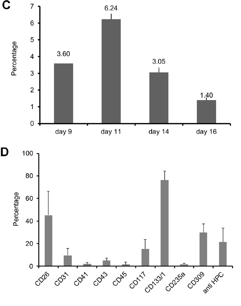
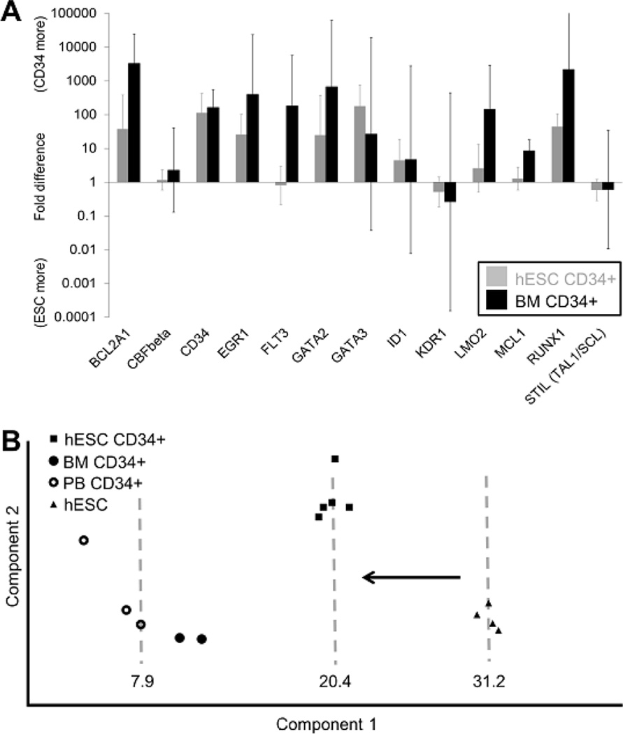
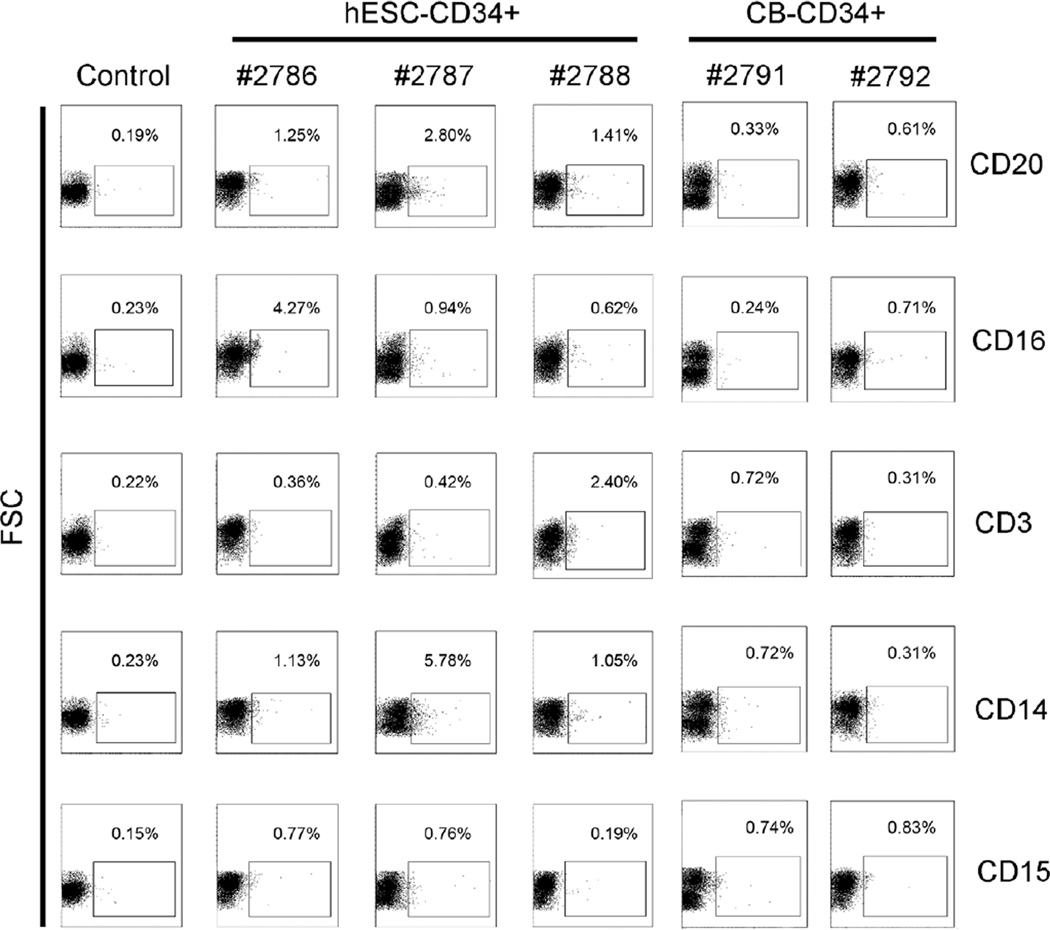
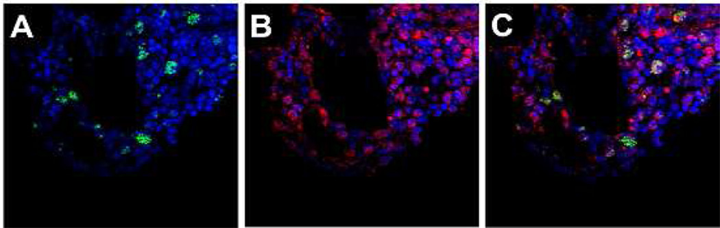
Until now, ex vivo generation of CD34(+) hematopoietic stem cells (HSCs) from human embryonic stem cells (hESCs) mostly involved use of feeder cells of nonhuman origin. Although they provided invaluable models to study hematopoiesis, in vivo engraftment of hESC-derived HSCs remains a challenging task. In this study, we used a novel coculture system composed of human bone marrow-derived mesenchymal stromal/stem cells (MSCs) and peripheral blood CD14(+) monocyte-derived macrophages to generate CD34(+) cells from hESCs in vitro. Human ESC-derived CD34(+) cells generated using this method expressed surface makers associated with adult human HSCs and upregulated hematopoietic stem cell genes comparable to human bone marrow-derived CD34(+) cells. Finally, transplantation of purified hESC-derived CD34(+) cells into the preimmune fetal sheep, primed with transplantation of MSCs derived from the same hESC line, demonstrated multilineage hematopoietic activity with graft presence up to 16 weeks after transplantation. This in vivo demonstration of engraftment and robust multilineage hematopoietic activity by hESC-derived CD34(+) cells lends credence to the translational value and potential clinical utility of this novel differentiation and transplantation protocol.
Copyright © 2013 ISEH - Society for Hematology and Stem Cells. Published by Elsevier Inc. All rights reserved.
Conflict of interest statement
Figures
Similar articles
-
Sustained, retransplantable, multilineage engraftment of highly purified adult human bone marrow stem cells in vivo.Blood. 1996 Dec 1;88(11):4102-9. Blood. 1996. PMID: 8943843
-
Functional assessment of hematopoietic niche cells derived from human embryonic stem cells.Stem Cells Dev. 2014 Jun 15;23(12):1355-63. doi: 10.1089/scd.2013.0497. Epub 2014 Mar 11. Stem Cells Dev. 2014. PMID: 24517837 Free PMC article.
-
Simultaneous generation of CD34+ primitive hematopoietic cells and CD73+ mesenchymal stem cells from human embryonic stem cells cocultured with murine OP9 stromal cells.Exp Hematol. 2007 Jan;35(1):146-54. doi: 10.1016/j.exphem.2006.09.003. Exp Hematol. 2007. PMID: 17198883
-
In vivo generation of beta-cell-like cells from CD34(+) cells differentiated from human embryonic stem cells.Exp Hematol. 2010 Jun;38(6):516-525.e4. doi: 10.1016/j.exphem.2010.03.002. Epub 2010 Mar 12. Exp Hematol. 2010. PMID: 20227460 Free PMC article.
-
Human CD34- hematopoietic stem cells: basic features and clinical relevance.Int J Hematol. 2002 May;75(4):370-5. doi: 10.1007/BF02982126. Int J Hematol. 2002. PMID: 12041666 Review.
Cited by
-
Development of Severe Combined Immunodeficient (SCID) Pig Models for Translational Cancer Modeling: Future Insights on How Humanized SCID Pigs Can Improve Preclinical Cancer Research.Front Oncol. 2018 Nov 30;8:559. doi: 10.3389/fonc.2018.00559. eCollection 2018. Front Oncol. 2018. PMID: 30560086 Free PMC article.
-
De Novo Generation of Human Hematopoietic Stem Cells from Pluripotent Stem Cells for Cellular Therapy.Cells. 2023 Jan 14;12(2):321. doi: 10.3390/cells12020321. Cells. 2023. PMID: 36672255 Free PMC article. Review.
-
Influence of a dual-injection regimen, plerixafor and CXCR4 on in utero hematopoietic stem cell transplantation and engraftment with use of the sheep model.Cytotherapy. 2014 Sep;16(9):1280-93. doi: 10.1016/j.jcyt.2014.05.025. Cytotherapy. 2014. PMID: 25108653 Free PMC article.
-
Experimental approaches to derive CD34+ progenitors from human and nonhuman primate embryonic stem cells.Am J Stem Cells. 2015 Mar 15;4(1):32-7. eCollection 2015. Am J Stem Cells. 2015. PMID: 25973329 Free PMC article. Review.
-
Fetal articular cartilage regeneration versus adult fibrocartilaginous repair: secretome proteomics unravels molecular mechanisms in an ovine model.Dis Model Mech. 2018 Jul 6;11(7):dmm033092. doi: 10.1242/dmm.033092. Dis Model Mech. 2018. PMID: 29991479 Free PMC article.
References
Publication types
MeSH terms
Substances
Grants and funding
LinkOut - more resources
Full Text Sources
Other Literature Sources
Research Materials
