Functional integrity of the T-tubular system in cardiomyocytes depends on p21-activated kinase 1
- PMID: 23612118
- PMCID: PMC3679655
- DOI: 10.1016/j.yjmcc.2013.04.014
Functional integrity of the T-tubular system in cardiomyocytes depends on p21-activated kinase 1
Abstract
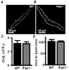
p21-activated kinase (Pak1), a serine-threonine protein kinase, regulates cytoskeletal dynamics and cell motility. Recent experiments further demonstrate that loss of Pak1 results in exaggerated hypertrophic growth in response to pathophysiological stimuli. Calcium (Ca) signaling plays an important role in the regulation of transcription factors involved in hypertrophic remodeling. Here we aimed to determine the role of Pak1 in cardiac excitation-contraction coupling (ECC). Ca transients were recorded in isolated, ventricular myocytes (VMs) from WT and Pak1(-/-) mice. Pak1(-/-) Ca transients had a decreased amplitude, prolonged rise time and delayed recovery time. Di-8-ANNEPS staining revealed a decreased T-tubular density in Pak1(-/-) VMs that coincided with decreased cell capacitance and increased dis-synchrony of Ca induced Ca release (CICR) at individual release units. These changes were not observed in atrial myocytes of Pak1(-/-) mice where the T-tubular system is only sparsely developed. Experiments in cultured rabbit VMs supported a role of Pak1 in the maintenance of the T-tubular structure. T-tubular density in rabbit VMs significantly decreased within 24h of culture. This was accompanied by a decrease of the Ca transient amplitude and a prolongation of its rise time. However, overexpression of constitutively active Pak1 in VMs attenuated the structural remodeling as well as changes in ECC. The results provide significant support for a prominent role of Pak1 activity not only in the functional regulation of ECC but for the structural maintenance of the T-tubular system whose remodeling is an integral feature of hypertrophic remodeling.
Copyright © 2013 Elsevier Ltd. All rights reserved.
Figures


Similar articles
-
p21-Activated kinase1 (Pak1) is a negative regulator of NADPH-oxidase 2 in ventricular myocytes.J Mol Cell Cardiol. 2014 Feb;67:77-85. doi: 10.1016/j.yjmcc.2013.12.017. Epub 2013 Dec 28. J Mol Cell Cardiol. 2014. PMID: 24380729 Free PMC article.
-
Pak1 is required to maintain ventricular Ca²⁺ homeostasis and electrophysiological stability through SERCA2a regulation in mice.Circ Arrhythm Electrophysiol. 2014 Oct;7(5):938-48. doi: 10.1161/CIRCEP.113.001198. Epub 2014 Sep 12. Circ Arrhythm Electrophysiol. 2014. PMID: 25217043 Free PMC article.
-
Expression of active p21-activated kinase-1 induces Ca2+ flux modification with altered regulatory protein phosphorylation in cardiac myocytes.Am J Physiol Cell Physiol. 2009 Jan;296(1):C47-58. doi: 10.1152/ajpcell.00012.2008. Epub 2008 Oct 15. Am J Physiol Cell Physiol. 2009. PMID: 18923061 Free PMC article.
-
PAK1 is a novel cardiac protective signaling molecule.Front Med. 2014 Dec;8(4):399-403. doi: 10.1007/s11684-014-0380-9. Epub 2014 Nov 22. Front Med. 2014. PMID: 25416031 Review.
-
p21-activated kinase 1 (PAK1) as a therapeutic target for cardiotoxicity.Arch Toxicol. 2022 Dec;96(12):3143-3162. doi: 10.1007/s00204-022-03384-1. Epub 2022 Sep 18. Arch Toxicol. 2022. PMID: 36116095 Review.
Cited by
-
P21-activated kinase in inflammatory and cardiovascular disease.Cell Signal. 2014 Sep;26(9):2060-9. doi: 10.1016/j.cellsig.2014.04.020. Epub 2014 May 2. Cell Signal. 2014. PMID: 24794532 Free PMC article. Review.
-
Colitis induced ventricular alternans increases the risk for ventricular arrhythmia.J Mol Cell Cardiol. 2025 Jul;204:68-78. doi: 10.1016/j.yjmcc.2025.05.004. Epub 2025 May 21. J Mol Cell Cardiol. 2025. PMID: 40409406 Free PMC article.
-
Cardiac deficiency of P21-activated kinase 1 promotes atrial arrhythmogenesis in mice following adrenergic challenge.Philos Trans R Soc Lond B Biol Sci. 2023 Jun 19;378(1879):20220168. doi: 10.1098/rstb.2022.0168. Epub 2023 May 1. Philos Trans R Soc Lond B Biol Sci. 2023. PMID: 37122217 Free PMC article.
-
Sheet-Like Remodeling of the Transverse Tubular System in Human Heart Failure Impairs Excitation-Contraction Coupling and Functional Recovery by Mechanical Unloading.Circulation. 2017 Apr 25;135(17):1632-1645. doi: 10.1161/CIRCULATIONAHA.116.024470. Epub 2017 Jan 10. Circulation. 2017. PMID: 28073805 Free PMC article.
-
Novel insights into mechanisms for Pak1-mediated regulation of cardiac Ca(2+) homeostasis.Front Physiol. 2015 Mar 17;6:76. doi: 10.3389/fphys.2015.00076. eCollection 2015. Front Physiol. 2015. PMID: 25852566 Free PMC article. Review.
References
-
- Clerk A, Sugden PH. Activation of p21-activated protein kinase alpha (alpha PAK) by hyperosmotic shock in neonatal ventricular myocytes. FEBS letters. 1997;403:23–5. - PubMed
-
- Edwards DC, Sanders LC, Bokoch GM, Gill GN. Activation of LIM-kinase by Pak1 couples Rac/Cdc42 GTPase signalling to actin cytoskeletal dynamics. Nature Cell Biology. 1999;1:253–9. - PubMed
-
- Jaffer ZM, Chernoff J. p21-activated kinases: three more join the Pak. The international journal of biochemistry & cell biology. 2002;34:713–7. - PubMed
-
- Szczepanowska J. Involvement of Rac/Cdc42/PAK pathway in cytoskeletal rearrangements. Acta Biochim Pol. 2009;56:225–34. - PubMed
Publication types
MeSH terms
Substances
Grants and funding
LinkOut - more resources
Full Text Sources
Other Literature Sources
Research Materials

