Review
. 2013 Oct;34(5):658-90.
doi: 10.1210/er.2012-1026. Epub 2013 Apr 23.

The osteocyte: an endocrine cell ... and more

Affiliations
Review

The osteocyte: an endocrine cell ... and more

Sarah L Dallas et al. Endocr Rev. 2013 Oct.

Abstract

Few investigators think of bone as an endocrine gland, even after the discovery that osteocytes produce circulating fibroblast growth factor 23 that targets the kidney and potentially other organs. In fact, until the last few years, osteocytes were perceived by many as passive, metabolically inactive cells. However, exciting recent discoveries have shown that osteocytes encased within mineralized bone matrix are actually multifunctional cells with many key regulatory roles in bone and mineral homeostasis. In addition to serving as endocrine cells and regulators of phosphate homeostasis, these cells control bone remodeling through regulation of both osteoclasts and osteoblasts, are mechanosensory cells that coordinate adaptive responses of the skeleton to mechanical loading, and also serve as a manager of the bone's reservoir of calcium. Osteocytes must survive for decades within the bone matrix, making them one of the longest lived cells in the body. Viability and survival are therefore extremely important to ensure optimal function of the osteocyte network. As we continue to search for new therapeutics, in addition to the osteoclast and the osteoblast, the osteocyte should be considered in new strategies to prevent and treat bone disease.


Figures
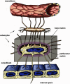
Figure 1.
Figure 1.
The osteocyte. Schematic representation of an embedded osteocyte located within its lacuna, illustrating its dendritic processes passing through the bone matrix (gray shading) within narrow tunnels termed canaliculi. The osteocyte's dendritic processes interconnect with other osteocytes as well as surface osteoblasts. Note that some osteocyte processes may extend beyond the osteoblast layer to potentially interact with cells in the marrow and that osteocyte dendrites are also in intimate contact with the vasculature. The composition of the perilacunar matrix immediately adjacent to the osteocyte (mauve shading) is different from that of the rest of the bone matrix (gray shading), which may influence the magnitude of mechanical strains perceived by the osteocyte.
Figure 2.
Figure 2.
Osteocyte morphology and interaction with the vasculature. A, Confocal fluorescence image of osteocytes in the cortex of an adult mouse femur stained with Alexa Fluor 488 phalloidin, which stains the actin cytoskeleton of the cell. Note the highly dendritic morphology of the osteocyte with many actin-positive dendrites extending from the cell in all directions. Note also the extensive dendritic interconnections between adjacent osteocytes. B, Confocal fluorescence image of the osteocyte lacunocanalicular system in mouse cortical bone as revealed by the tracer dye, procion red. This dye is dispersed throughout the lacunocanalicular system within only 5 minutes of iv injection, demonstrating the intimate connection of the canalicular fluid with the circulation (as modified from Ref. 231). C, Scanning electron micrograph of an acid-etched resin-casted mouse cortical bone specimen, revealing the 3-dimensional organization of the osteocyte lacunocanalicular system and its interaction with a blood vessel (BV). Note the 2 osteocyte lacunae (OCY) and extensive canaliculi, which are intimately connected to the surface of the blood vessel. Also note the extensive, interconnected canaliculi throughout the bone matrix.
Figure 3.
Figure 3.
Osteocyte differentiation. A, Schematic diagram depicting the transitional stages that occur as osteoblasts differentiate into mature osteocytes. During this process, the volume of the cell body and the number of cell organelles decreases. 1 = preosteoblast; 2 = osteoblast; 3 = embedding osteoblast; 4 = osteoid osteocyte; 5 = mineralizing osteocyte; 6 and 7 = mature osteocytes. B, Tetrachrome-stained section of an adult mouse tibia illustrating several of the osteoblast-osteocyte transitional stages depicted schematically in panel A (bar = 25 μm). C, Table illustrating the relative temporal expression of various osteogenic markers during the transition from osteoblast to osteocyte as depicted in panels A and B. Runt-related transcription factor 2 (RUNX2) directs early osteoblast differentiation and is expressed in both preosteoblasts and osteoblasts. Osteocalcin (OCN) is expressed by mature osteoblasts and early osteocytes. E11 is the earliest osteocyte marker to be expressed during differentiation but is not found in mature osteocytes in vivo. DMP1, CapG, and MEPE expression is observed in mineralizing and mature osteocytes, whereas sclerostin expression is confined to mature osteocytes. ORP150 is also only found in mature osteocytes within the hypoxic environment of the mineralized bone matrix.
Figure 4.
Figure 4.
Osteocyte regulation of bone remodeling. Osteocytes express RANKL and macrophage-colony stimulating factor (M-CSF) to promote, and OPG and NO to inhibit, osteoclast formation and activity. Osteocytes also regulate bone formation via the secretion of modulators of the Wnt signaling pathway. PGE2, NO, and ATP act to activate Wnt signaling, whereas sclerostin, DKK1, and SFRP1 all inhibit Wnt signaling and subsequent osteoblast activity. Maintenance of this balance between resorption and formation by the osteocyte is essential for bone homeostasis.
Figure 5.
Figure 5.
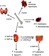
Endocrine signaling by osteocytes. FGF23 secreted by the osteocyte regulates serum phosphate (Pi) by down-regulating the expression of sodium/phosphate cotransporters in the kidney. There is also a decrease in 1-α hydroxylase production by the kidney, resulting in decreased NaPi-IIb in the intestine and reduced Pi uptake. High serum FGF23 has also been linked with an increased risk of left ventricular hypertrophy in the heart, atherosclerosis, and vascular calcification. However, vascular and soft tissue calcification is increased in Fgf23-null mouse models, suggesting that FGF23 can promote and inhibit vascular calcification. FGF23 is also known to negatively regulate the secretion of PTH via the parathyroid.

References

    1. Beno T, Yoon YJ, Cowin SC, Fritton SP. Estimation of bone permeability using accurate microstructural measurements. J Biomech. 2006;39(13):2378–2387 - PubMed
    1. Franz-Odendaal TA, Hall BK, Witten PE. Buried alive: how osteoblasts become osteocytes. Dev Dyn. 2006;235(1):176–190 - PubMed
    1. Bonewald LF. The amazing osteocyte. J Bone Miner Res. 2011;26(2):229–238 - PMC - PubMed
    1. Dallas SL, Bonewald LF. Dynamics of the transition from osteoblast to osteocyte. Ann NY Acad Sci. 2010;1192(1):437–443 - PMC - PubMed
    1. Manolagas SC. Birth and death of bone cells: basic regulatory mechanisms and implications for the pathogenesis and treatment of osteoporosis. Endocr Rev. 2000;21(2):115–137 - PubMed

Publication types