. 2013:4:1761.
doi: 10.1038/ncomms2763.

Dual role of myosin II during Drosophila imaginal disc metamorphosis

Affiliations

Dual role of myosin II during Drosophila imaginal disc metamorphosis

Silvia Aldaz et al. Nat Commun. 2013.

Abstract

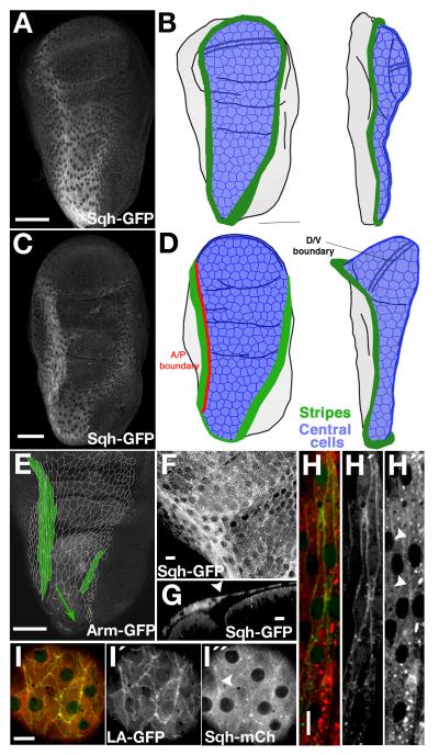
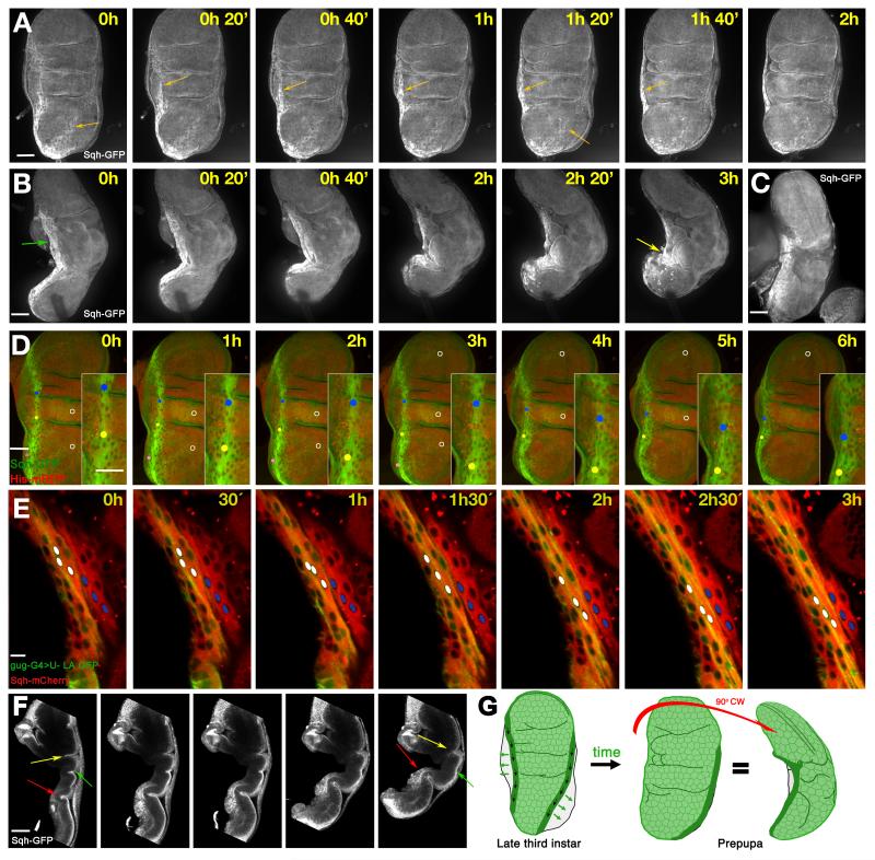
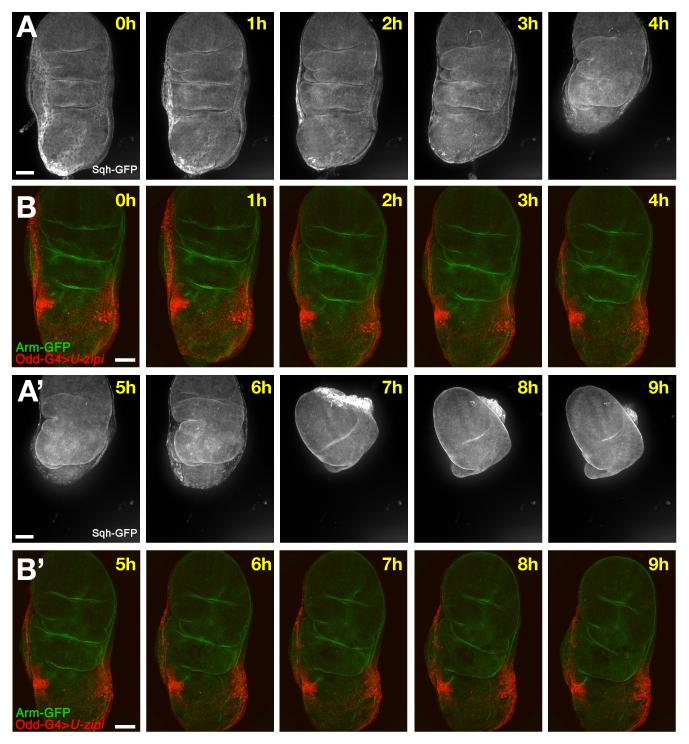
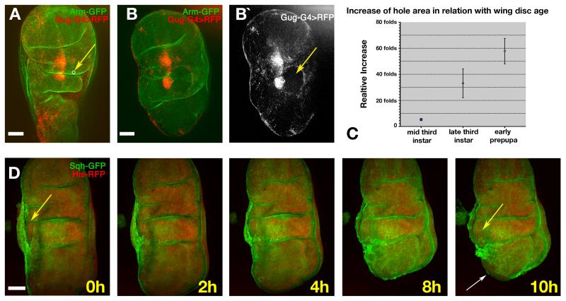
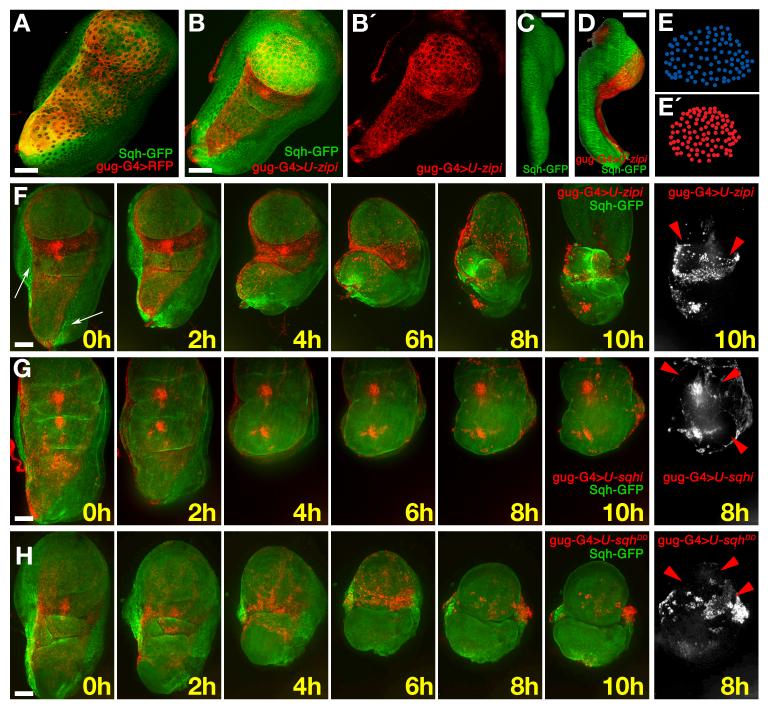
The motor protein non-muscle myosin II is a major driver of the movements that sculpt three-dimensional organs from two-dimensional epithelia. The machinery of morphogenesis is well established but the logic of its control remains unclear in complex organs. Here we use live imaging and ex vivo culture to report a dual role of myosin II in regulating the development of the Drosophila wing. First, myosin II drives the contraction of a ring of cells that surround the squamous peripodial epithelium, providing the force to fold the whole disc through about 90°. Second, myosin II is needed to allow the squamous cells to expand and then retract at the end of eversion. The combination of genetics and live imaging allows us to describe and understand the tissue dynamics, and the logic of force generation needed to transform a relatively simple imaginal disc into a more complex and three-dimensional adult wing.


Figures

Fig. 1
Fig. 1. Myosin II expression in the peripodial epithelium of the wing imaginal disc
A) Sqh-GPF expression in a live third instar wing imaginal disc in sqh null mutant background. (B) Scheme showing relevant domains of the peripodial epithelium in a frontal (left) and a lateral (right) view at the same stage. The green regions are the stripes of cuboidal cells with the highest expression of myosin II. The blue region corresponds to the squamous central cells. C) Sqh-GPF expression in a live pre-pupal wing disc in sqh null mutant background. Folds in the discs and a double line representing the D/V boundary are also shown in all the cartoons. D) Scheme showing the stripes (green) and the central cells (blue) at late-third instar stage. One of the stripes runs along the A/P boundary (red line) of the peripodial epithelium. E) Cell outlines of the central and stripe cells traced from different confocal planes of an Arm-GFP unfixed wing disc. The stripe cells (green) are elongated parallel to the A/P axis. F) Magnification of a Sqh-GFP disc at the border (green arrowheads) between stripe and central cells, near the hinge region. G) Saggital view of the same disc at the position of the dotted line in panel F. The central PE cells are flatter than the stripe cells (white arrowhead marks the border between the cell types). H, I) High resolution view of the expression of myosin II (red, Sqh-mCherry) and actin (green) in the stripe cells (H), labelled with odd-G4>UAS-lifeact-GFP, and the central cells (I), labelled with Sqh-mCherry/Gug-G4> UAS-lifeact-GFP. Arrowheads in right side panels of I and H show regions of higher accumulation of Myosin II, that co-localize with Lifeact expression. Note that sqh-mcherry forms aggregates seen as bright puncta that are artefacts of the fluorescence protein. Scale bars: A, C, E, 50μm; F, G, H, I, 10μm.
Fig. 2
Fig. 2. Myosin II and the movements of the stripe cells during the eversion
A) Frames from Supplementary movie 1: frontal view of a prepupal imaginal disc during the first steps of eversion. The disc is labelled with Sqh-GFP in a sqh null mutant background, and shows the movement of the stripes (orange arrows). B) Frames from Supplementary movie 2: lateral view of a Sqh-GFP imaginal disc in a sqh null mutant background. The stripe moves parallel to the A/P border (green arrow) towards a region at the lateral face (yellow arrow), from where later retraction of the PE will start. C) A live Sqh-GFP wing imaginal disc dissected from an early pupa showing similar movement of the A/P stripe in vivo. D) Frames from Supplementary movie 3: the movement of stripe cells in a prepupal imaginal disc (green: Sqh-GFP; red, Histone2A-RFP). Dots track three individual cells, and white circles label three central cells. The dots move towards the tip of the disc, while the circles stay relatively stationary. E) Frames from Supplementary movie 4: the movement of stripe cells along the hinge lateral region. Stripe cells are labelled in green with odd-Gal4>UAS-lifeactin; myosin II labelled in red with Sqh-mcherry. White dots mark three stripe cell nuclei which move relative to three cells outside the stripe marked with blue dots. F) Section of a lateral view of a folding disc showing the movements of the disc proper. The middle fold (yellow arrow) disappears, the most proximal fold (green arrow) becomes deeper, and the notum (red arrow) becomes more three-dimensional. G) Stripe movements during the first steps of eversion. The PE stripe cells move proximally and laterally (black and green arrows, respectively). In the prepupa the A/P stripe is not visible in a frontal view of the disc; in a lateral view, the highest accumulation of myosin II is concentrated in a shorter stripe at the lateral face of the now more three-dimensional disc. Note that in all figures time is approximate, and time 0 corresponds to the starting point of image capture, not to a specific developmental stage. Scale bars: A, B, C, D, F, 50μm; E 10μm.
Fig. 3
Fig. 3. Effect of the loss of function of myosin II in the stripes cells
A, C) Frames from Supplementary movie 1 showing the complete process of eversion including folding and the retraction. B,D) Frames from Supplementary movie 5 showing an Arm-GFP (green) disc where myosin II function has been reduced in the stripes (odd-Gal4>UAS-zip-RNAi, red). The stripes do not move laterally as they do in the control. The whole disc is it not able to fold properly, although the disc proper continues the normal morphological changes such as the apposition of wing dorsal and ventral compartments, and the deep increase of some folds. Scale bars: 50μm.
Fig. 4
Fig. 4. Effect of laser ablation in the central cells during the eversion
A) Late third instar disc prior to ablation (Arm-GFP, green; scattered central cells are labelled in red by gug-Gal4>UAS-RFP). The white circle indicated by the yellow arrow shows the size and location of the laser-cut hole. B) The same disc 30 minutes after ablation. The hole (indicated by absence of red fluorescence, yellow arrow in B (right), has expanded dramatically). C) Graph showing the fold-increase of hole area over approximately 30 minutes versus age of the disc when the ablation was made. n=4 at each time point. Error bars represent s.d D) Frames from Supplementary movie 6 showing a hole cut near to the A/P stripe (yellow arrow). After two hours the wound healing process has reduced the area of the hole, but later the hole expands again and the folding of the whole disc is impaired. At ten hours the disc proper can be seen emerging from the hole; the opening of the stalk is also visible at this stage (white arrow). Scale bars: 50μm.
Fig. 5
Fig. 5. Myosin II is required for the expansion of the central cells
A) Prepupal disc; gug-Gal4>UAS-RFP (red) is expressed in central squamous cells; a few stripe cells (Sqh-GFP, green) also express gug-Gal4 (yellow). B, B′) A disc from the same stage where myosin II has been reduced in the central cells (gug-Gal4>UAS-zip-RNAi). The area of the red central cells is reduced. C) Lateral view of a wild-type third instar wing disc showing that the peripodial epithelium covers the whole distal/proximal axis. D) Lateral view of a gug-Gal4>UAS-zip-RNAi/UAS-RFP disc. The reduction of myosin II in the central cells causes misfolding of the disc; the wing pouch and notum are closer together. E) Tracing of nuclei of the PM in the pouch region of the disc in panel A. E (bottom)) Tracing of nuclei of the PM in the pouch region of the disc in panel B. F) Frames from Supplementary movie 7 of a disc where myosin II (green) has been strongly reduced (gug-Gal4>UAS-zip-RNAi) in the central cells of the PE (red). The white arrow indicates the remaining stripes. The disc folds in the opposite direction to normal (i.e. the top surfaces of the notum and wing pouch move together). In the 8h frame, the wing pouch is seen to rupture the central cells of the PE; the remaining central cells stay attached to the disc proper (red arrowheads). G) Frames from Supplementary movie 8, where myosin II expression has been moderately reduced (gug-Gal4>UAS-sqh-RNAi). The area covered by the central cells (red) is similar to WT and the disc folds correctly, but retraction does not occur and several holes appear in the final frames (red arrowheads). H) Frames from Supplementary movie 10 showing a gug-Gal4>UAS-sqhDD/UAS-RFP disc. Strong activation of myosin II in the central cells (red) reduces their area. The disc proper shows some extra folds in the 0 hour frame. The disc folds in the opposite way to WT, similar to the example in panel E. The retraction is aberrant and multiple holes are formed in the PE (red arrowheads). Scale bars: 50μm.
Fig. 6
Fig. 6. Model of myosin II function during eversion
A) In the third instar, the cuboidal stripe cells (dark green, indicating very high myosin II levels) surround the squamous central cells of the peripodial epithelium. B) In the late third instar, the central cells (light green) start expanding to cover the larger surface created after the protrusion of the wing pouch. C) The early prepupa develops a more three-dimensional wing disc. The contraction of the stripes together with the protrusion of the wing pouch induces the expansion of the central cells to cover the frontal and lateral faces of the wing disc, in addition to the large ventral surface of the wing. D) The folding of the disc induced by the contraction of the myosin II stripes produces further expansion of the central cells. E) The final stage of folding coincides with the start of the retraction, which starts at the stalk region, shown in grey near the bottom of the diagram. This is the moment of maximum expansion of the central cells and the maximum contraction of the stripes is reached.

Similar articles

Cited by

References

    1. Escudero LM, Bischoff M, Freeman M. Myosin II regulates complex cellular arrangement and epithelial architecture in Drosophila. Dev Cell. 2007;13:717–729. - PubMed
    1. Bertet C, Sulak L, Lecuit T. Myosin-dependent junction remodelling controls planar cell intercalation and axis elongation. Nature. 2004;429:667–671. - PubMed
    1. Simoes Sde M, et al. Rho-kinase directs Bazooka/Par-3 planar polarity during Drosophila axis elongation. Dev Cell. 2010;19:377–388. - PMC - PubMed
    1. Young PE, Richman AM, Ketchum AS, Kiehart DP. Morphogenesis in Drosophila requires nonmuscle myosin heavy chain function. Genes Dev. 1993;7:29–41. - PubMed
    1. Karess RE, et al. The regulatory light chain of nonmuscle myosin is encoded by spaghetti-squash, a gene required for cytokinesis in Drosophila. Cell. 1991;65:1177–1189. - PubMed

Publication types

MeSH terms

LinkOut - more resources