. 2013:4:1766.
doi: 10.1038/ncomms2731.

The four-transmembrane protein IP39 of Euglena forms strands by a trimeric unit repeat

Affiliations
Free PMC article

The four-transmembrane protein IP39 of Euglena forms strands by a trimeric unit repeat

Hiroshi Suzuki et al. Nat Commun. 2013.
Free PMC article

Abstract

Euglenoid flagellates have striped surface structures comprising pellicles, which allow the cell shape to vary from rigid to flexible during the characteristic movement of the flagellates. In Euglena gracilis, the pellicular strip membranes are covered with paracrystalline arrays of a major integral membrane protein, IP39, a putative four-membrane-spanning protein with the conserved sequence motif of the PMP-22/EMP/MP20/Claudin superfamily. Here we report the three-dimensional structure of Euglena IP39 determined by electron crystallography. Two-dimensional crystals of IP39 appear to form a striated pattern of antiparallel double-rows in which trimeric IP39 units are longitudinally polymerised, resulting in continuously extending zigzag-shaped lines. Structural analysis revealed an asymmetric molecular arrangement in the trimer, and suggested that at least four different interactions between neighbouring protomers are involved. A combination of such multiple interactions would be important for linear strand formation of membrane proteins in a lipid bilayer.


Figures
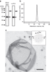
Figure 1
Figure 1. Euglena IP39 protein was purified and reconstituted into lipid for the 2D crystal.
(a) Purified IP39 was separated by SDS–PAGE and analysed by silver staining (left) or western blotting probed with anti-phosphotyrosine antibody (right). The apparent molecular weight of the major band (single arrowhead) is ~39 kDa, consistent with previous studies. The faint bands at the higher molecular weight (double arrowhead) correspond to dimers. (b) Size-exclusion chromatography of the purified IP39 protein shows a monodisperse peak. (c) A negatively stained image of the vesicular 2D crystal of IP39, observed by conventional electron microscopy. The black bar represents 400 nm. (Inset) Fourier transform of the negatively stained 2D crystal. The two independent lattices (a1* b1* and a2* b2*) indicate overlapping of the upper and lower side crystals in the vesicle.
Figure 2
Figure 2. 3D reconstructed structure of the Euglena IP39 crystal.
(a) 3D map of the IP39 crystal lattice viewed parallel to the two-fold axis from the luminal side of vesicular membrane. The grey mesh represents the EM density map contoured at 1.3σ. The internal molecular surfaces coloured blue and red represent two distinct pseudo-symmetric strands of the densities contoured at 3σ. The black box and closed ovals indicate a unit cell and two-fold symmetry axes, respectively. The right-angled arrows indicate the directions of the a- and b-axes. The black bar represents 50 Å. (b) Three-dimensional map viewed parallel to the membrane plane. Colour representations of the EM density map are the same as in a. The light grey bands represent the location of the lipid bilayer with a thickness of 35 Å. The asterisks indicate the pseudo two-fold axes along the b-axis. The numbers beside the double-headed arrows indicate the dimensions of a unit cell.
Figure 3
Figure 3. Structural comparison of strands in the 2D crystal.
(a,b) Molecular surfaces represent the isolated density maps of strand A viewed from the carbon side (a) and strand B viewed from the luminal side (b), contoured at 1.5σ. (c,d) EM density maps of strand A (blue surface) and strand B (red mesh) are superimposed and viewed perpendicular to the membrane plane from the luminal side (c) and the carbon side (d) for strand B. Colour codes are the same as in Fig. 2. The black bars represent 20 Å.
Figure 4
Figure 4. Structural comparison of IP39 protomers.
(a) EM density map containing approximately two unit cells is shown by mesh representation at 1.4σ, viewed from the luminal side. Unique molecular densities in strand B are coloured magenta (Mol1), cyan (Mol2) and yellow (Mol3). The molecules shown with the same colours are related by crystallographic symmetry. The black bar represents 20 Å. (b,c) Only the EM density of the three coloured molecules in a are shown by removing the other densities for clarity and viewed nearly parallel to the membrane plane (b). The three isolated molecules are superimposed (c) and the two sides of the densities are indicated as ’Nock‘ and ’Point‘. The black bars represent 10 Å. (d,e) Ribbon models of a four-helical bundle protein (PDB: 2UUI) are fitted to the EM density map (light grey mesh contoured at 1.3σ, putative transmembrane region only) of the three molecules in strand B. The fitted model helices are represented by magenta, cyan and yellow ribbons superimposed on Mol1, 2 and 3, respectively, viewed perpendicular (d, from the same direction with a) and parallel (e, the upper side is the ’Nock‘ side) to the membrane plane. The white bars represent 10 Å. (f) A schematic diagram representing the arrangement, directions and spacing of the IP39 protomers in a single strand viewed perpendicular to the membrane plane by the black and white circles. The black semicircular side indicates the large protruding side in the cytosolic region of the IP39 proteins.
Figure 5
Figure 5. Anti-phosphotyrosine Fab antibody distinguishes the intracellular side of the IP39 structures.
(a,b) Projection maps merged from the non-tilted images of IP39 crystals (a) and IP39 crystals with Fab fragments of anti-phosphotyrosine antibody (b). Red arrows indicate the prominent disappearance of the lines of the projection densities with the addition of Fab. The black bars represent 40 Å. (c,d) Three-dimensional maps reconstituted from 0–45° images of IP39 crystals (grey mesh) and IP39 crystals with Fab fragments of anti-phosphotyrosine antibody (green surface) are superimposed. The resolution limits of the two maps are cutoff at 10 Å. The EM maps are viewed perpendicular (c, from the luminal side) and parallel (d) to the membrane plane. Red and black arrows indicate the regions of the bridge-like protrusion in strands A and B, respectively. The black bars represent 40 Å.
Figure 6
Figure 6. Combination of four different interaction modes forms a linear strand.
(a) A schematic diagram showing the arrangement of the IP39 protomers as shown in Fig. 4f. The four coloured rectangles distinguish the different pairs of interacting molecules. (be) The respective pairs of molecules shown in a are extracted (upper panels) and the corresponding EM density maps are represented by transparent surfaces with the fitted model helices superimposed (lower panels). The maps of Mol1-Mol2 (b), Mol2-Mol3 (c) and Mol3-Mol1 (d) are viewed perpendicular to the membrane planes from their intracellular sides, and the map of Mol1-Mol1 (e) is viewed slightly diagonally from the intracellular side. The black open arrow indicates the overlaid area of the Mol3 protrusion on the cytosolic region of Mol2. The surface colour codes are the same as in Fig. 4a. The black bars represent 10 Å.

References

    1. Van Itallie C. M. & Anderson J. M. Claudins and epithelial paracellular transport. Annu. Rev. Physiol. 68, 403–429 (2006) . - PubMed
    1. Taylor V., Welcher A. A., Program A. E. & Suter U. Epithelial membrane protein-1, peripheral myelin protein 22, and lens membrane protein 20 define a novel gene family. J. Biol. Chem. 270, 28824–28833 (1995) . - PubMed
    1. Van Itallie C. M. & Anderson J. M. The molecular physiology of tight junction pores. Physiology 19, 331–338 (2004) . - PubMed
    1. Notterpek L. et al.. Peripheral myelin protein 22 is a constituent of intercellular junctions in epithelia. Proc. Natl Acad. Sci. USA 98, 14404–14409 (2001) . - PMC - PubMed
    1. Grey A. C., Jacobs M. D., Gonen T., Kistler J. & Donaldson P. J. Insertion of MP20 into lens fibre cell plasma membranes correlates with the formation of an extracellular diffusion barrier. Exp. Eye Res. 77, 567–574 (2003) . - PubMed

Publication types

MeSH terms