. 2013 Jun 21;15(23):8952-61.
doi: 10.1039/c3cp50721a. Epub 2013 Apr 24.

Amyloid-β-neuropeptide interactions assessed by ion mobility-mass spectrometry

Affiliations

Amyloid-β-neuropeptide interactions assessed by ion mobility-mass spectrometry

Molly T Soper et al. Phys Chem Chem Phys. .

Abstract

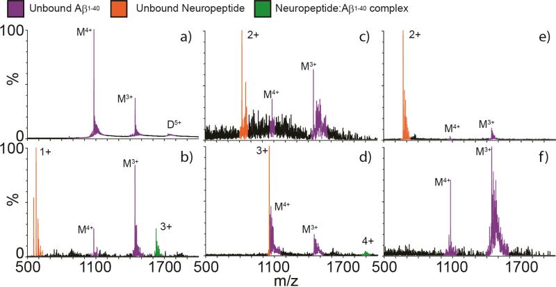
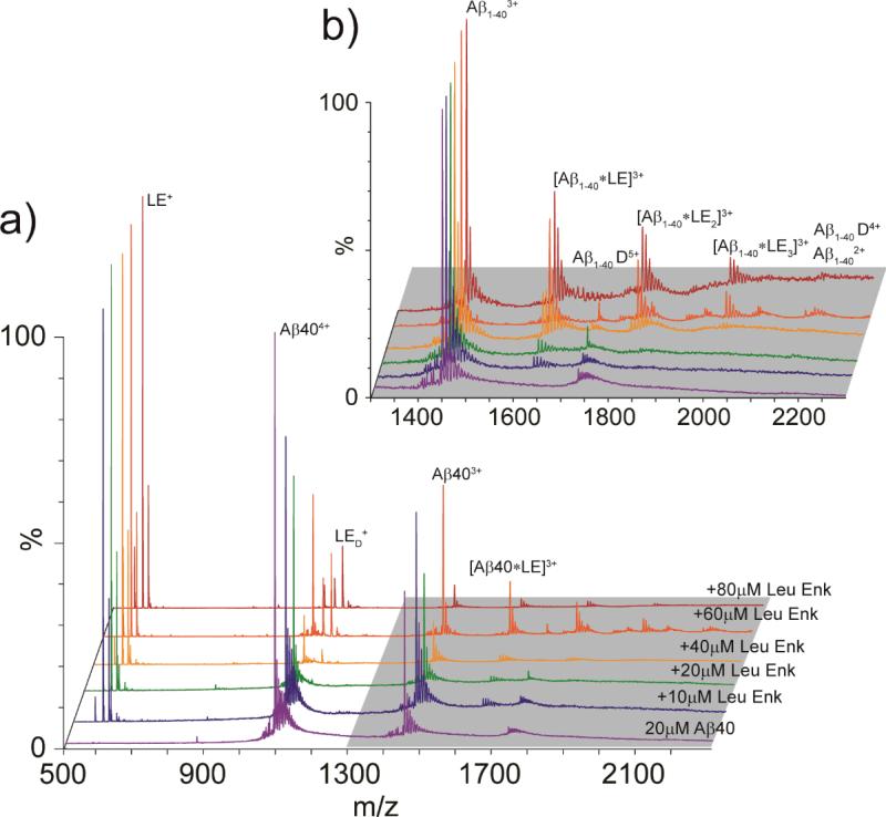
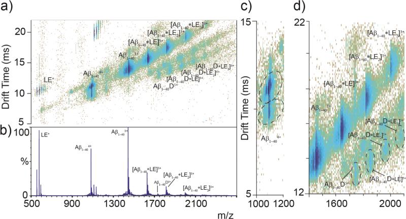
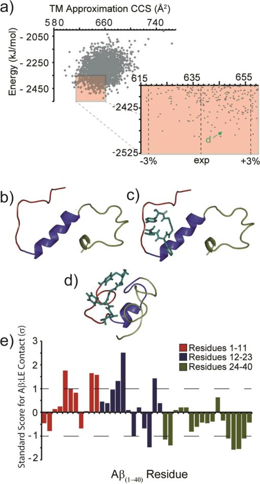
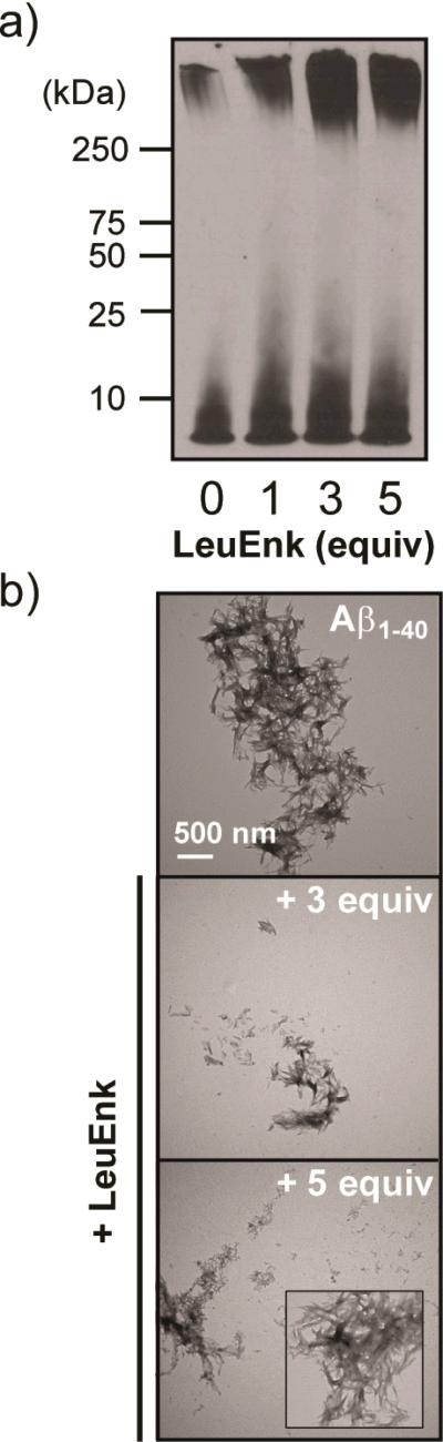
Recently, small peptides have been shown to modulate aggregation and toxicity of the amyloid-β protein (Aβ). As such, these new scaffolds may help discover a new class of biotherapeutics useful in the treatment of Alzheimer's disease. Many of these inhibitory peptide sequences have been derived from natural sources or from Aβ itself (e.g., C-terminal Aβ fragments). In addition, much earlier work indicates that tachykinins, a broad class of neuropeptides, display neurotrophic properties, presumably through direct interactions with either Aβ or its receptors. Based on this work, we undertook a limited screen of neuropeptides using ion mobility-mass spectrometry to search for similar such peptides with direct Aβ binding properties. Our results reveal that the neuropeptides leucine enkephalin (LE) and galanin interact with both the monomeric and small oligomeric forms of Aβ(1-40) to create a range of complexes having diverse stoichiometries, while some tachyknins (i.e., substance P) do not. LE interacts with Aβ more strongly than galanin, and we utilized ion mobility-mass spectrometry, molecular dynamics simulations, gel electrophoresis/Western blot, and transmission electron microscopy to study the influence of this peptide on the structure of Aβ monomer, small Aβ oligomers, as well as the eventual formation of Aβ fibrils. We find that LE binds selectively within a region of Aβ between its N-terminal tail and hydrophobic core. Furthermore, our data indicate that LE modulates fibril generation, producing shorter fibrillar aggregates when added in stoichiometric excess relative to Aβ.


Figures

Figure 1
Figure 1
Analyses of Aβ1-40 incubated with one equivalent of each neuropeptide by nESI-IM-MS. (A) Mass spectrum of Aβ1-40 only, with signals corresponding to monomeric and dimeric peptides marked with ‘M’ and ‘D’, respectively. Satellite peaks observed correspond to alkali metal adducts commonly observed in nESI-MS. Aβ1-40 was then mixed with equivalent amounts of (B) LE (free [M+H]+ at m/z = 556.6), (C) somatostatin (free [M+2H]2+ at m/z = 820.5), (D) galanin (free [M+3H]3+ at m/z = 1053.8), (E) substance P (free [M+2H]2+ at m/z = 675.5) and (F) neurotensin (no free signal detected, peptide mass = 1674.0). LE and galanin are the two neuropeptides in this set where we detect complexes with Aβ1-40 (complexes signals in green), while the other neuropeptides screened result in signals for unbound Aβ1-40 (purple) and unbound neuropeptide (orange). Poorer signal intensities are recorded in panels C and F due to signal suppression surrounding the addition of the neuropeptides indicated.
Figure 2
Figure 2
(A) MS spectra for Aβ1-40 acquired at 20 μM (purple). Aβ1-40 was then incubated with increasing concentrations of LE, ranging from 10-80 μM (stoichiometric ratios from 0.5 to 4). At sufficiently high LE concentrations, Aβ1-40 is seen in complex with LE, producing Aβ:LE complexes ranging from 1:1: to 1:3. (B) A magnified region of the spectrum shown in A (grey highlight), where signals corresponding to Aβ:LE complexes are labeled.
Figure 3
Figure 3
(A) IM-MS data for Aβ1-40 (20 μM) incubated with LE (60 μM) for 1 h on ice, where Aβ:LE complexes are labeled, along with free peptide. IM separation allows for the identification of Aβ dimer complexes previously undetected by MS alone. Aβ1-40 is seen in complex with LE at stoichiometric ratios up to 1:3 Aβ1-40:LE and 2:3 Aβ1-40:LE. (B) MS dataset for the IM-MS plot shown in A. (C) Two main conformations of Aβ1-404+ are identified, with a third minor conformer, as observed previously. (D) A magnified region of the IM-MS data shown in A, showing detail on Aβ1-404+: LE complexes. Monomer complexes are observed in greater relative abundance than those related to dimeric Aβ.
Figure 4
Figure 4
(A) Output from all molecular dynamics simulations. The lowest energy 201 structures with CCS values within ±3% of experiment are highlighted (red box). (B) The structure of Aβ monomer (PDB 2LFM). (C) A docked structure of Aβ (PDB 2LFM) with LE using AutoDock Vina. (D) A representative low energy model (indicated in A) from the main structural family identified from our MD simulations, in agreement with experimental CCS values. Colors represent the N-terminus (red), core/helix region (blue), and the C-terminus (green). (E) A plot of the standard score (Z-score) for Aβ residues within 4 Å of the bound LE. Larger values denote contacts of greater significance on the standard deviation (σ) scale. Negative values denote contacts of reduced significance.
Figure 5
Figure 5
Influence of LE on Aβ1-40 aggregation in vitro. (A) Visualization of Aβ species generated in the absence and presence of LE by gel electrophoresis and Western blotting (6E10). Experimental conditions: [Aβ1-40] = 25 μM; [LE] = 0, 25, 75, or 125 μM; 100 mM ammonium acetate, pH 6.9; 37 °C; 24 h; agitation. (B) TEM images of Aβ species in the absence and presence of LE (3 and 5 equiv) from samples in A. The scale bar depicts 500 nm.

References

    1. Brookmeyer R, Johnson E, Ziegler-Graham K, Arrighi HM. Alzheimers Dement. 2007;3:186–191. - PubMed
    1. Alzheimer's Association Alzehimers Dement. 2012;8:131–168. - PubMed
    1. Selkoe DJ. Phys. Rev. 2001;81:741–766. - PubMed
    1. O'Brien RJ, Wong PC. Annu. Rev. Neurosci. 2011;34:185–204. - PMC - PubMed
    1. Harper JD, Lansbury PT., Jr Annu. Rev. Biochem. 1997;66:385–407. - PubMed

Publication types

MeSH terms