. 2013 May 15;32(10):1409-24.
doi: 10.1038/emboj.2013.90. Epub 2013 Apr 23.

Stabilization of integrin-linked kinase by the Hsp90-CHIP axis impacts cellular force generation, migration and the fibrotic response

Affiliations

Stabilization of integrin-linked kinase by the Hsp90-CHIP axis impacts cellular force generation, migration and the fibrotic response

Korana Radovanac et al. EMBO J. .

Abstract

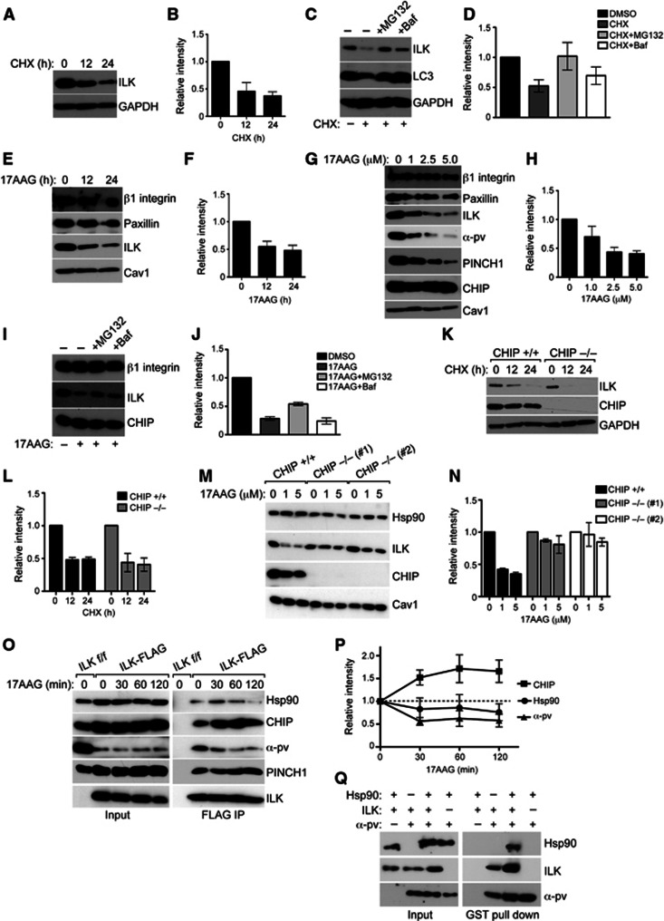
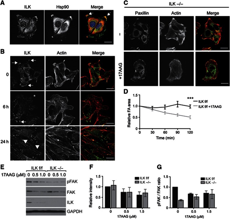
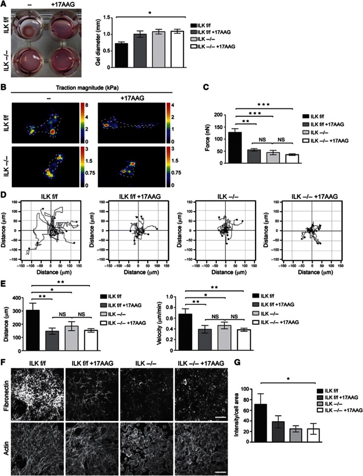
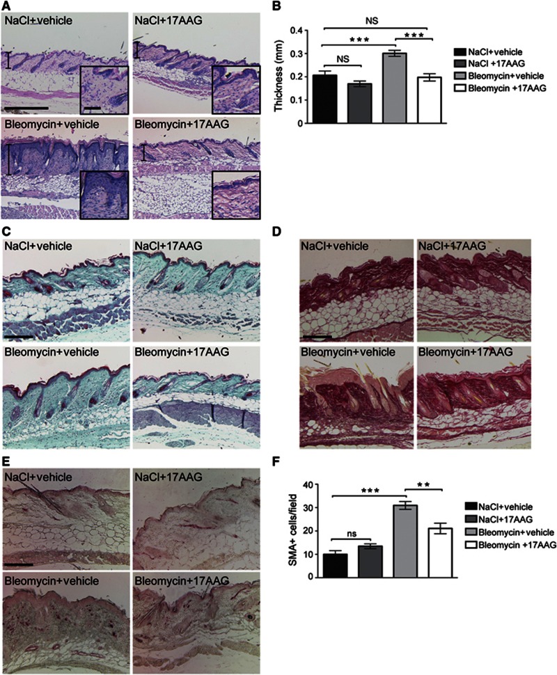
Integrin-linked kinase (ILK) is an adaptor protein required to establish and maintain the connection between integrins and the actin cytoskeleton. This linkage is essential for generating force between the extracellular matrix (ECM) and the cell during migration and matrix remodelling. The mechanisms by which ILK stability and turnover are regulated are unknown. Here we report that the E3 ligase CHIP-heat shock protein 90 (Hsp90) axis regulates ILK turnover in fibroblasts. The chaperone Hsp90 stabilizes ILK and facilitates the interaction of ILK with α-parvin. When Hsp90 activity is blocked, ILK is ubiquitinated by CHIP and degraded by the proteasome, resulting in impaired fibroblast migration and a dramatic reduction in the fibrotic response to bleomycin in mice. Together, our results uncover how Hsp90 regulates ILK stability and identify a potential therapeutic strategy to alleviate fibrotic diseases.


Conflict of interest statement

The authors declare that they have no conflict of interest.

Figures

Figure 1
Figure 1
ILK is modified by polyubiquitination. (A) Silver staining of FLAG immunoprecipitates from ILK-FLAG expressing fibroblasts. ILK f/f cells were used as a negative control. Arrows indicate bands analysed by mass spectrometry; asterisk indicates the expected molecular weight of unmodified ILK. ILK and ubiquitin were identified from all bands analysed. (B) Western blot analysis of FLAG immunoprecipitates from ILK−/− fibroblasts expressing ILK-FLAG and HA-tagged ubiquitin or ubiquitin mutants containing a single lysine K48 or K63. Both K48- and K63-linked ubiquitin chains are detected. Source data for this figure is available on the online supplementary information page.
Figure 2
Figure 2
ILK interacts with the E3 ligase CHIP. (A) A volcano plot representing results of label-free FLAG pull downs of ILK. The differences in t-test of ILK f/f control versus ILK-FLAG are plotted against negative logarithmic P-values of the t-test performed from triplicates. The curve separates proteins specifically enriched in either condition (blue dots) from background (grey dots). Proteins specifically enriched in ILK-FLAG pull downs are found in the upper left quadrant of the plot. Known interacting partners of ILK as well as novel interacting partners CHIP, Hsc70 and Hsp90, are marked in purple. Names of all proteins specifically interacting with ILK-FLAG are reported in Supplementary Table S1. (B) FLAG pull down from ILK−/− fibroblasts expressing ILK-FLAG. CHIP is detected in the ILK immunoprecipitate. (C) Lysates from ILK f/f cells were subjected to immunoprecipitation with antibodies against CHIP. Endogenous ILK is detected in CHIP immunoprecipitates. Rabbit IgG was used as a negative control. (D) Schematic illustration of various EGFP-CHIP constructs. (E) GFP pull down from fibroblasts expressing EGFP-CHIP or mutants. Endogenous ILK is detected in immunoprecipitates of full-length EGFP-CHIP and EGFP-U-Box. K30A mutation of CHIP abrogates ILK binding. (F) Western blot of His pull down with recombinant His-tagged CHIP and ILK. ILK co-precipitates with CHIP and presence of Hsc70 enhances this interaction. (G) FLAG pull down from ILK−/− fibroblasts expressing ILK-FLAG, ANK-FLAG, and KD-FLAG. CHIP interacts with ANK-FLAG, whereas Hsp90 binds the KD. Source data for this figure is available on the online supplementary information page.
Figure 3
Figure 3
CHIP ubiquitinates ILK in vitro and in vivo. (A) Western blot of in vitro ubiquitination assay. Addition of recombinant CHIP induces multiple higher molecular weight bands recognized by ILK antibody (arrows, bracket) in a dose-dependent manner. ILK band of expected molecular weight is marked by asterisk. (B) Western blot of in vitro ubiquitination assay. CHIP-WT but not ligase-dead H260Q mutant CHIP ubiquitinates ILK. (C) Western blot analysis of FLAG immunoprecipitates from CHO cells expressing ILK-FLAG, CHIP, CHIP-H260Q, and ubiquitin-HA. Expression of CHIP but not ligase-dead H260Q mutant enhances ubiquitination of ILK. Source data for this figure is available on the online supplementary information page.
Figure 4
Figure 4
Interaction of ILK and CHIP is modulated by the cytoskeleton. (A) Immunofluorescence analysis of endogenous CHIP, ILK, and phalloidin to stain the actin cytoskeleton in ILK f/f and ILK−/− fibroblasts. Right panels show an enlargement of the area indicated by the white rectangle. Note co-localization in of ILK and CHIP in focal adhesions (arrows) and localization of CHIP along actin stress fibres (arrowheads). CHIP co-localizes with actin in the cell periphery in the absence of ILK. Scale bars 20 μm. (B) Immunofluorescence analysis of endogenous CHIP, paxillin, and phalloidin to stain for the actin cytoskeleton in ILK f/f fibroblasts during 30, 90, and 120 min of cell spreading. CHIP localizes to focal adhesions during late stages of cell spreading. Right panels show an enlargement of the area indicated by the white rectangle. Arrowheads indicate immature focal complexes lacking CHIP and actin stress fibres. Arrows indicate co-localization of CHIP, paxillin, and actin in focal adhesions. Scale bars 30 μm. (C) FLAG pull down from ILK−/− fibroblasts expressing ILK-FLAG and treated with Y27632 or Lat. Increased levels of CHIP are detected in immunoprecipitates treated with Y27632 or Lat compared to control. Source data for this figure is available on the online supplementary information page.
Figure 5
Figure 5
Hsp90 stabilizes ILK and protects it from CHIP-mediated degradation. (A) Western blot of fibroblasts treated with CHX to stop protein synthesis for time points indicated. Majority of ILK is degraded in 24 h. GAPDH is used as loading control. (B) Quantification of ILK levels from experiments from panel A. Data are presented as mean±s.e.m., n=3. (C) Western blot of fibroblasts treated with CHX to stop protein synthesis for time points indicated. Inhibition of proteasome with MG132 or inhibition of lysosome with Baf retards ILK degradation. LC3 is a positive control for inhibition of lysosome; GAPDH is used as loading control. (D) Quantification of ILK levels from experiments from C. Data are presented as mean±s.e.m., n=3. (E) Western blot of fibroblasts treated with 5 μM 17AAG to inhibit Hsp90 for time points indicated. Inhibition of Hsp90 leads to downregulation of ILK levels. Other focal adhesion proteins such as paxillin or β1 integrin are unaffected. Caveolin1 (Cav1) is used as a loading control. (F) Quantification of ILK levels from experiments from E. Data are presented as mean±s.e.m., n=3. (G) Western blot of fibroblasts treated with increasing concentrations of 17AAG. Inhibition of Hsp90 leads to dose-dependent downregulation of ILK levels together with PINCH1 and α-parvin. Paxillin or β1 integrin are unaffected. (H) Quantification of ILK levels from experiments from G. Data are presented as mean±s.e.m., n=3. (I) Western blot of fibroblasts treated with 17AAG in the presence of MG132 or Baf. Blocking the proteasome with MG132 inhibits 17AAG-induced degradation of ILK. (J) Quantification of ILK levels from experiments from I. Data are presented as mean±s.e.m., n=3. (K) Western blot of CHIP+/+ and CHIP−/− fibroblasts treated with CHX for time points indicated. ILK levels are reduced to a comparable extent in CHIP+/+ and CHIP−/− fibroblasts. (L) Quantification of ILK levels from experiments from K. Data are presented as mean±s.e.m., n=3. (M) Western blot of CHIP+/+ and CHIP−/− fibroblasts from two independent isolations treated with 17AAG. Inhibition of Hsp90 leads to downregulation of ILK levels. This effect is attenuated in CHIP−/− cells. (N) Quantification of ILK levels from experiments from M. Data are presented as mean±s.e.m., n=3. (O) Western blot of FLAG immunoprecipitates from ILK-FLAG expressing cells treated with 17AAG. Inhibition of Hsp90 leads to an increase in the interaction of ILK with CHIP and a dissociation of ILK from Hsp90 and α-parvin. Interaction with PINCH1 is not affected. (P) Quantification of experiments from O. Data are presented as mean±s.e.m., n=3. (Q) Western blot of GST pull down with recombinant GST-tagged α-parvin together with recombinant ILK and Hsp90. ILK co-precipitates with α-parvin, whereas Hsp90 co-precipitates with α-parvin only in the presence of ILK. Presence of Hsp90 enhances the interaction between ILK and α-parvin. Source data for this figure is available on the online supplementary information page.
Figure 6
Figure 6
Inhibition of Hsp90 leads to reorganization of the F-actin cytoskeleton and focal adhesions. (A) Immunofluorescence analysis of ILK and Hsp90 in fibroblasts. Note co-localization of ILK and Hsp90 in large peripheral focal adhesions (arrows), but not in cytoplasmic fibrillar adhesions or focal complexes. Scale bar, 20 μm. (B) Immunofluorescence analysis of ILK and actin in fibroblasts treated with 17AAG. Note loss of ILK from focal adhesions (arrows, asterisk), an increase in fibrillar adhesions (arrowheads), and reorganization of the actin cytoskeleton. Scale bars 20 μm. (C) ILK−/− fibroblasts were treated with 17AAG after which they were fixed and stained with antibodies against paxillin and with phalloidin to visualize the actin cytoskeleton. No major change in focal adhesion or the actin cytoskeleton was observed upon 17AAG treatment. Scale bars 20 μm. (D) Quantification of relative focal adhesion area from live-cell imaging experiments on cells treated with 17AAG. Note that focal adhesions are disassembled as indicated by a decrease in focal adhesion area in 17AAG-treated cells. Data are presented as mean±s.e.m., n=20 movies from three independent experiments, ***P≤0.0001 (Student’s t-test). (E) Western blot of ILK f/f and ILK−/− fibroblasts treated with 17AAG. 17AAG treatment leads to a decrease in total FAK levels in ILK f/f and ILK−/− cells, and to a decrease in FAK phosphorylation (pFAK) in ILK f/f cells. ILK−/− cells show reduced basal levels of pFAK. (F) Quantification of total FAK levels from experiments shown in E. Data are presented as mean±s.e.m., n=3. (G) Quantification of pFAK to total FAK ratio from experiments shown in E. Data are presented as mean±s.e.m., n=3. Source data for this figure is available on the online supplementary information page.
Figure 7
Figure 7
Hsp90-mediated stabilization of ILK promotes force generation, matrix assembly, and migration. (A) Gel contraction assay with ILK f/f and−/− fibroblasts treated with 17AAG. 17AAG or deletion of ILK impairs the ability of fibroblasts to contract collagen gels. 17AAG in ILK−/− cells does not induce an additive effect. Data are presented as mean±s.e.m., n=3, *P=0.0174 (Friedman’s ANOVA). (B) Heat-scale map of traction stress magnitudes. The colour code indicates local traction in kPa. Cell outlines are indicated by dotted lines. (C) Quantification of total cellular traction force. Data are presented as mean±s.e.m., n>30, ***P≤0.0001 (Kruskal–Wallis), NS=not significant. (D) Tracks of ILK f/f and−/− fibroblasts migrating in 3D collagen. 17AAG or deletion of ILK impairs the ability of fibroblasts to migrate. (E) Quantification of migration distance (left panel) and velocity (right panel) from 3D migration assays. Data are presented as mean±s.e.m., n=5, **P=0.002 (repeated measures ANOVA), NS=not significant. (F) Immunofluorescence staining of the fibronectin matrix and the actin cytoskeleton (phalloidin) to visualize cell area. Note decreased matrix deposition in ILK f/f cells treated with 17AAG, and in ILK−/− cells. Scale bars 100 μm. (G) Quantification of the integrated intensity of the fibronectin matrix staining normalized to total cell surface area. Data are presented as mean±s.e.m., n=4, *P=0.0214 (Kruskal–Wallis).
Figure 8
Figure 8
Bleomycin-induced skin fibrosis is blocked by inhibition of Hsp90. (A) Haematoxylin/eosin staining of bleomycin-treated skin shows characteristic fibrosis of the skin with dermal thickening. This effect is not visible in mice treated with 17AAG together with bleomycin. Scale bar, 150 μm (large panel), 20 μm (inset). (B) Quantification of dermal thickness from bleomycin-treated mice. Data are presented as mean±s.e.m., n=5/5/8/5, ***P=0.0004 (ANOVA), NS=not significant. (C) Trichrome staining of bleomycin-treated skin shows increased density of the extracellular matrix in the dermis. This effect is not visible in mice treated with 17AAG together with bleomycin. Scale bar, 200 μm. (D) Picosirius red staining of bleomycin-treated skin shows enhanced abundance of collagenous matrix in the dermis and subcutaneous fat. This effect is not visible in mice treated with 17AAG together with bleomycin. Scale bar, 150 μm. (E) α-SMA staining identifies elevated numbers of activated fibroblasts in the dermis of bleomycin-treated mice. This effect is attenuated in mice treated with 17AAG together with bleomycin. (F) Quantification of α-SMA-positive cells. Data are presented as mean±s.e.m., n=5/5/8/5, ***P<0.0001, **P<0.005 (ANOVA), NS=not significant.

Comment in

References

    1. Aoyagi Y, Fujita N, Tsuruo T (2005) Stabilization of integrin-linked kinase by binding to Hsp90. Biochem Biophys Res Commun 331: 1061–1068 - PubMed
    1. Arndt V, Dick N, Tawo R, Dreiseidler M, Wenzel D, Hesse M, Furst DO, Saftig P, Saint R, Fleischmann BK, Hoch M, Höhfeld J (2010) Chaperone-assisted selective autophagy is essential for muscle maintenance. Curr Biol 20: 143–148 - PubMed
    1. Azimifar SB, Böttcher RT, Zanivan S, Grashoff C, Kruger M, Legate KR, Mann M, Fässler R (2012) Induction of membrane circular dorsal ruffles requires co-signalling of integrin-ILK-complex and EGF receptor. J Cell Sci 125: 435–448 - PubMed
    1. Ballinger CA, Connell P, Wu Y, Hu Z, Thompson LJ, Yin LY, Patterson C (1999) Identification of CHIP, a novel tetratricopeptide repeat-containing protein that interacts with heat shock proteins and negatively regulates chaperone functions. Mol Cell Biol 19: 4535–4545 - PMC - PubMed
    1. Blumbach K, Zweers MC, Brunner G, Peters AS, Schmitz M, Schulz JN, Schild A, Denton CP, Sakai T, Fässler R, Krieg T, Eckes B (2010) Defective granulation tissue formation in mice with specific ablation of integrin-linked kinase in fibroblasts—role of TGFbeta1 levels and RhoA activity. J Cell Sci 123: 3872–3883 - PubMed

Publication types

MeSH terms