Review
. 2013 Apr 23;6(272):re2.
doi: 10.1126/scisignal.2003832.

Signaling mechanisms in mammalian myoblast fusion

Affiliations
Review

Signaling mechanisms in mammalian myoblast fusion

Sajedah M Hindi et al. Sci Signal. .

Abstract

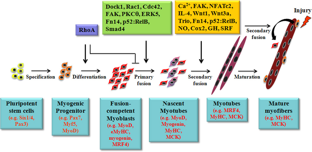
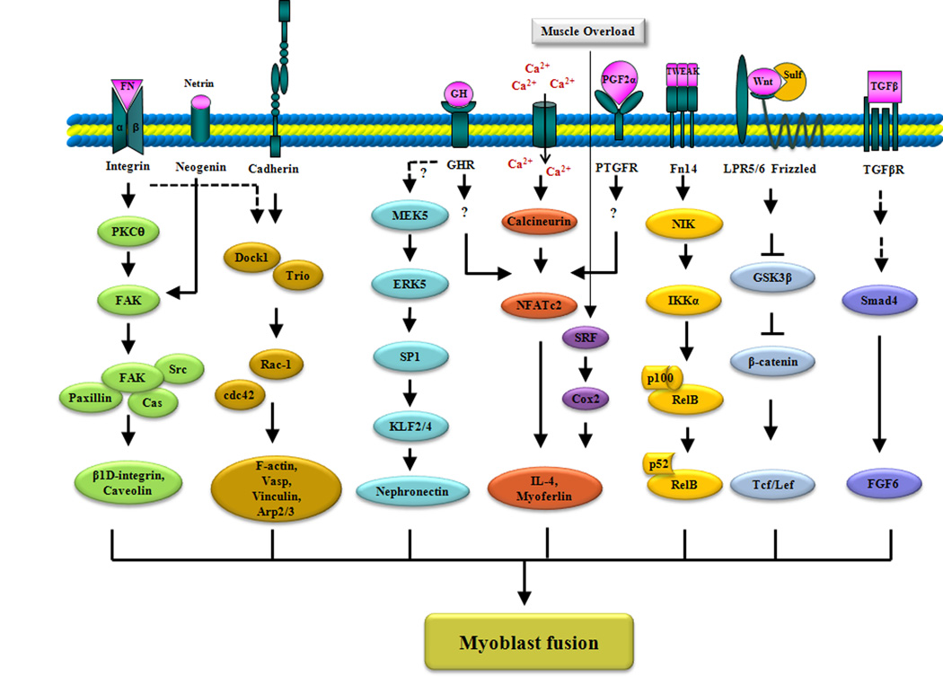
Myoblast fusion is a critical process that contributes to the growth of muscle during development and to the regeneration of myofibers upon injury. Myoblasts fuse with each other as well as with multinucleated myotubes to enlarge the myofiber. Initial studies demonstrated that myoblast fusion requires extracellular calcium and changes in cell membrane topography and cytoskeletal organization. More recent studies have identified several cell-surface and intracellular proteins that mediate myoblast fusion. Furthermore, emerging evidence suggests that myoblast fusion is also regulated by the activation of specific cell-signaling pathways that lead to the expression of genes whose products are essential for the fusion process and for modulating the activity of molecules that are involved in cytoskeletal rearrangement. Here, we review the roles of the major signaling pathways in mammalian myoblast fusion.


Figures

Fig. 1
Fig. 1. The roles of different signaling molecules in primary and secondary myoblast fusion during myogenesis
Muscle progenitor cells first undergo myogenic commitment and differentiation to become fusion-competent myoblasts. The initial commitment to differentiation requires the activity of the RhoA GTPase. Active RhoA interferes with myoblast fusion, and so it is deactivated before fusion occurs. A number of signaling molecules and pathways are activated in fusion-competent myoblasts that regulate primary myoblast fusion, which results in nascent myotubes. Additional signaling molecules are then recruited, which lead to fusion of additional mononucleated myoblasts with nascent myotubes. Secondary fusion also plays a critical role in the regeneration of injured myofibers. Major signaling molecules involved in primary and secondary myoblast fusion based on experimental evidence are depicted along the top. Specific myogenic markers expressed at different stages in cells of myogenic lineage during myogenesis are noted along the bottom.
Fig. 2
Fig. 2. The signaling mechanisms that stimulate myoblast fusion
Emphasis is placed on ligands and receptors that activate pathways, the main effectors of each pathway, and their target molecules. The interactions of integrins with their ligands, such as fibronectin (FN), causes the autophosphorylation of FAK by PKC-θ, which leads to the activation of downstream signaling that results in the increased expression of the genes encoding caveolin-3 and the β1D integrin. Neogenin-netrin signaling also causes FAK activation. Ligation of M-cadherin activates Rac1 in a Trio- and Dock1-dependent manner. Cdc42 and Rac1 are major activators of vinculin, F-actin, Vasp, and the Arp2/3 complex for the cytoskeletal remodeling that occurs before myoblast fusion. The MEK5-dependent activation of ERK5 promotes binding of the transcription factor SP1 to the promoter of the genes encoding the transcription factors Klf2 and Klf4, leading to their increased abundance. Subsequently, Klf2 and Klf4 bind to the Npnt promoter and induce the production of nephronectin during myoblast fusion. Increases in intracellular calcium concentration lead to activation of the serine-threonine protein phosphatase calcineurin, which dephosphorylates and activates the transcription factor NFATc2. Activated NFATc2 stimulates myoblast fusion through the increased production of IL-4 and myoferlin. Possible stimuli for NFATc2 activation are PGF2α and GH. In response to mechanical overload, the transcription factor SRF becomes activated, which leads to the increased abundance of IL-4 though COX2-dependent mechanisms. Low amounts of the cytokine TWEAK activate the noncanonical NF-κB pathway through activation of NIK and IKKα. IKKα-mediated phosphorylation of p100 results in its proteolytic processing to generate the p52 subunit and the subsequent nuclear accumulation of p52-RelB heterodimers. Binding of Wnt to Frizzled and LRP5/6 disrupts the binding of GSK-3β to β-catenin, thus preventing the phosphorylation and degradation of β-catenin. β-catenin then translocates to the nucleus and acts as a transcriptional coactivator for Tcf/Lef target genes. Recruitment of TGF-β to its cell-surface receptor leads to the activation of Smad4, which translocates to the nucleus and recruits other transcription factors to collectively induce the expression of target genes, such as Fgf6. The dashed arrows indicate that direct interactions have not yet been shown.

References

    1. Abmayr SM, Pavlath GK. Myoblast fusion: Lessons from flies and mice. Development. 2012;139:641–656. - PMC - PubMed
    1. Rochlin K, Yu S, Roy S, Baylies MK. Myoblast fusion: When it takes more to make one. Dev. Biol. 2010;341:66–83. - PMC - PubMed
    1. Pavlath GK. Spatial and functional restriction of regulatory molecules during mammalian myoblast fusion. Exp. Cell Res. 2010;316:3067–3072. - PMC - PubMed
    1. Krauss RS. Regulation of promyogenic signal transduction by cell-cell contact and adhesion. Exp. Cell Res. 2010;316:3042–3049. - PMC - PubMed
    1. Schwander M, Leu M, Stumm M, Dorchies OM, Ruegg UT, Schittny J, Müller U. Beta1 integrins regulate myoblast fusion and sarcomere assembly. Dev. Cell. 2003;4:673–685. - PubMed

Publication types