

Review of the inhibition of biological activities of food-related selected toxins by natural compounds
- PMID: 23612750
- PMCID: PMC3705290
- DOI: 10.3390/toxins5040743
Review of the inhibition of biological activities of food-related selected toxins by natural compounds
Abstract
There is a need to develop food-compatible conditions to alter the structures of fungal, bacterial, and plant toxins, thus transforming toxins to nontoxic molecules. The term 'chemical genetics' has been used to describe this approach. This overview attempts to survey and consolidate the widely scattered literature on the inhibition by natural compounds and plant extracts of the biological (toxicological) activity of the following food-related toxins: aflatoxin B1, fumonisins, and ochratoxin A produced by fungi; cholera toxin produced by Vibrio cholerae bacteria; Shiga toxins produced by E. coli bacteria; staphylococcal enterotoxins produced by Staphylococcus aureus bacteria; ricin produced by seeds of the castor plant Ricinus communis; and the glycoalkaloid α-chaconine synthesized in potato tubers and leaves. The reduction of biological activity has been achieved by one or more of the following approaches: inhibition of the release of the toxin into the environment, especially food; an alteration of the structural integrity of the toxin molecules; changes in the optimum microenvironment, especially pH, for toxin activity; and protection against adverse effects of the toxins in cells, animals, and humans (chemoprevention). The results show that food-compatible and safe compounds with anti-toxin properties can be used to reduce the toxic potential of these toxins. Practical applications and research needs are suggested that may further facilitate reducing the toxic burden of the diet. Researchers are challenged to (a) apply the available methods without adversely affecting the nutritional quality, safety, and sensory attributes of animal feed and human food and (b) educate food producers and processors and the public about available approaches to mitigating the undesirable effects of natural toxins that may present in the diet.
Figures


Similar articles
-
Antitoxins: novel strategies to target agents of bioterrorism.Nat Rev Microbiol. 2004 Sep;2(9):721-6. doi: 10.1038/nrmicro977. Nat Rev Microbiol. 2004. PMID: 15372082 Review.
-
Vaccines against the category B toxins: Staphylococcal enterotoxin B, epsilon toxin and ricin.Adv Drug Deliv Rev. 2005 Jun 17;57(9):1424-39. doi: 10.1016/j.addr.2005.01.017. Adv Drug Deliv Rev. 2005. PMID: 15935880 Review.
-
Evaluation of β-D-glucan biopolymer as a novel mycotoxin binder for fumonisin and deoxynivalenol in soybean feed.Foodborne Pathog Dis. 2014 Jun;11(6):433-8. doi: 10.1089/fpd.2013.1711. Epub 2014 Mar 24. Foodborne Pathog Dis. 2014. PMID: 24660841
-
Fusarial toxins: secondary metabolites of Fusarium fungi.Rev Environ Contam Toxicol. 2014;228:101-20. doi: 10.1007/978-3-319-01619-1_5. Rev Environ Contam Toxicol. 2014. PMID: 24162094 Review.
-
Intracellular transport and processing of protein toxins produced by enteric bacteria.Adv Exp Med Biol. 1997;412:225-32. doi: 10.1007/978-1-4899-1828-4_34. Adv Exp Med Biol. 1997. PMID: 9192018 Review.
Cited by
-
Plant breeding for harmony between sustainable agriculture, the environment, and global food security: an era of genomics-assisted breeding.Planta. 2023 Oct 12;258(5):97. doi: 10.1007/s00425-023-04252-7. Planta. 2023. PMID: 37823963 Review.
-
Dietary Modulation of Bacteriophages as an Additional Player in Inflammation and Cancer.Cancers (Basel). 2021 Apr 23;13(9):2036. doi: 10.3390/cancers13092036. Cancers (Basel). 2021. PMID: 33922485 Free PMC article. Review.
-
Antibacterial Activity of Juglone against Staphylococcus aureus: From Apparent to Proteomic.Int J Mol Sci. 2016 Jun 18;17(6):965. doi: 10.3390/ijms17060965. Int J Mol Sci. 2016. PMID: 27322260 Free PMC article.
-
Assessment of genetic integrity, splenic phagocytosis and cell death potential of (Z)-4-((1,5-dimethyl-3-oxo-2-phenyl-2,3dihydro-1H-pyrazol-4-yl) amino)-4-oxobut-2-enoic acid and its effect when combined with commercial chemotherapeutics.Genet Mol Biol. 2018 Jan-Mar;41(1):154-166. doi: 10.1590/1678-4685-GMB-2017-0091. Epub 2018 Feb 19. Genet Mol Biol. 2018. PMID: 29473933 Free PMC article.
-
Degradation of Toxins Derived from Foodborne Pathogens by Atmospheric-Pressure Dielectric-Barrier Discharge.Int J Mol Sci. 2024 May 30;25(11):5986. doi: 10.3390/ijms25115986. Int J Mol Sci. 2024. PMID: 38892174 Free PMC article.
References
-
- Cousin M.A., Riley R.T., Pestka J.J. Foodborne Mycotoxins: Chemistry, Biology, Ecology, and Toxicology. In: Fratamico P.M., Bhunia A.K., Smith J.L., editors. Foodborne Pathogens-Microbiology and Molecular Biology. Caister Academic Press; Norfolk, UK: 2005. pp. 163–226.
-
- Friedman M. The Chemistry and Biochemistry of the Sulfhydryl Group in Amino Acids, Peptides, and Proteins. Pergamon Press; Oxford, UK: 1973. p. 499.
-
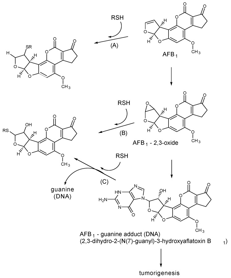
- Friedman M., Wehr C.M., Schade J.E., MacGregor J.T. Inactivation of aflatoxin B1 mutagenicity by thiols. Food Chem. Toxicol. 1982;20:887–892. - PubMed
-
- Friedman M. Sulfhydryl Groups and Food Safety. In: Friedman M., editor. Nutritional and Toxicological Aspects of Food Safety (Advances in Experimental Medicine and Biology) Vol. 177. Plenum Press; New York, NY, USA: 1984. pp. 31–63. - PubMed
Publication types
MeSH terms
Substances
LinkOut - more resources
Full Text Sources
Other Literature Sources
Medical
