. 2013 Jun 7;288(23):16247-16261.
doi: 10.1074/jbc.M112.435545. Epub 2013 Apr 23.

Diverse sequence determinants control human and mouse receptor interacting protein 3 (RIP3) and mixed lineage kinase domain-like (MLKL) interaction in necroptotic signaling

Affiliations

Diverse sequence determinants control human and mouse receptor interacting protein 3 (RIP3) and mixed lineage kinase domain-like (MLKL) interaction in necroptotic signaling

Wanze Chen et al. J Biol Chem. .

Abstract

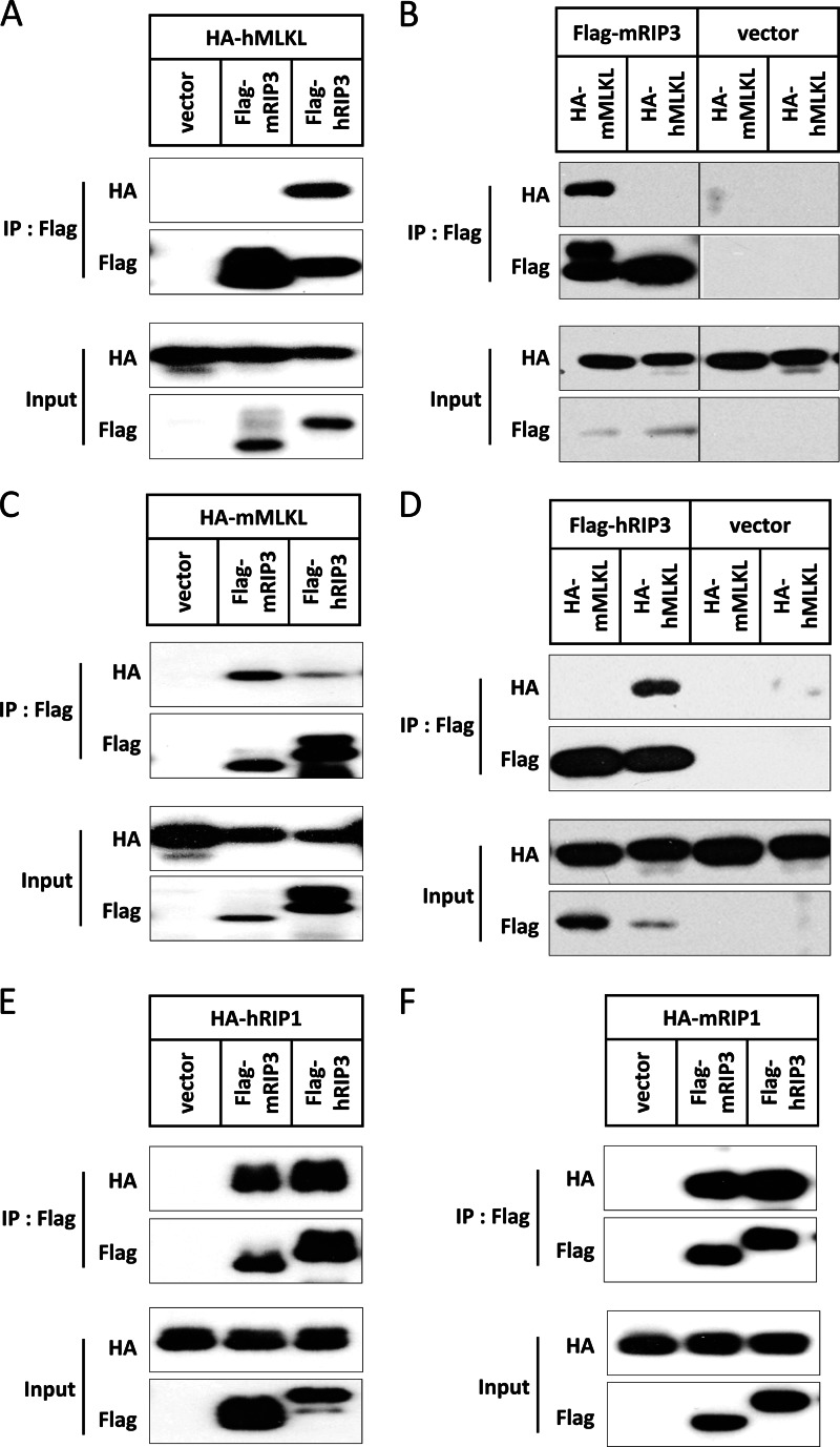
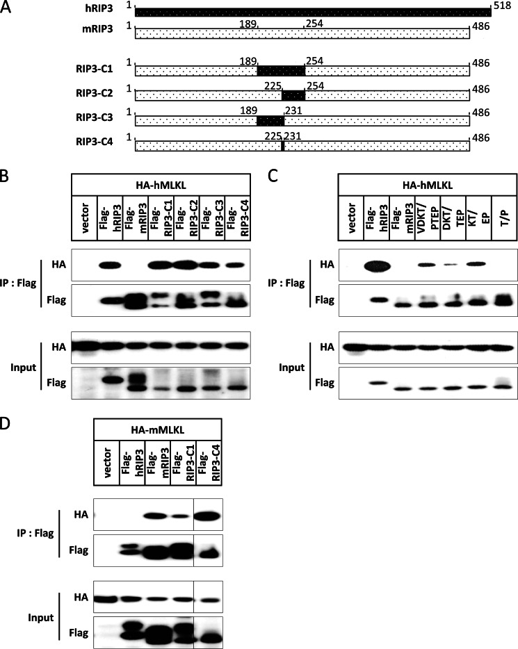
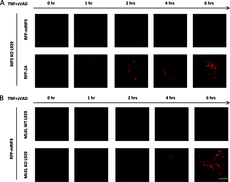
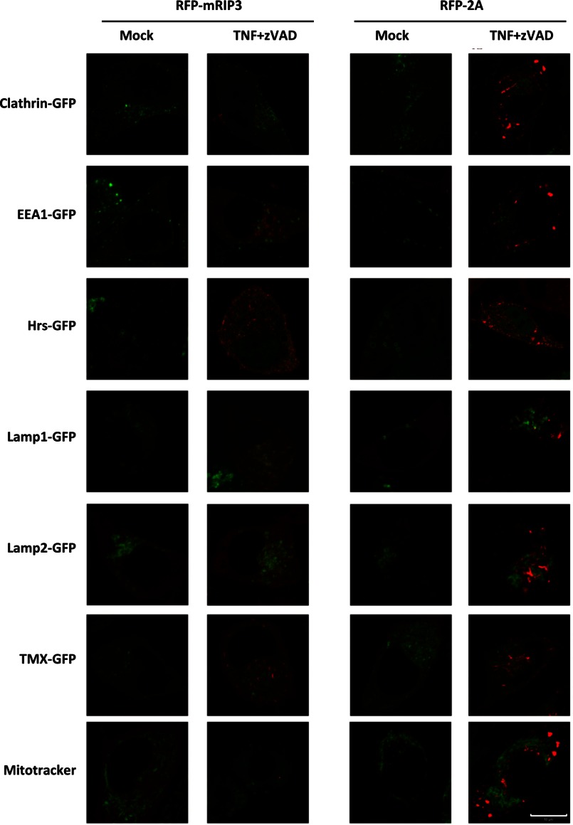
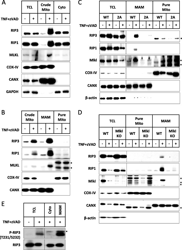
Receptor interacting protein 3 (RIP3) is a protein kinase essential for TNF-induced necroptosis. Phosphorylation on Ser-227 in human RIP3 (hRIP3) is required for its interaction with human mixed lineage kinase domain-like (MLKL) in the necrosome, a signaling complex induced by TNF stimulation. RIP1 and RIP3 mediate necrosome aggregation leading to the formation of amyloid-like signaling complexes. We found that TNF induces Thr-231 and Ser-232 phosphorylation in mouse RIP3 (mRIP3) and this phosphorylation is required for mRIP3 to interact with mMLKL. Ser-232 in mRIP3 corresponds to Ser-227 in hRIP3, whereas Thr-231 is not conserved in hRIP3. Although the RIP3-MLKL interaction is required for necroptosis in both human and mouse cells, hRIP3 does not interact with mMLKL and mRIP3 cannot bind to hMLKL. The species specificity of the RIP3-MLKL interaction is primarily determined by the sequence differences in the phosphorylation sites and the flanking sequence around the phosphorylation sites in hRIP3 and mRIP3. It appears that the RIP3-MLKL interaction has been selected as an evolutionarily conserved mechanism in mediating necroptosis signaling despite that differing structural and mechanistic bases for this interaction emerged simultaneously in different organisms. In addition, we further revealed that the interaction of RIP3 with MLKL prevented massive abnormal RIP3 aggregation, and therefore should be crucial for formation of the amyloid signaling complex of necrosomes. We also found that the interaction between RIP3 and MLKL is required for the translocation of necrosomes to mitochondria-associated membranes. Our data demonstrate the importance of the RIP3-MLKL interaction in the formation of functional necrosomes and suggest that translocation of necrosomes to mitochondria-associated membranes is essential for necroptosis signaling.

Keywords: Cell Death; MLKL; Necroptosis; Necrosis (Necrotic Death); Protein Kinases; Protein Phosphorylation; Protein-Protein Interactions; RIP; RIP3; Tumor Necrosis Factor (TNF).


Figures

FIGURE 1.
FIGURE 1.
Identification of phosphorylation sites in mRIP3. A, L929 and NIH3T3-N cells were treated with TNF (10 ng/ml) plus Z-VAD (20 μm) for the indicated time points. Cells were then harvested and the cell lysates were subjected to SDS-PAGE. Endogenous mRIP3 and GAPDH were detected by immunoblotting with mRIP3 and GAPDH antibodies. B, NIH3T3-A cells stably expressing FLAG-mRIP3 were treated with TNF (30 ng/ml) plus Z-VAD for the indicated time points. Cells were then harvested and the cell lysates were subjected to immunoblotting. C, FLAG-mRIP3-expressing NIH3T3-A cells were lysed and immunoprecipitated with anti-FLAG antibody-conjugated beads. The immunoprecipitates were treated with or without λ-phosphatase (1000 units/mg of total protein) for 30 min at 30 °C and then subjected to immunoblotting with anti-FLAG antibody. D, HT29 cells were treated with human TNF (30 nm) plus Z-VAD (20 μm) plus cycloheximide (CHX) (2 μg/ml) for the indicated time points. Cells were lysed with lysis buffer without phosphatase inhibitor, and treated with or without λ-phosphatase. Endogenous RIP3 was examined by immunoblotting. E, FLAG-mRIP3-expressing NIH3T3-A cells were treated with TNF plus Z-VAD for 2 h. Cells were then harvested and lysed before FLAG-mRIP3 was immunoprecipitated with anti-FLAG beads. After purification, FLAG-mRIP3 was digested with trypsin and the phosphopeptides were enriched by immobilized metal ion affinity chromatography, and then analyzed by mass spectrometry (see ”Experimental Procedures“). 12 potential phosphorylation sites of mRIP3 were identified. F, 12 mRIP3 mutants were created by mutating each of the 12 potential phosphorylation sites, Thr or Ser to Ala. All the mutants were introduced into NIH3T3-A cells by lentiviral vector. 48 h post-infection, cells were treated with TNF plus Z-VAD for 4 h and cell viability (PI negative) was analyzed by flow cytometer. The expression of these mutants was analyzed by immunoblotting with anti-FLAG antibody. GAPDH was analyzed as control. The data were shown as mean ± S.D. of triplicate samples. G, the 12 mRIP3 mutants were expressed in 293T cells by transient transfection. Cells were harvested and the cell lysates were then subjected to immunoblotting with anti-FLAG antibody. Samples in panels A–C and G were subjected to SDS-PAGE for a longer period time to separate shifted and non-shifted mRIP3 bands.
FIGURE 2.
FIGURE 2.
Phosphorylation of mRIP3 on Thr-231 and Ser-232 is essential for mRIP3 to mediate necroptosis. A, HeLa cells were infected with lentivirus encoding hRIP3-WT or hRIP3-S227A. 48 h later, cells were treated with human TNF (30 ng/ml) plus Z-VAD (20 μm) plus Smac mimetic (10 nm) for 24 h. Cell viability was then analyzed. NIH3T3-A cells were infected with lentivirus encoding mRIP3-WT or mRIP3-S232A. 48 h later, cells were treated with mouse TNF plus Z-VAD for 4 h followed by cell viability analysis. S227A mutation completely abolished the function of hRIP3 in mediating cell death but the S232A mutation in mRIP3 only partly impaired its function. B, alignment of the kinase domain of mRIP3 and hRIP3. Identical amino acid residues were shown in black background. Similar amino acid residues were shown in gray background. The solid triangle denotes the phosphorylation sites. C, a mRIP3 mutant, mRIP3–12M, in which the 12 potential phosphorylation sites were mutated to Ala was introduced into NIH3T3-A cells. The TNF plus Z-VAD-induced cell death was analyzed as described in panel A. D and E, mRIP3 mutants as indicated in the figure were introduced into NIH3T3-A cells. Cell viability was analyzed as in panel A. The simultaneous mutation of Thr-231 and Ser-232 to Ala (2A mutant) has similar effects as the mutation of all 12 potential phosphorylation sites in mRIP3. The data were shown as mean ± S.D. of triplicate samples. F, expression vector encoding nothing (empty vector), mRIP3-WT, D143N (kinase-dead), T231A, S232A, or 2A was transfected into 293T cells. 24 h after transfection, cells were harvested and the cell lysates were immunoblotted with mRIP3 antibody or anti-phosphorylated mRIP3 antibody. The anti-phospho-mRIP3 antibody detects Thr-231 and Ser-232 dual phosphorylated RIP3. G, L929 cells were treated with or without TNF plus Z-VAD for different periods of time. Cells were lysed and the lysates were subjected to immunoblotting with anti-phospho-Thr-231/Ser-232-mRIP3 and anti-RIP3 antibodies.
FIGURE 3.
FIGURE 3.
T231A/S232A double mutation disrupts the interaction of mRIP3 with mMLKL without affecting its kinase activity. A, empty vector, FLAG-mRIP3 wild-type, or T231A, S232A, 2A mutants were co-transfected with HA-mMLKL in 293T cells, respectively. 18 h later, cells were lysed and the lysates were subjected to immunoprecipitation with anti-FLAG beads. The input and immunoprecipitates (IP) were immunoblotted with anti-HA and anti-FLAG antibodies. B, the experiments were performed as in A except that HA-mMLKL was replaced by HA-mRIP1. C, 293T cells were transfected with empty vector, FLAG-mRIP3 wild-type, or FLAG-mRIP3 mutants as indicated. 36 h later, FLAG-mRIP3 wild-type and mutants from cell lysates were immunoprecipitated with anti-FLAG beads. The immunoprecipitates were subjected to in vitro kinase assay with [γ-32P]ATP using MBP as substrate. The amount of input RIP3 protein was analyzed by immunoblotting with anti-FLAG antibody. mRIP3 kinase-dead mutant D143N was included as a negative control.
FIGURE 4.
FIGURE 4.
Thr-231 and Ser-232 phosphorylation in mRIP3 is essential for the recruitment of mMLKL to necrosomes. A, cell lysates from RIP3 wild-type and RIP3 knock-out (KO) L929 cells were immunoblotted with mRIP3 antibody. GAPDH was immunoblotted as the loading control. B, L929 RIP3-KO cells were reconstituted with empty vector, FLAG-mRIP3 wild-type, and 2A mutant by lentiviral vector. 48 h later, cells were treated with TNF plus Z-VAD for 4 h and cell viability was analyzed. The data were shown by mean ± S.D. of triplicate samples. L929 parental (RIP3 wild-type) cells were included as control. C and D, RIP3 KO L929 cells stably expressing FLAG-mRIP3, 2A mutant, or empty vector were treated with TNF plus Z-VAD for the indicated time points. Cells were lysed with lysis buffer containing 1% Triton X-100. Supernatants (input) were subjected to RIP3 complex immunoprecipitation (IP) using anti-FLAG beads. The pellets (Triton X-100-insoluble fractions) were mixed with SDS sample buffer, ultrasonicated, and boiled. The immunoprecipitates, pellets, and inputs were immunoblotted with antibodies as indicated. Vimentin and GAPDH were immunoblotted as loading control of the pellet and total cell lysate, respectively.
FIGURE 5.
FIGURE 5.
The interaction of RIP3 and MLKL is species specific. A, empty vector, FLAG-mRIP3, and FLAG-hRIP3 were co-transfected with HA-hMLKL. 18 h later, cells were harvested and the cell lysates were then subjected to immunoprecipitation (IP) with anti-FLAG beads. Both immunoprecipitates and total cell lysates (input) were immunoblotted with anti-HA and anti-FLAG antibodies. B, HA-mMLKL and HA-hMLKL were co-transfected with FLAG-mRIP3 or empty vector. 18 h later, cells were harvested and the cell lysates were subjected to immunoprecipitation with anti-FLAG beads. Both immunoprecipitates and total cell lysate were immunoblotted with anti-HA and anti-FLAG antibodies. C, the same as in A except that HA-hMLKL was replaced by HA-mMLKL. D, the same as in B except that FLAG-mRIP3 was replaced by FLAG-hRIP3. E, the same as in A except that HA-hMLKL was replaced by HA-hRIP1. F, the same as in A except that HA-hMLKL was replaced by HA-mRIP1.
FIGURE 6.
FIGURE 6.
The sequence flanking the phosphorylation sites determines the species-specific interaction between RIP3 and MLKL. A, schematic presentation of RIP3 chimeric constructs. RIP3-C1 was made using mRIP3 as the backbone and amino acids 189–254 were replaced with hRIP3 aa 184–249. Similarly, RIP3-C2, -C3, and -C4 were made by replacing mouse 225–254 aa with human 220–249 aa, mouse 189–231 aa with human 184–226 aa, and mouse 225–231 aa with human 220–226 aa, respectively. B, empty vector and FLAG-hRIP3, FLAG-mRIP3, RIP3-C1, -C2, -C3, and -C4 were co-transfected with HA-hMLKL into 293T cells. 18 h after transfection, cells were harvested and the cell lysates were subjected to immunoprecipitation (IP) with anti-FLAG beads. Both immunoprecipitates and total cell lysates were immunoblotted with anti-HA and anti-FLAG antibodies. C, RIP3-VDKT/PTEP, RIP3-DKT/TEP, RIP3-KT/EP, and RIP3-T/P were made by using mRIP3 as backbone and the amino acids 228VDKT231 were replaced with hRIP3 223PTEP226, 229DKT231 with hRIP3 224TEP226, 230KT231 with hRIP3 225EP226, and Thr-231 with hRIP3 Pro-226, respectively. These constructs together with FLAG-hRIP3 and FLAG-mRIP3 were co-transfected with HA-hMLKL and the results analyzed as in B. D, empty vector and FLAG-hRIP3, FLAG-mRIP3, RIP3-C1, and -C4 were co-transfected with HA-mMLKL into 293T cells and the results analyzed as in B.
FIGURE 7.
FIGURE 7.
Loss of interaction with MLKL leads to massive aggregation of RIP3 in TNF-stimulated L929 cells. A, RIP3 KO L929 cells were infected with lentivirus encoding RFP-mRIP3 or RFP-mRIP3–2A. The infected cells were then treated with TNF plus Z-VAD for different periods of time as indicated and live cell imaging was recorded. B, the same as in A except that MLKL wild-type and KO L929 cells were infected with lentivirus encoding RFP-mRIP3. Scale bar, 10 μm.
FIGURE 8.
FIGURE 8.
RIP3 KO L929 cells were reconstituted with RFP-mRIP3 or RFP-mRIP3–2A. The cells were then either transfected with GFP-clathrin (marker of clathrin-dependent endocytosis vesicles), GFP-EEA1 (marker of early endosomes), GFP-Hrs (marker of multivesicular bodies), GFP-TMX (ER marker), GFP-Lamp1 (Lysosome marker), GFP-Lamp2 (Lysosome marker), or stained with MitoTracker. The cells were treated with or without TNF plus Z-VAD for 3 h and live cell imaging was recorded. Scale bar, 10 μm.
FIGURE 9.
FIGURE 9.
The interaction of RIP3 with MLKL leads to translocation of necrosomes to MAM. A, L929 cells were fractionated before and after 3 h of TNF plus Z-VAD treatment. The total cell lysates (TCL), crude mitochondrial (Crude Mito), and cytosolic (Cyto) fractions were immunoblotted with antibodies against RIP3, RIP1, MLKL, COX-IV (mitochondrial marker), CANX (ER marker, enriched in MAM), and GAPDH. B, the crude mitochondria-containing fraction was further fractionated into highly pure mitochondria (Pure Mito) and MAM. The crude mitochondria, MAM, and pure mitochondria were immunoblotted with antibodies against RIP3, RIP1, MLKL, COX-IV, and CANX. C, RIP3 KO L929 cells were reconstituted with wild-type RIP3 or RIP3–2A. The cells were treated with or without TNF plus Z-VAD for 3 h and the cell lysates were then fractionated. The TCL, MAM, and pure mitochondria were immunoblotted with antibodies against RIP3, RIP1, MLKL, COX-IV, CANX, and actin. D, the same as in C except that MLKL wild-type and KO L929 cells were used here. E, L929 cells were treated with or without TNF plus Z-VAD for 3 h and then fractionated. The TCL, Cyto, and MAM were immunoblotted with an antibody against Thr-231 and Ser-232 dual-phosphorylated mRIP3 (P-RIP3). Total RIP3 was immunoblotted as loading controls. Asterisks indicate nonspecific bands.

Similar articles

Cited by

References

    1. Degterev A., Huang Z., Boyce M., Li Y., Jagtap P., Mizushima N., Cuny G. D., Mitchison T. J., Moskowitz M. A., Yuan J. (2005) Chemical inhibitor of nonapoptotic cell death with therapeutic potential for ischemic brain injury. Nat. Chem. Biol. 1, 112–119 - PubMed
    1. Cho Y. S., Challa S., Moquin D., Genga R., Ray T. D., Guildford M., Chan F. K. (2009) Phosphorylation-driven assembly of the RIP1-RIP3 complex regulates programmed necrosis and virus-induced inflammation. Cell 137, 1112–1123 - PMC - PubMed
    1. Upton J. W., Kaiser W. J., Mocarski E. S. (2010) Virus inhibition of RIP3-dependent necrosis. Cell Host Microbe 7, 302–313 - PMC - PubMed
    1. Upton J. W., Kaiser W. J., Mocarski E. S. (2012) DAI/ZBP1/DLM-1 complexes with RIP3 to mediate virus-induced programmed necrosis that is targeted by murine cytomegalovirus vIRA. Cell Host Microbe 11, 290–297 - PMC - PubMed
    1. Robinson N., McComb S., Mulligan R., Dudani R., Krishnan L., Sad S. (2012) Type I interferon induces necroptosis in macrophages during infection with Salmonella enterica serovar Typhimurium. Nat. Immunol. 13, 954–962 - PMC - PubMed

Publication types

MeSH terms