. 2013 Jun 7;288(23):16862-16871.
doi: 10.1074/jbc.M113.464750. Epub 2013 Apr 23.

Batroxobin binds fibrin with higher affinity and promotes clot expansion to a greater extent than thrombin

Affiliations

Batroxobin binds fibrin with higher affinity and promotes clot expansion to a greater extent than thrombin

Trang T Vu et al. J Biol Chem. .

Abstract

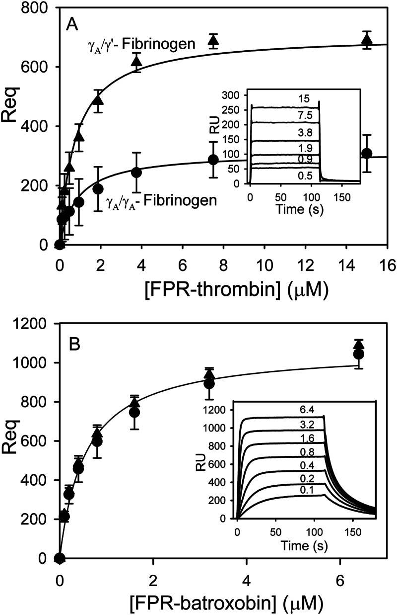
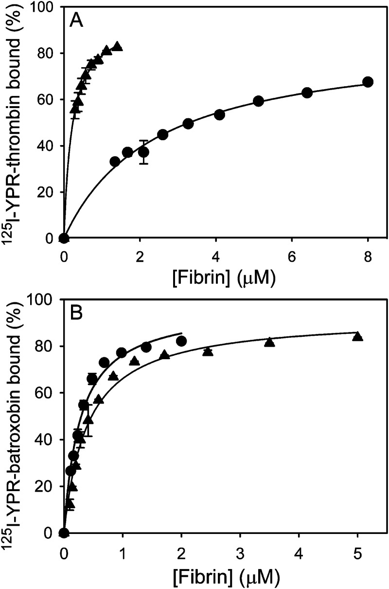
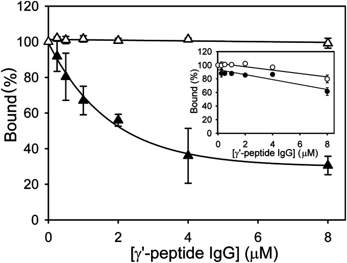
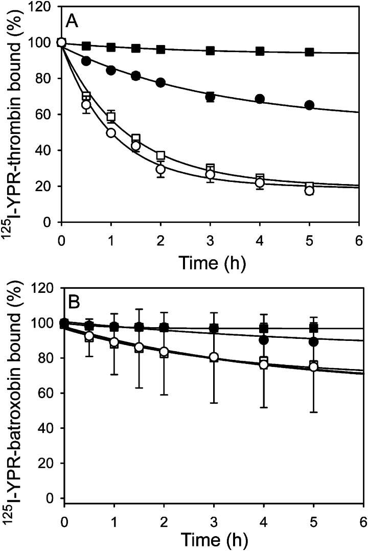
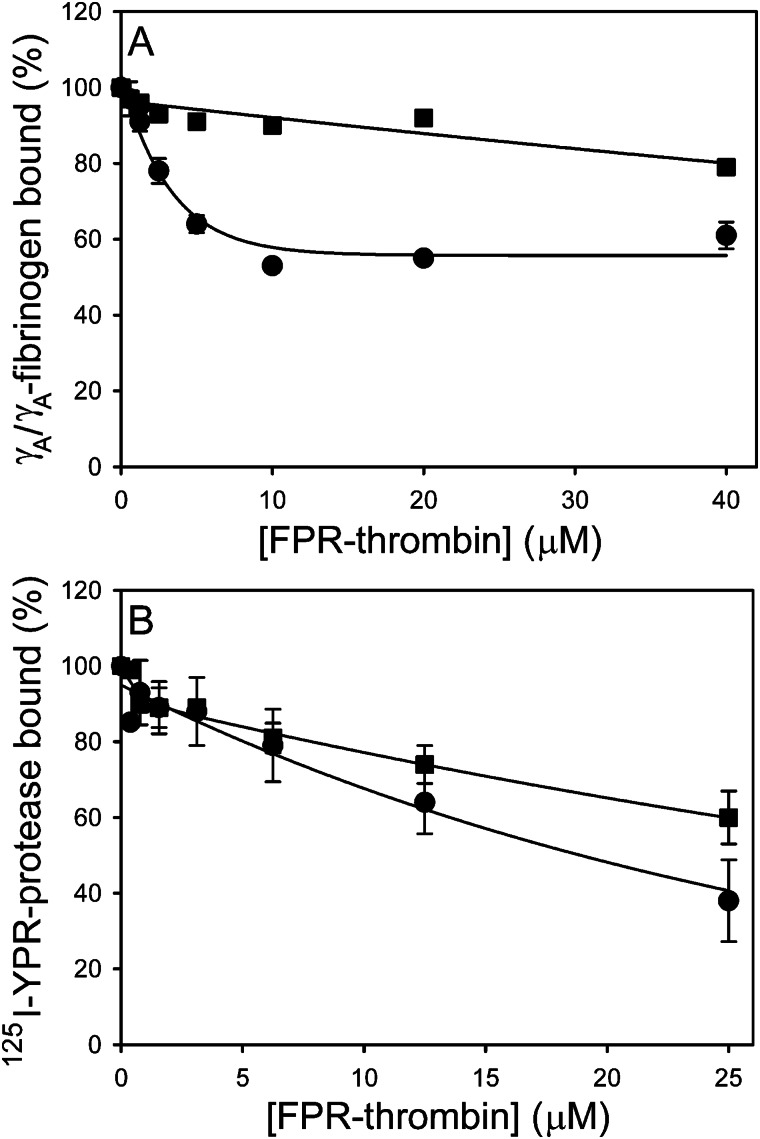
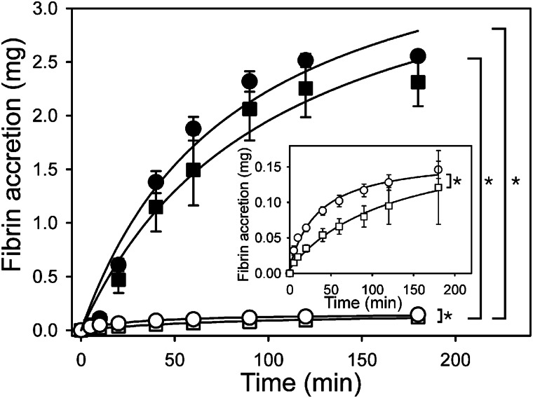
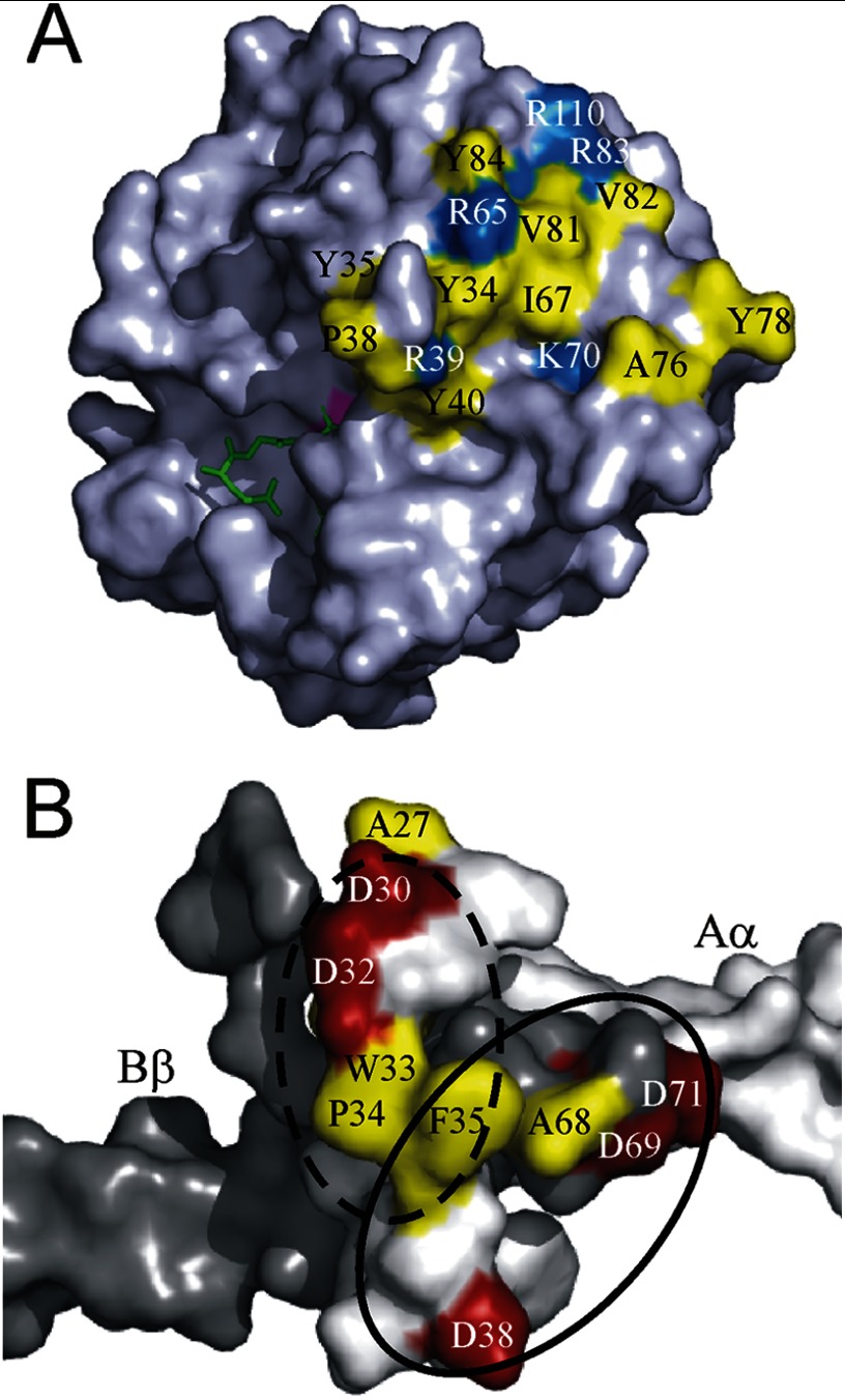
Batroxobin is a thrombin-like serine protease from the venom of Bothrops atrox moojeni that clots fibrinogen. In contrast to thrombin, which releases fibrinopeptide A and B from the NH2-terminal domains of the Aα- and Bβ-chains of fibrinogen, respectively, batroxobin only releases fibrinopeptide A. Because the mechanism responsible for these differences is unknown, we compared the interactions of batroxobin and thrombin with the predominant γA/γA isoform of fibrin(ogen) and the γA/γ' variant with an extended γ-chain. Thrombin binds to the γ'-chain and forms a higher affinity interaction with γA/γ'-fibrin(ogen) than γA/γA-fibrin(ogen). In contrast, batroxobin binds both fibrin(ogen) isoforms with similar high affinity (Kd values of about 0.5 μM) even though it does not interact with the γ'-chain. The batroxobin-binding sites on fibrin(ogen) only partially overlap with those of thrombin because thrombin attenuates, but does not abrogate, the interaction of γA/γA-fibrinogen with batroxobin. Furthermore, although both thrombin and batroxobin bind to the central E-region of fibrinogen with a Kd value of 2-5 μM, the α(17-51) and Bβ(1-42) regions bind thrombin but not batroxobin. Once bound to fibrin, the capacity of batroxobin to promote fibrin accretion is 18-fold greater than that of thrombin, a finding that may explain the microvascular thrombosis that complicates envenomation by B. atrox moojeni. Therefore, batroxobin binds fibrin(ogen) in a manner distinct from thrombin, which may contribute to its higher affinity interaction, selective fibrinopeptide A release, and prothrombotic properties.

Keywords: Batroxobin; Blood Coagulation Factors; Fibrin; Fibrinogen; Fibrinopeptides; Snake Venom; Thrombin.


Figures

FIGURE 1.
FIGURE 1.
Integrity of thrombin and batroxobin as assessed by SDS-PAGE analysis. Thrombin (lane 2) and batroxobin (lane 4) were subjected to SDS-PAGE analysis on a 4–15% polyacrylamide gradient gel under nonreducing conditions. The molecular weights of the mobility markers are shown on the left (lane 1).
FIGURE 2.
FIGURE 2.
SPR analysis of the interaction of FPR-batroxobin and FPR-thrombin with immobilized γAA- or γA/γ′-fibrinogen. γAA- (●) and γA/γ′-fibrinogen (▴) were adsorbed on individual flow cells to ∼6000–8000 RU. Increasing concentrations (0–15 μm) of FPR-thrombin (A) or FPR-batroxobin (B) were successively injected into flow cells for 2 min, followed by a 4-min wash to monitor dissociation. The amount of protease bound at equilibrium (Req) after background correction is plotted against the input FPR-protease concentration. The insets show representative sensorgrams for the interaction of FPR-thrombin (A) and FPR-batroxobin (B) with γAA-fibrinogen in concentrations up to 15 and 6.4 μm, respectively. Data points represent the mean ± S.D. of 2- 3 experiments, and the lines represent nonlinear regression analyses.
FIGURE 3.
FIGURE 3.
Binding of 125I-YPR-batroxobin and 125I-YPR-thrombin to γAA- or γA/γ′-fibrin clots. 20 nm 125I-YPR-thrombin (A) or 40 nm 125I-YPR-batroxobin (B) was added to microcentrifuge tubes containing 0–8 μm γAA- (●) or γA/γ′-fibrinogen (▴), and 10 nm α-thrombin was used to initiate clotting. After incubation for 60 min, clots were pelleted by centrifugation, and free 125I-YPR-protease in the supernatant was used to calculate the bound fraction. Data are plotted as the percentage of 125I-YPR-protease bound to fibrin clots versus the fibrinogen concentration. Data points represent the mean ± S.D. of two experiments, each performed in duplicate, and the lines represent nonlinear regression analyses.
FIGURE 4.
FIGURE 4.
Effect of γ′-peptide-directed IgG on 125I-YPR-batroxobin and 125I-YPR-thrombin binding to γAA- or γA/γ′-fibrin clots. The binding of 40 nm 125I-YPR-thrombin (●/▴) or 125I-YPR-batroxobin (○/Δ) to clots formed from 1 μm γAA- (circles) or γA/γ′-fibrinogen (triangles) was assessed in the absence or presence of γ′-peptide-directed IgG up to 8 μm. Clots were generated with thrombin and pelleted, and clot-bound 125I-YPR-protease was determined. The percentage of 125I-YPR-protease bound in the absence of antibody is plotted versus antibody concentration. The main figure illustrates the percentage of 125I-YPR-protease bound to γA/γ′-fibrin clots, and the inset shows the percentage of 125I-YPR-protease bound to γAA-fibrin clots. Symbols represent the mean ± S.D. of two experiments, each performed in duplicate, and the lines represent nonlinear regression analyses of the data.
FIGURE 5.
FIGURE 5.
Diffusion of 125I-YPR-batroxobin and 125I-YPR-thrombin from γAA- or γA/γ′-fibrin clots. Clots were formed around plastic loops by incubating 9 μm γAA- (●/○) or γA/γ′-fibrinogen (■/□) with 100 nm thrombin and 30 nm fXIII in the presence of 20 nm 125I-YPR-thrombin (A) or 40 nm 125I-YPR-batroxobin (B) and then immersed in solutions containing TBS-Ca (●/■) or 2 m NaCl and 5 mm EDTA (○/□). At the indicated time points, 0.5-ml aliquots of the bathing solutions were removed and counted for radioactivity to quantify bound 125I-YPR-protease. Data are plotted as the percentage of clot-bound 125I-YPR-protease versus time, and lines represent nonlinear regression analyses. Data points represent the mean ± S.D. of three separate determinations.
FIGURE 6.
FIGURE 6.
Effect of FPR-thrombin on batroxobin binding to γAA-fibrinogen or γAA-fibrin clots. A, effect of FPR-thrombin on γAA-fibrinogen binding to immobilized FPR-batroxobin was determined using SPR. γAA-fibrinogen (2.5 μm) was incubated with FPR-thrombin concentrations up to 40 μm in the absence (●) or presence (■) of 80 μm hirudin, and the mixture was then injected into flow cells containing immobilized FPR-batroxobin. Binding in the presence of competitors was normalized relative to that determined in their absence. Data are plotted as the percentage of γAA-fibrinogen bound versus the FPR-thrombin concentration. B, binding of 125I-YPR-batroxobin (■) and 125I-YPR-thrombin (●) to γAA-fibrin clots was measured in the absence or presence of FPR-thrombin concentrations up to 25 μm. Clots were formed by incubating 2 or 0.06 μm γAA- fibrinogen with 20 nm 125I-YPR-thrombin or 40 nm 125I-YPR-batroxobin, respectively, and 10 nm thrombin. After 60 min, clots were counted for radioactivity to quantify bound 125I-YPR-protease. Data are plotted as the percentage of 125I-YPR-protease bound versus the FPR-thrombin concentration. Data points represent the mean ± S.D. of 3–4 experiments, and the lines represent nonlinear regression analyses.
FIGURE 7.
FIGURE 7.
Clot accretion induced by batroxobin or thrombin bound to γAA- or γA/γ′-fibrin clots. Clots were formed around plastic loops by incubating 8.3 μm γAA-fibrinogen (●/○) or γA/γ′-fibrinogen (■/□) with 76.5 units/ml of batroxobin (●/■) or 45 nm thrombin (○/□) in the presence of 2 mm CaCl2, 30 nm fXIII, and 500 cpm of 125I-fibrinogen for 30 min at 37 °C. After determining radioactivity, clots were incubated in 1 ml of citrated human plasma containing ∼800,000 cpm/ml of 125I-fibrinogen. At intervals, clots were washed twice with 1 ml of HBS and again counted for radioactivity so that fibrin accretion could be determined. The inset provides an expanded view of clot accretion induced by thrombin. Data points represent the mean ± S.D. of three experiments, each performed in triplicate, and the lines represent nonlinear regression analyses. (*, p < 0.001).
FIGURE 8.
FIGURE 8.
Models of binding sites on batroxobin and fibrinogen. A, crystal structure of TSV-PA (Protein Data Bank accession number 1BQY) in complex with Glu-Gly-Arg chloromethyl ketone (green) is shown in space-filling format using PyMOL. The image is rotated clockwise ∼90° from standard view with the active site Ser-195 (magenta) facing left. Hydrophobic (yellow) and basic (blue) residues in batroxobin that constitute a putative exosite are mapped onto the TSV-PA structure. Residues are identified using chymotrypsin numbering system. B, crystal structure of the NH2 termini of fibrinogen α- and β-chains forming the thrombin-binding site (Protein Data Bank accession number 2A45) is shown in space-filling format. Both the γ-chains and the reciprocal α- and β-chains are omitted for clarity. The α- and β-chain surfaces are white and gray, respectively. Thrombin binding residues α-chain Phe-35 and Asp-38 and β-chain Ala-68, Asp-69, and Asp-71 are shown within a solid black oval with hydrophobic residues in yellow and acidic residues in red. Residues comprising the putative batroxobin-binding site include α-chain Asp-30, Asp-32, Trp-33, Pro-34, and Phe-35 (hatched black oval). The site where the unmapped α1–26 segment exits the α-chain at Αla-27 indicates the vicinity of FpA.

Similar articles

Cited by

References

    1. Wolberg A. S. (2012) Determinants of fibrin formation, structure, and function. Curr. Opin. Hematol. 19, 349–356 - PubMed
    1. Mosesson M. W. (2005) Fibrinogen and fibrin structure and functions. J. Thromb. Haemost. 3, 1894–1904 - PubMed
    1. Wolfenstein-Todel C., Mosesson M. W. (1980) Human plasma fibrinogen heterogeneity: evidence for an extended carboxyl-terminal sequence in a normal γ chain variant (γ′). Proc. Natl. Acad. Sci. U.S.A. 77, 5069–5073 - PMC - PubMed
    1. Chung D. W., Davie E. W. (1984) γ and γ′ chains of human fibrinogen are produced by alternative mRNA processing. Biochemistry 23, 4232–4236 - PubMed
    1. Fornace A. J., Jr., Cummings D. E., Comeau C. M., Kant J. A., Crabtree G. R. (1984) Structure of the human γ-fibrinogen gene. Alternate mRNA splicing near the 3′ end of the gene produces γA and γB forms of γ-fibrinogen. J. Biol. Chem. 259, 12826–12830 - PubMed

Publication types