. 2013 Jun 14;288(24):17225-37.
doi: 10.1074/jbc.M112.421685. Epub 2013 Apr 23.

The DNAJB6 and DNAJB8 protein chaperones prevent intracellular aggregation of polyglutamine peptides

Affiliations

The DNAJB6 and DNAJB8 protein chaperones prevent intracellular aggregation of polyglutamine peptides

Judith Gillis et al. J Biol Chem. .

Abstract

Fragments of proteins containing an expanded polyglutamine (polyQ) tract are thought to initiate aggregation and toxicity in at least nine neurodegenerative diseases, including Huntington's disease. Because proteasomes appear unable to digest the polyQ tract, which can initiate intracellular protein aggregation, preventing polyQ peptide aggregation by chaperones should greatly improve polyQ clearance and prevent aggregate formation. Here we expressed polyQ peptides in cells and show that their intracellular aggregation is prevented by DNAJB6 and DNAJB8, members of the DNAJ (Hsp40) chaperone family. In contrast, HSPA/Hsp70 and DNAJB1, also members of the DNAJ chaperone family, did not prevent peptide-initiated aggregation. Intriguingly, DNAJB6 and DNAJB8 also affected the soluble levels of polyQ peptides, indicating that DNAJB6 and DNAJB8 inhibit polyQ peptide aggregation directly. Together with recent data showing that purified DNAJB6 can suppress fibrillation of polyQ peptides far more efficiently than polyQ expanded protein fragments in vitro, we conclude that the mechanism of DNAJB6 and DNAJB8 is suppression of polyQ protein aggregation by directly binding the polyQ tract.

Keywords: Confocal Microscopy; DNAJB; Huntington's Disease; Molecular Chaperone; Peptides; Polyglutamine Disease.


Figures

FIGURE 1.
FIGURE 1.
DNAJB6 and DNAJB8 reduce aggregation of expanded polyQ peptides. A, schematic representation of the cleavage of GFP-Ub-polyQ constructs by Ub C-terminal hydrolases directly after Ub, thereby separating GFP-Ub and polyQ. B, the percentage of the fluorescent HEK293 cells that contained fluorescent aggregates at 72 h after transfection with GFP-Ub-Q104 in combination with DNAJB6a, DNAJB6b, DNAJB8, HSPA1A, or DNAJB1, respectively. Data are mean ± S.E. of three independent experiments (two-tailed unpaired Student's t test). *, p < 0.05; ***, p < 0.001; ns, not significant. C, SDS-insoluble fractions prepared 72 h after transfection with the indicated constructs and analyzed by filter trap analysis. Blots were stained for polyQ using 1C2 antibody. Data are mean ± S.E. of three independent experiments (two-tailed unpaired Student's t test). **, p < 0.01; ***, p < 0.001. D, expression levels of the GFP or dsRED-tagged chaperones coexpressed with Ub-Q104 in HEK293 cells for 72 h. No molecular weight marker is present because the fluorescence of the proteins was measured directly. E, upper panel, knockdown of DNAJB6 increases aggregation in cells expressing Ub-Q104, as analyzed by filter trap analysis 72 h after transfection. Lower panel, knockdown of DNAJB6 by siRNA leads to a reduction in endogenous DNAJB6 levels. Data are mean ± S.E. of three independent experiments (two-tailed unpaired Student's t test). *, p < 0.05; ***, p < 0.001.
FIGURE 2.
FIGURE 2.
DNAJB6 and DNAJB8 reduce insoluble levels of expanded polyQ peptides and Htt-exon-1. A, SDS-soluble and SDS-insoluble fractions prepared 24 h after transfection with GFP-Ub-Q104 in combination with DNAJB6b and DNAJB8 and analyzed by Western blotting. Blots were stained for polyQ using 1C2 antibody and anti-actin antibody. The arrows indicate specific Glu-104 peptide bands that are quantified together in the lower panel. Data are mean ± S.E. of three independent experiments (two-tailed unpaired Student's t test). **, p < 0.01; ***, p < 0.001; ns, not significant. B, SDS-soluble and SDS-insoluble fractions prepared 72 h after transfection with Htt-exon-1-Q103-GFP in combination with DNAJB6b and DNAJB8 and analyzed by Western blotting. The blots were stained for polyQ using 1C2 antibody (upper panel), anti-GFP antibody (center panel), and anti-actin antibody (lower panel).
FIGURE 3.
FIGURE 3.
Tetracysteine (C4)-tagged polyQ peptides behave similarly as non-labeled polyQ peptides. A, the percentage of HEK293 cells containing aggregates 72 h after transfection with GFP-Ub-Q16/Q104 or C4-tagged GFP-Ub-Q17/Q99. Data are mean ± S.E. of three independent experiments. Similar amounts of aggregates were detected with either non-tagged or C4-tagged polyQ peptides. B, SDS-soluble and SDS-insoluble fractions prepared 24 h after transfection of HEK293 cells with Ub-Q99-C4 in combination with DNAJB6b and DNAJB8 and analyzed by Western blotting. The blots were stained for polyQ (1C2) and actin. The arrows indicate specific Glu-104 peptide bands. C, SDS-insoluble fractions of HEK293 cells expressing GFP-Ub-Q16/Q104 or C4-tagged GFP-Ub-Q17/Q99 prepared 24 h after transfection were stained for polyQ. D, confocal microscopy images of ReAsH-labeled HeLa cells expressing C4-tagged GFP-Ub-Q17 or GFP-Ub-Q99 48 h after transfection. ReAsH-positive cells were detected only when polyQ-expanded C4-tagged Glu-99 peptides were expressed (lower panel). The inset shows a GFP-Ub-decorated aggregate. Scale bar = 10 μm.
FIGURE 4.
FIGURE 4.
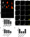
The chaperones DNAJB6b and DNAJB8 interact with aggregated polyQ peptides. A, FLIM analysis of FlAsH in HeLa cells expressing Ub-Q99-C4 labeled with either FlAsH alone or both with FlAsH and ReAsH showing in each column, from left to right, representative graphs of fluorescence intensity, a false-color map of the fluorescence lifetime calculated from the phase shift (τϕ), and a histogram of the lifetime distribution with the same false-color scale as the lifetime map. The histogram represents average phase modulation lifetimes. Only aggregated polyQ peptides showed high FRET-FLIM efficiencies. ns, not significant. Data are mean ± S.E. (two-tailed unpaired Student's t test). ***, p < 0.001; ns, not significant. B, FLIM analysis of GFP in HeLa cells expressing ReAsH-labeled Ub-Q99-C4 together with the indicated GFP-tagged constructs 48 h after transfection. The histogram represents average phase modulation lifetimes. FLIM analysis resulted in high FRET-FLIM efficiencies between aggregated Glu-99-C4 peptides and the proteasomal subunit β7. Data are mean ± S.E. (two-tailed unpaired Student's t test). *, p < 0.05; **, p < 0.01; ***, p < 0.001; ns, not significant. C, confocal microscopy images of HeLa cells expressing ReAsH-labeled Glu-99-C4 in combination with GFP-tagged DNAJB6a, DNAJB6b, or DNAJB8, which are all present in the core of the aggregate. Red fluorescent protein-tagged DNAJB1 is not recruited into aggregates, whereas red fluorescent protein-tagged HSPA1A is only recruited to the outside of the aggregate. Scale bar = 10 μm. D, FLIM analysis of GFP in cells expressing ReAsH-labeled Ub-Q99-C4 together with indicated GFP-tagged chaperones 48 h after transfection. FLIM analysis resulted in high FRET-FLIM efficiencies between aggregated polyQ peptides and DNAJB6b and DNAJB8. No FRET-FLIM was detected between aggregated polyQ peptides and Hsp70. Data are mean ± S.E. (two-tailed unpaired Student's t test). ***, p < 0.001; ns, not significant. Scale bar = 10 μm.
FIGURE 5.
FIGURE 5.
The serine-rich region within DNAJB6 and DNAJB8 is essential for reduction of polyQ peptide-induced aggregation. A, schematic representation of domains present in DNAJB6a, DNAJB6b, and DNAJB8. NLS; nuclear localization signal. B, the percentage of fluorescent HEK293 cells that contained aggregates 72 h after transfection with GFP-Ub-Q104 in combination with DNAJB6b or DNAJB8 and their J domain (H31Q) and serine-rich region mutants (ΔSSF-TST and ΔSSF-SST, respectively). Data are mean ± S.E. of three independent experiments (two-tailed unpaired Student's t test). *, p < 0.05; **, p < 0.01; ***, p < 0.001. An inactive J domain induced a small increase in aggregation, whereas deletion of the serine-rich region severely increased aggregation of Glu-104 peptides. C, SDS-insoluble fractions prepared 72 h after transfection of HEK293 cells with he indicated constructs and analyzed by filter trap analysis. The blots were stained for polyQ using 1C2 antibody. Data are mean ± S.E. of three independent experiments (two-tailed unpaired Student's t test). **, p < 0.01; ***, p < 0.001; ns, not significant. D, Western blot analysis of the SDS-insoluble fraction of HEK cells expressing GFP-Ub-Q104 in combination with DNAJB6b or DNAJB8 and their mutants for 24 h. Western blot analyses were stained for polyQ with 1C2 antibody. The arrows indicate specific Glu-104 peptide bands. E, Western blot analysis of the SDS-soluble fraction of HEK293 cells expressing GFP-Ub-Q104 in combination with DNAJB6b or DNAJB8 and their mutants for 24 h. Western blot analyses were stained for polyQ with 1C2 antibody, anti-actin antibody (center panel), or anti-DNAJB6b/8 antibody (lower panel). The arrows indicate specific Glu-104 peptide bands. F, FLIM analysis of GFP-tagged DNAJB6b and the serine-rich region mutant (upper panel) and GFP-tagged DNAJB8 and the serine-rich region mutant (lower panel) in cells expressing ReAsH-labeled Ub-Q99-C4 48 h after transfection shows high FRET-FLIM efficiencies between aggregated polyQ peptides and DNAJB6b or DNAJB8, but much less FRET-FLIM was detected between aggregated polyQ peptides and their serine-rich region mutant counterparts. Data are mean ± S.E. (two-tailed unpaired Student's t test). *, p < 0.05; ***, p < 0.001. Scale bar = 10 μm. G, DNAJB6 or DNAJB8 do not reduce intermolecular interactions between aggregated polyQ peptides. The histogram represents average phase modulation lifetimes of FlAsH in HeLa cells expressing Ub-Q99-C4 together with the indicated constructs and labeled with both FlAsH and ReAsH 48 h after transfection.
FIGURE 6.
FIGURE 6.
DNAJB6 and DNAJB8 prevent aggregation of Glu-104 peptides by keeping them soluble. A, confocal microscopy images of HeLa cells transfected with DNAJB6a, DNAJB6b, or DNAJB8, respectively, stained with Hoechst, and fixed 72 h after transfection. Scale bar = 10 μm. B, the ratio of aggregates present in the cytoplasm, nucleus, or both compartments 72 h after transfection with GFP-Ub-Q104 in combination with DNAJB6a, DNAJB6b, or DNAJB8, respectively, in Mel JuSo cells. Data are mean ± S.E. of three independent experiments. Chaperones did not specifically reduce aggregation in the compartment where they reside. C, fluorescent recovery after photobleaching analysis shows that DNAJB6a, DNAJB6b, and DNAJB8 did not recover after photobleaching, whereas HSPA1A fluorescent levels recovered. D, fluorescence loss of aggregates was measured using fluorescence loss in photobleaching analysis by repetitive photobleaching of another part of the cytoplasm. Fluorescence loss of HSPA1A was observed, whereas fluorescent levels of DNAJB6a, DNAJB6b, and DNAJB8 were not affected.

Similar articles

Cited by

References

    1. Orr H. T., Zoghbi H. Y. (2007) Trinucleotide repeat disorders. Annu. Rev. Neurosci. 30, 575–621 - PubMed
    1. DiFiglia M., Sapp E., Chase K. O., Davies S. W., Bates G. P., Vonsattel J. P., Aronin N. (1997) Aggregation of huntingtin in neuronal intranuclear inclusions and dystrophic neurites in brain. Science 277, 1990–1993 - PubMed
    1. Goti D., Katzen S. M., Mez J., Kurtis N., Kiluk J., Ben-Haïem L., Jenkins N. A., Copeland N. G., Kakizuka A., Sharp A. H., Ross C. A., Mouton P. R., Colomer V. (2004) A mutant ataxin-3 putative-cleavage fragment in brains of Machado-Joseph disease patients and transgenic mice is cytotoxic above a critical concentration. J. Neurosci. 24, 10266–10279 - PMC - PubMed
    1. Lunkes A., Lindenberg K. S., Ben-Haïem L., Weber C., Devys D., Landwehrmeyer G. B., Mandel J. L., Trottier Y. (2002) Proteases acting on mutant huntingtin generate cleaved products that differentially build up cytoplasmic and nuclear inclusions. Mol. Cell 10, 259–269 - PubMed
    1. Cooper J. K., Schilling G., Peters M. F., Herring W. J., Sharp A. H., Kaminsky Z., Masone J., Khan F. A., Delanoy M., Borchelt D. R., Dawson V. L., Dawson T. M., Ross C. A. (1998) Truncated N-terminal fragments of huntingtin with expanded glutamine repeats form nuclear and cytoplasmic aggregates in cell culture. Hum. Mol. Genet. 7, 783–790 - PubMed

Publication types