. 2013 May 31;288(22):15687-98.
doi: 10.1074/jbc.M112.431056. Epub 2013 Apr 23.

Mammalian target of rapamycin complex 1 (mTORC1) enhances bortezomib-induced death in tuberous sclerosis complex (TSC)-null cells by a c-MYC-dependent induction of the unfolded protein response

Affiliations

Mammalian target of rapamycin complex 1 (mTORC1) enhances bortezomib-induced death in tuberous sclerosis complex (TSC)-null cells by a c-MYC-dependent induction of the unfolded protein response

Justin T Babcock et al. J Biol Chem. .

Abstract

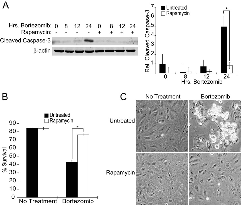
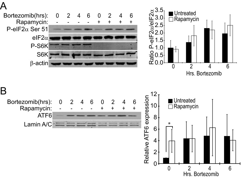
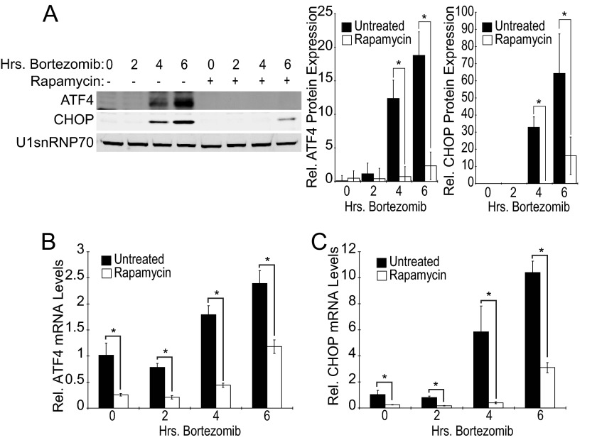
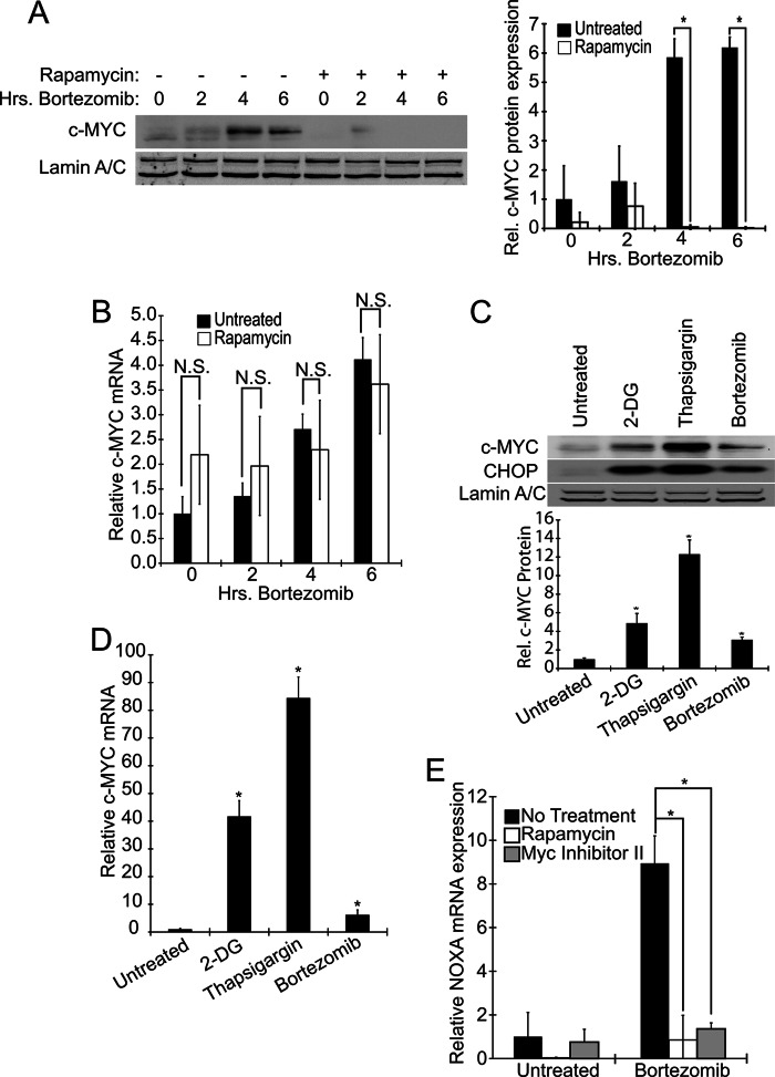
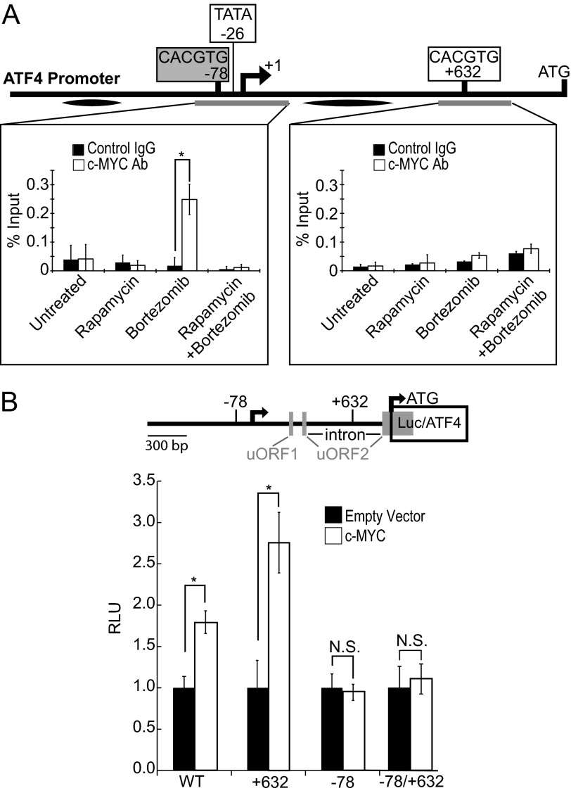
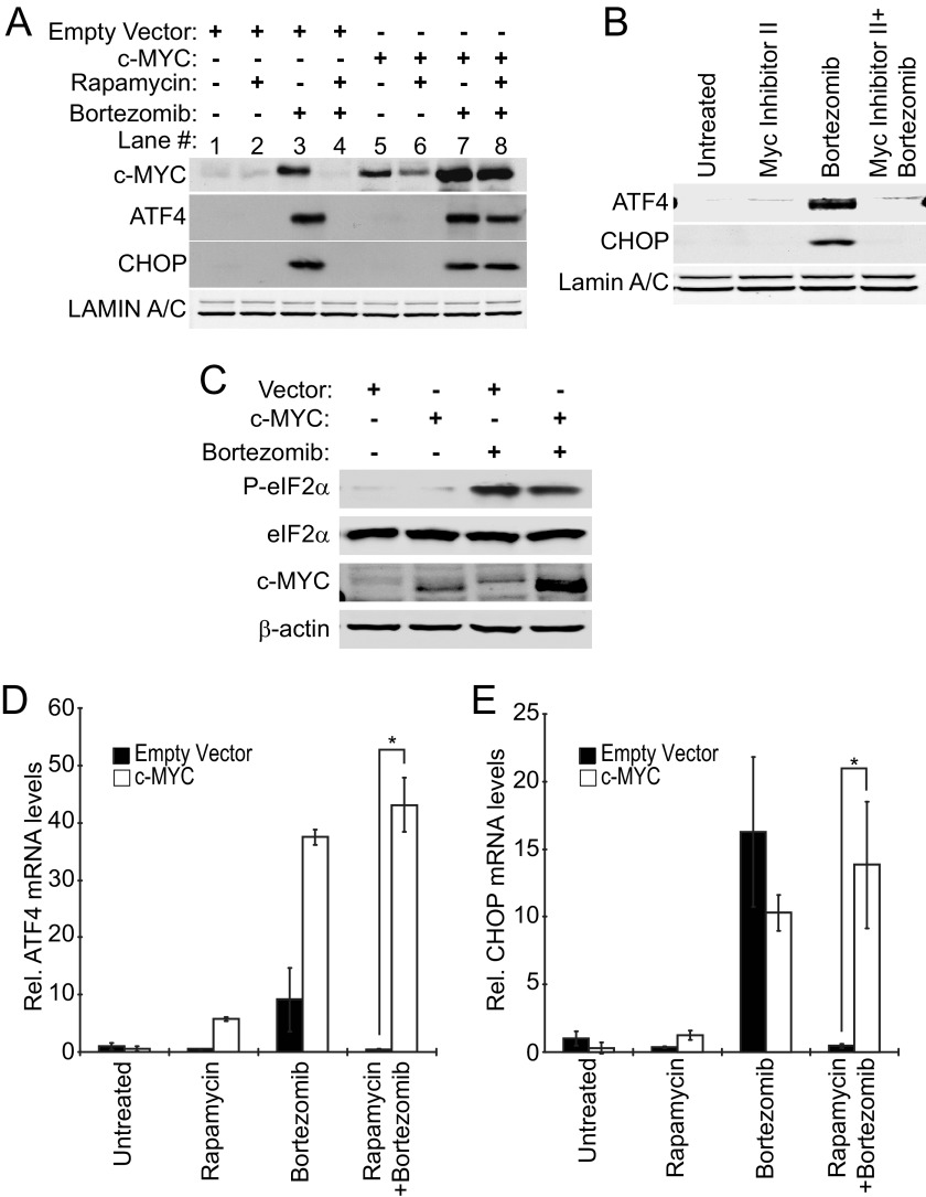
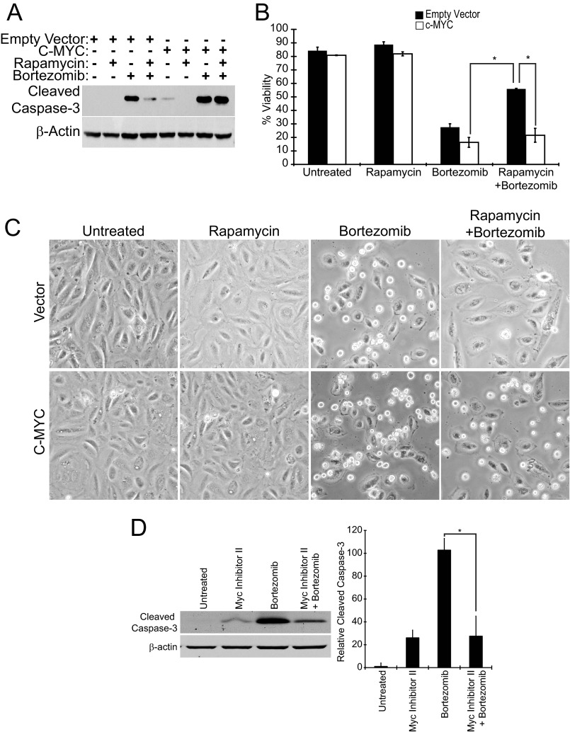
Many factors, including duration and intensity of the unfolded protein response (UPR), dictate whether cells will adapt to endoplasmic reticulum stress or undergo apoptosis. In tuberous sclerosis (TSC), elevation of mammalian target of rapamycin complex 1 (mTORC1) activity has been proposed to compound the induction of UPR transcription factors ATF4 and CHOP, suggesting that the UPR could be targeted to eradicate TSC1/2-null cells during patient therapy. Here we report that control of c-MYC translation by mTORC1 plays a key role in determining whether TSC2-null Elt3 rat leiomyoma cells apoptose in response to UPR induction by the proteasome inhibitor bortezomib. Although bortezomib induces eukaryotic initiating factor 2α phosphorylation, mTORC1 activity was also required for downstream induction of the UPR transcription factors ATF4 and CHOP by a mechanism involving increased expression of c-MYC. Although bortezomib-induced c-MYC transcription was resistant to rapamycin treatment, mTORC1 activity was required for efficient c-MYC translation. c-MYC subsequently bound to the ATF4 promoter, suggesting direct involvement of an mTORC1/c-MYC-driven signaling pathway in the activation of the UPR. Consistent with this notion, exogenously expressed c-MYC reversed the ability of rapamycin to prevent bortezomib-induced CHOP and ATF4 expression as well as apoptosis. These findings indicate that the induction of ATF4/CHOP expression occurs via mTORC1 regulation of c-MYC and that this signaling pathway is a major determinant in the ability of bortezomib to induce apoptosis.

Keywords: ATF4; Apoptosis; Bortezomib; Myc; Rapamycin; Tuberous Sclerosis (Tsc); Unfolded Protein Response; mTOR.


Figures

FIGURE 1.
FIGURE 1.
Elt3 cells undergo rapamycin-sensitive apoptosis when treated with bortezomib. A, Elt3 cells were pretreated with 50 nm rapamycin or vehicle control for 24 h before being exposed to 20 nm bortezomib for an additional 8, 12, or 24 h. Lysates were prepared after these drug treatments and analyzed by Western blot using antibodies selective for cleaved caspase-3 or β-actin. B, trypan blue staining of Elt3 cells exposed to bortezomib for 24 h in the presence or absence of rapamycin pretreatment was used to quantitate cell death. *, p < 0.05. C, shown is phase contrast microscopy of the Elt3 cells treated with rapamycin and bortezomib as indicated.
FIGURE 2.
FIGURE 2.
Early UPR markers are induced by bortezomib but unaffected by rapamycin treatment of Elt3 cells. Elt3 cells were pretreated with 50 nm rapamycin for 24 h before being exposed to 20 nm bortezomib for the times shown. A, whole cell lysates were prepared, and the indicated proteins were measured by Western blot using specific antibodies. B, nuclear lysates were prepared and probed for the nuclear cleaved fragment of ATF6 using Lamin A/C as a loading control.
FIGURE 3.
FIGURE 3.
Rapamycin prevents induction of downstream UPR markers. Elt3 cells were pretreated with 50 nm rapamycin or vehicle control for 24 h before being exposed to 20 nm bortezomib for an additional 2, 4, or 6 h. A, nuclear lysates were prepared and subjected to SDS-PAGE. ATF4, CHOP, and U1snRNP70 were measured by Western blot. Relative levels of ATF4 and CHOP proteins in each treatment group are presented as histograms on the right side of the panel. B and C, shown are quantitative real-time-PCR measurement of ATF4 and CHOP mRNA levels in Elt3 cells pretreated with rapamycin (24 h) followed by exposure to bortezomib for up to 6 h, as indicated (*, p < 0.05).
FIGURE 4.
FIGURE 4.
Elevation of ATF4 and CHOP protein levels by bortezomib requires the synthesis of new mRNA and protein. A, Elt3 cells were treated with 20 nm bortezomib for 2, 4, and 6 h in the presence or absence of 5 μg/ml actinomycin D. ATF4, CHOP, and lamin A/C levels in nuclear lysates were determined by Western blot. B, Elt3 cells were treated as in A. Phosphorylation of eIF2α, total eIF2α, the cleaved amino-terminal fragment of ATF6, and β-actin in cell lysates were determined by Western blot. C, Elt3 cells were treated with 20 nm bortezomib in the presence or absence of 100 μg/ml cycloheximide. Protein levels were measured as in A. D, Elt3 cells were treated as in C. Protein levels were measured as in B.
FIGURE 5.
FIGURE 5.
Bortezomib and other ER stressors induce expression and activity of c-MYC in a rapamycin-sensitive manner. Elt3 cells were pretreated with 50 nm rapamycin or vehicle control for 24 h before exposure to 20 nm bortezomib for 2, 4, or 6 h, as indicated. A, lysates were then prepared, and the levels of c-MYC and lamin A/C were measured by Western blot. B, levels of c-MYC mRNA were measured under the same conditions as A. C and D, 10 mm 2-deoxyglucose (2-DG), 1 μm thapsigargin, or 20 nm bortezomib were separately used to induce ER stress for 6 h. c-MYC and CHOP protein and c-MYC mRNA levels were then measured. N.S., not significant. E, basal- and bortezomib (50 nm, 6 h)-induced NOXA mRNA levels were measured after pretreatment with rapamycin (24 h) or MYC inhibitor II (2 h). *, p < 0.05.
FIGURE 6.
FIGURE 6.
c-MYC binds to and stimulates the ATF4 promoter. A, Elt3 cells were treated with 50 nm rapamycin for 24 h and 20 nm bortezomib for 4 h where indicated. Cells were then fixed and analyzed by ChIP for c-MYC binding to the E-boxes at positions −78 and +632 of the rat ATF4 promoter. Graphs represent the mean ± S.D. of three independent ChIP experiments. Gray bars below the gene indicate the regions amplified. Black ovals indicate additional control sequences found not to be amplified following c-MYC immunoprecipitation.4 Ab, antibody. B, 1.5 kb of the ATF4 gene (5′ of rat ATF4 coding region) were cloned upstream of the luciferase gene. The E-boxes located at −78 and +632 were mutated singly or together to prevent c-MYC binding and activation of luciferase transcription. These reporters were then transfected into 293T cells with empty vector or c-MYC, and luciferase activity was determined 48 h later. *, p < 0.05. RLU, relative light units.
FIGURE 7.
FIGURE 7.
Overexpression of c-MYC rescues rapamycin-mediated suppression of ATF4 and CHOP expression in cells exposed to bortezomib. Lentivirus was used to stably express c-MYC in Elt3 cells. A, vector- and c-MYC-expressing cells were then pretreated with vehicle control or 50 nm rapamycin (24 h) before treatment with bortezomib for 6 h. Nuclear lysates were prepared, and levels of ATF4, CHOP, and Lamin A/C proteins were measured by Western blot. B, Elt3 cells were pretreated with Myc inhibitor II for 2 h before treatment with bortezomib for an additional 6 h. Western blots were executed as in A. C, Elt3 cells overexpressing c-MYC were treated with bortezomib for 6 h. Phosphorylation of eIF2α, total eIF2α, c-MYC, and β-actin were measured by Western blot. D and E, under the same conditions, ATF4 and CHOP mRNAs were measured by quantitative real-time-PCR. *, p < 0.05.
FIGURE 8.
FIGURE 8.
Overexpression of c-MYC restores bortezomib-induced apoptosis. Elt3 cells overexpressing c-MYC or containing empty vector were incubated for 24 h with 20 nm bortezomib with or without an additional 24-h rapamycin pretreatment. A, cleaved caspase-3 and β-actin were detected in cell lysates by Western blotting. B, trypan blue staining was carried out to measure the survival of similarly treated Elt3 cells. C, photographs of the two cell lines after 24 h bortezomib exposure in the presence or absence of rapamycin pretreatment are shown. D. Elt3 cells were pretreated with MYC Inhibitor II for 2 h before the addition of bortezomib for a further 24 h. Cleaved caspase-3 and β-actin were detected in cell lysates by Western blotting. Levels of cleavage caspase-3 from three independent experiments are shown in the histogram to the right of these Western blots. *, p < 0.05.

Similar articles

Cited by

References

    1. Crino P. B., Nathanson K. L., Henske E. P. (2006) The tuberous sclerosis complex. New Eng. J. Med. 355, 1345–1356 - PubMed
    1. Castro A. F., Rebhun J. F., Clark G. J., Quilliam L. A. (2003) Rheb binds tuberous sclerosis complex 2 (TSC2) and promotes S6 kinase activation in a rapamycin- and farnesylation-dependent manner. J. Biol. Chem. 278, 32493–32496 - PubMed
    1. Inoki K., Li Y., Xu T., Guan K. L. (2003) Rheb GTPase is a direct target of TSC2 GAP activity and regulates mTOR signaling. Genes Dev. 17, 1829–1834 - PMC - PubMed
    1. Zhang Y., Gao X., Saucedo L. J., Ru B., Edgar B. A., Pan D. (2003) Rheb is a direct target of the tuberous sclerosis tumour suppressor proteins. Nat. Cell Biol. 5, 578–581 - PubMed
    1. Tee A. R., Fingar D. C., Manning B. D., Kwiatkowski D. J., Cantley L. C., Blenis J. (2002) Tuberous sclerosis complex-1 and 2 gene products function together to inhibit mammalian target of rapamycin (mTOR)-mediated downstream signaling. Proc. Natl. Acad. Sci. U.S.A. 99, 13571–13576 - PMC - PubMed

Publication types

MeSH terms

Substances