

Leptin-sensitive sensory nerves innervate white fat
- PMID: 23612999
- PMCID: PMC3680695
- DOI: 10.1152/ajpendo.00021.2013
Leptin-sensitive sensory nerves innervate white fat
Abstract
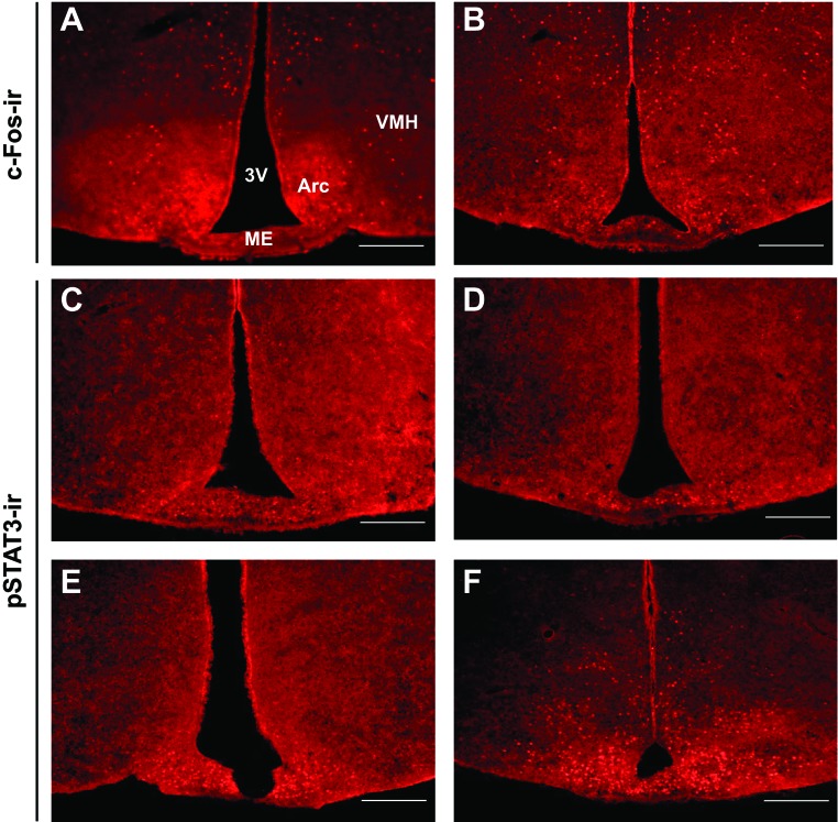
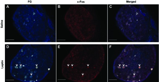
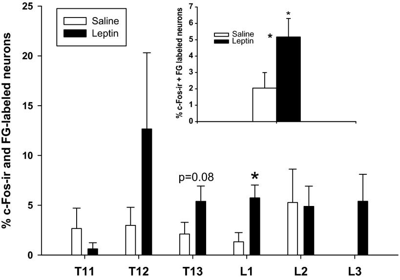
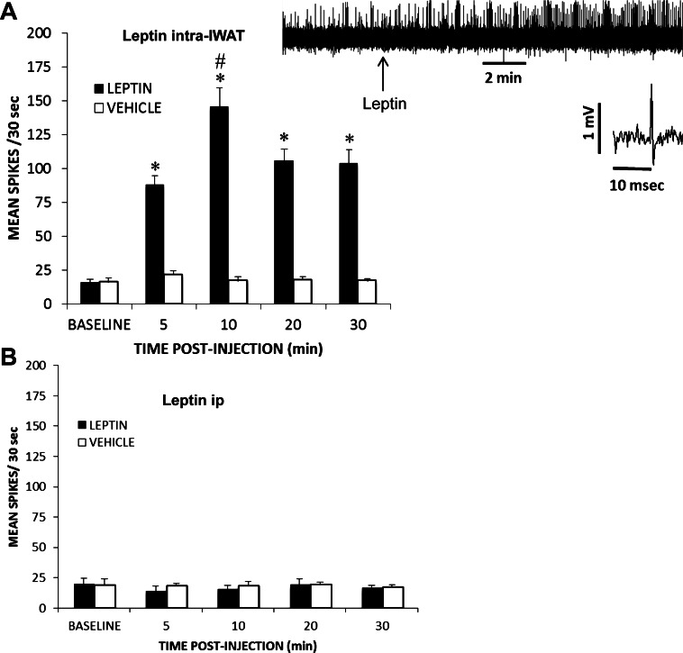
Leptin, the primary white adipose tissue (WAT) adipokine, is thought to convey lipid reserve information to the brain via the circulation. Because WAT responds to environmental/internal signals in a fat pad-specific (FPS) manner, systemic signals such as leptin would fail to communicate such distinctive information. Saturation of brain leptin transport systems also would fail to convey increased lipid levels beyond that point. WAT possesses sensory innervation exemplified by proven sensory-associated peptides in nerves within the tissue and by viral sensory nerve-specific transneuronal tract tracer, H129 strain of herpes simplex virus 1 labeling of dorsal root ganglia (DRG) pseudounipolar neurons, spinal cord and central sensory circuits. Leptin as a paracrine factor activating WAT sensory innervation could supply the brain with FPS information. Therefore, we tested for and found the presence of the long form of the leptin receptor (Ob-Rb) on DRG pseudounipolar neurons immunohistochemically labeled after injections of Fluorogold, a retrograde tract tracer, into inguinal WAT (IWAT). Intra-IWAT leptin injections (300 ng) significantly elevated IWAT nerve spike rate within 5 min and persisted for at least 30 min. Intra-IWAT leptin injections also induced significant c-Fos immunoreactivity (ir), indicating neural activation across DRG pseudounipolar sensory neurons labeled with Fluorogold IWAT injections. Intraperitoneal leptin injection did not increase c-Fos-ir in DRG or the arcuate nucleus, nor did it increase arcuate signal transducer and activator of transcription 3 phosphorylation-ir. Collectively, these results strongly suggest that endogenous leptin secreted from white adipocytes functions as a paracrine factor to activate spinal sensory nerves innervating the tissue.
Keywords: dorsal root ganglia; electrophysiology; tract tracing; white adipose tissue.
Figures

Similar articles
-
Anterograde transneuronal viral tract tracing reveals central sensory circuits from white adipose tissue.Am J Physiol Regul Integr Comp Physiol. 2009 Mar;296(3):R501-11. doi: 10.1152/ajpregu.90786.2008. Epub 2008 Dec 24. Am J Physiol Regul Integr Comp Physiol. 2009. PMID: 19109367 Free PMC article.
-
Short and long sympathetic-sensory feedback loops in white fat.Am J Physiol Regul Integr Comp Physiol. 2014 Jun 15;306(12):R886-900. doi: 10.1152/ajpregu.00060.2014. Epub 2014 Apr 9. Am J Physiol Regul Integr Comp Physiol. 2014. PMID: 24717676 Free PMC article.
-
Bidirectional crosstalk between the sensory and sympathetic motor systems innervating brown and white adipose tissue in male Siberian hamsters.Am J Physiol Regul Integr Comp Physiol. 2017 Mar 1;312(3):R324-R337. doi: 10.1152/ajpregu.00456.2015. Epub 2017 Jan 11. Am J Physiol Regul Integr Comp Physiol. 2017. PMID: 28077392 Free PMC article.
-
Sensory and sympathetic nervous system control of white adipose tissue lipolysis.Mol Cell Endocrinol. 2010 Apr 29;318(1-2):34-43. doi: 10.1016/j.mce.2009.08.031. Epub 2009 Sep 10. Mol Cell Endocrinol. 2010. PMID: 19747957 Free PMC article. Review.
-
Brain-adipose tissue cross talk.Proc Nutr Soc. 2005 Feb;64(1):53-64. doi: 10.1079/pns2004409. Proc Nutr Soc. 2005. PMID: 15877923 Review.
Cited by
-
Neural control of white, beige and brown adipocytes.Int J Obes Suppl. 2015 Aug;5(Suppl 1):S35-9. doi: 10.1038/ijosup.2015.9. Epub 2015 Aug 4. Int J Obes Suppl. 2015. PMID: 27152173 Free PMC article. Review.
-
Deconstruction of a hypothalamic astrocyte-white adipocyte sympathetic axis that regulates lipolysis in mice.Nat Commun. 2022 Dec 7;13(1):7536. doi: 10.1038/s41467-022-35258-6. Nat Commun. 2022. PMID: 36477150 Free PMC article.
-
Central sympathetic innervations to visceral and subcutaneous white adipose tissue.Am J Physiol Regul Integr Comp Physiol. 2014 Mar 15;306(6):R375-86. doi: 10.1152/ajpregu.00552.2013. Epub 2014 Jan 22. Am J Physiol Regul Integr Comp Physiol. 2014. PMID: 24452544 Free PMC article.
-
Molecular pathways linking adipose innervation to insulin action in obesity and diabetes mellitus.Nat Rev Endocrinol. 2019 Apr;15(4):207-225. doi: 10.1038/s41574-019-0165-y. Nat Rev Endocrinol. 2019. PMID: 30733616 Free PMC article. Review.
-
Adiponectin receptor-mediated signaling ameliorates cerebral cell damage and regulates the neurogenesis of neural stem cells at high glucose concentrations: an in vivo and in vitro study.Cell Death Dis. 2015 Aug 6;6(8):e1844. doi: 10.1038/cddis.2015.220. Cell Death Dis. 2015. PMID: 26247729 Free PMC article.
References
-
- Ahima RS, Prabakaran D, Mantzoros CS, Qu D, Lowell BB, Maratos-Flier E, Flier JS. Role of leptin in the neuroendocrine response to fasting. Nature 382: 250–252, 1996 - PubMed
-
- Bado A, Levasseur S, Attoub S, Kermorgant S, Laigneau JP, Bortoluzzi MN, Moizo L, Lehy T, Guerre-Millo M, Le Marchand-Brustel Y, Lewin MJ. The stomach is a source of leptin. Nature 394: 790–793, 1998 - PubMed
-
- Bamshad M, Aoki VT, Adkison MG, Warren WS, Bartness TJ. Central nervous system origins of the sympathetic nervous system outflow to white adipose tissue. Am J Physiol Regul Integr Comp Physiol 275: R291–R299, 1998 - PubMed
-
- Bartness TJ, Bamshad M. Innervation of mammalian white adipose tissue: Implications for the regulation of total body fat. Am J Physiol Regul Integr Comp Physiol 275: R1399–R1411, 1998 - PubMed
Publication types
MeSH terms
Substances
Grants and funding
LinkOut - more resources
Full Text Sources
Other Literature Sources
Miscellaneous