. 2013 Jul;62(7):1161-73.
doi: 10.1007/s00262-013-1411-0. Epub 2013 Apr 24.

Efficient ex vivo induction of T cells with potent anti-tumor activity by protein antigen encapsulated in nanoparticles

Affiliations

Efficient ex vivo induction of T cells with potent anti-tumor activity by protein antigen encapsulated in nanoparticles

Rodney A Rosalia et al. Cancer Immunol Immunother. 2013 Jul.

Abstract

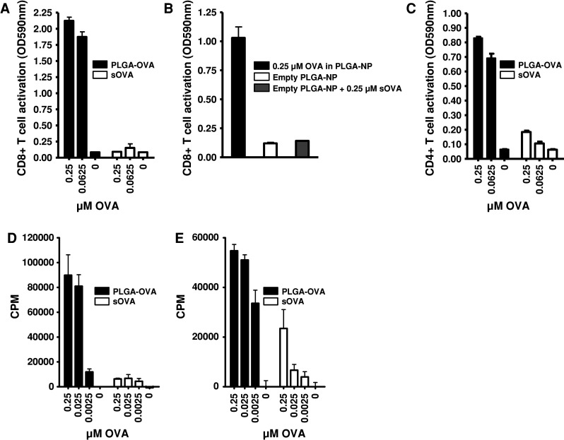
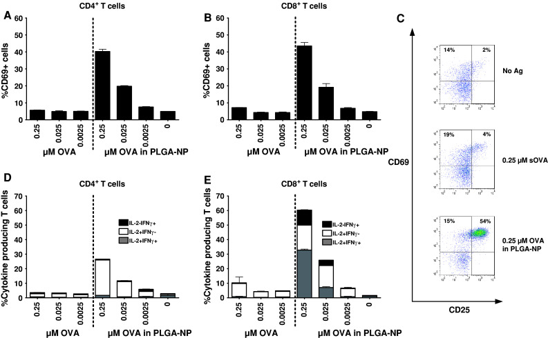
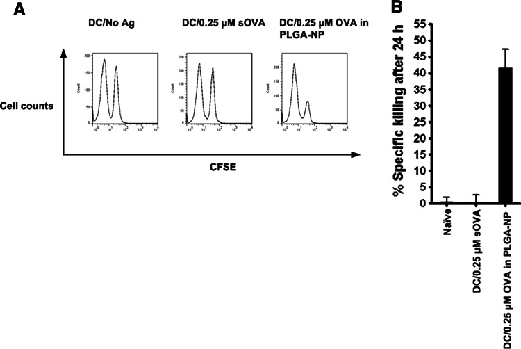
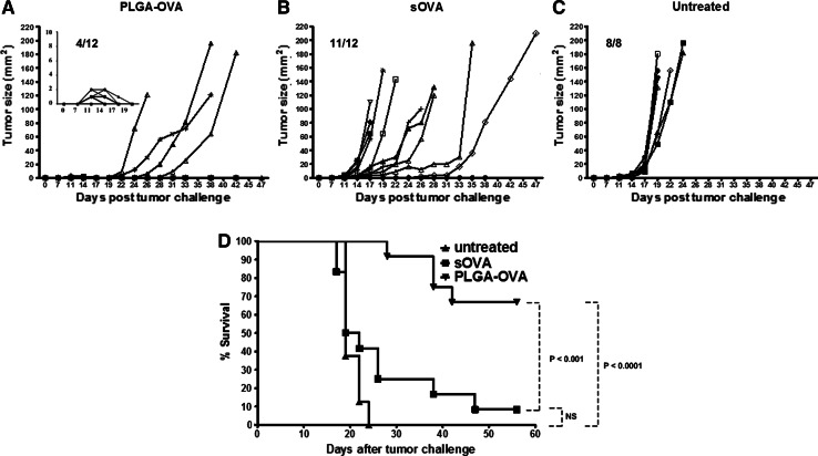
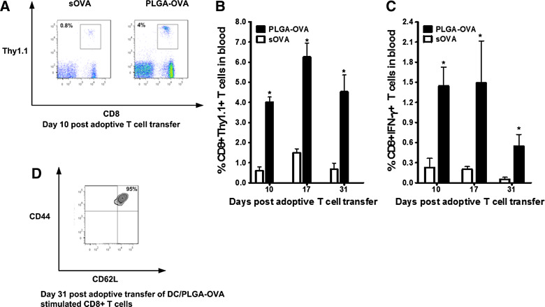
Protein antigen (Ag)-based immunotherapies have the advantage to induce T cells with a potentially broad repertoire of specificities. However, soluble protein Ag is generally poorly cross-presented in MHC class I molecules and not efficient in inducing robust cytotoxic CD8(+) T cell responses. In the present study, we have applied poly(lactic-co-glycolic acid) (PLGA) nanoparticles (NP) which strongly improve protein Ag presentation by dendritic cells (DC) in the absence of additional Toll-like receptor ligands or targeting devices. Protein Ag-loaded DC were used as antigen presenting cells to stimulate T cells in vitro and subsequently analyzed in vivo for their anti-tumor effect via adoptive transfer, a treatment strategy widely studied in clinical trials as a therapy against various malignancies. In a direct comparison with soluble protein Ag, we show that DC presentation of protein encapsulated in plain PLGA-NP results in efficient activation of CD4(+) and CD8(+) T cells as reflected by high numbers of activated CD69(+) and CD25(+), interferon (IFN)-γ and interleukin (IL)-2-producing T cells. Adoptive transfer of PLGA-NP-activated CD8(+) T cells in tumor-bearing mice displayed good in vivo expansion capacity, potent Ag-specific cytotoxicity and IFN-γ cytokine production, resulting in curing mice with established tumors. We conclude that delivery of protein Ag through encapsulation in plain PLGA-NP is a very efficient and simple procedure to stimulate potent anti-tumor T cells.


Conflict of interest statement

The authors declare that they have no conflict of interest.

Figures

Fig. 1
Fig. 1
Efficient MHC class I and class II presentation of OVA Ag incorporated in PLGA-NP. a D1 cells were pulsed for 24 h with titrated amounts (μM) of OVA, either in soluble form sOVA or encapsulated in PLGA-NP (PLGA-OVA). MHC class I presentation was detected by co-culture with H-2Kb/SIINFEKL-specific B3Z CD8+ T cells; b D1 cells were pulsed for 24 h with 0.25 μM OVA in PLGA-OVA, empty PLGA-NP or a mixture of empty PLGA-NP with 0.25 μM sOVA washed and co-cultured with B3Z CD8+ T cells to assess MHC class I Ag presentation; c D1 cells were pulsed for 24 h with titrated amounts of PLGA-OVA or sOVA, washed and co-cultured with I-Ab/ISQAVHAAHAEINEAGR-specific OT-IIZ CD4+ T cells to assess MHC class II Ag presentation. BMDC were loaded with titrated amounts of PLGA-OVA or sOVA. Ag-loaded DC were subsequently used to activate naïve OT-I (d) or OT-II T (e) cells for 72 h. T cell proliferation was measured in triplicate by 3[H]-thymidine uptake. Data shown are means of triplicate measurements ± SD from one representative example out of at least three independent experiments
Fig. 2
Fig. 2
DC pulsed with PLGA-OVA, but not sOVA, induce strong activation of T cells. D1 cells were pulsed for 24 h with titrated amounts of sOVA or PLGA-OVA. Ag-loaded DC were washed to remove excess Ag, and co-cultured for an additional 24 h with OT-I or OT-II splenocytes. Cells were harvested and analyzed by flow cytometry for the cell surface expression of CD69 on CD8+ T (a) cells and CD4+ T cells (b); c Expression of CD25 and CD69 was analyzed on CD8+ T cells which were stimulated for 24 h with DC which were loaded with either PLGA-OVA or sOVA. Immature DC without Ag served as negative control. Intracellular production of IL-2 and IFN-γ by CD8+ T cells (d); and CD4+ T (e) cells was analyzed by flow cytometry after 24 h stimulation with DC pulsed with titrated amounts of PLGA-OVA or sOVA. One representative experiment out of three independent experiments is shown. Data shown are means of triplicate measurements ± SD
Fig. 3
Fig. 3
Enhanced in vivo cytotoxicity of ex vivo PLGA-OVA-stimulated CD8+ T cells. a Mice were transferred with purified CD8+ OT-I T cells which were in vitro stimulated with D1 cells loaded with PLGA-OVA and sOVA. Differentially, CFSE-labeled SIINFEKL-peptide loaded and control-target cells were i.v. administered. After 18 h, the spleens from recipient animals were harvested and analyzed by flow cytometry for percentage of specific killing of target cells; b experiment was performed twice and averages ± SEM of n = 7 mice for each condition are shown in bar graphs
Fig. 4
Fig. 4
CD8+ T cells primed by PLGA-OVA-loaded DC eradicate established tumors. Animals were inoculated s.c. on the right flank with 2 × 105 B16-OVA tumor cells and rested for 1 week followed by a single i.v. injection of 2.5 × 106 purified CD8+ T cells which were ex vivo stimulated with Ag-loaded DC as described above. Tumor growth was monitored in individual animals treated with DC/PLGA-OVA-induced CD8+ T cells (a; n = 12 animals), DC/sOVA-induced CD8+ T cells (b; n = 12 animals) or in untreated animals (c; n = 8 animals). Insert in (a) represents tumor growth curves in the initial 19 days after tumor challenge; d Animal survival per group was assessed, and differences between the different groups were calculated using Log-rank (Mantel-Cox) test. p < 0.001 for animals treated with DC/PLGA-OVA compared to DC/sOVA-induced CD8+ T cells. p < 0.0001 for animals treated with DC/PLGA-OVA compared to untreated animals. NS = p > 0.05 for animals treated with DC/sOVA compared to untreated animals
Fig. 5
Fig. 5
PLGA-OVA stimulated CD8+ T cells expand and persist in the peripheral blood and have higher capacity to produce IFN-γ. a Tumor-bearing animals received a single i.v. injection of CD8+ T cells stimulated with PLGA-OVA-loaded DC. Tail vein blood samples were taken on day 10 after adoptive transfer of the CD8+ T cells, and numbers of CD8+Thy1.1+ T cells were measured by flow cytometry; b In vivo persistence of i.v. transferred CD8+Thy1.1+ T cells in tumor-bearing animals was monitored for 4 weeks in blood on days 17 and 31; c Intracellular IFN-γ production by CD8+ T cells was analyzed on days 10, 17 and 31 after adoptive transfer; d Memory phenotype of transferred DC/PLGA-OVA in vitro stimulated CD8+Thy1.1+ T cells was determined by analysis of CD44 and CD62L surface expression. Results shown are averages ± SEM from n = 3–12 mice per group, dependent on the number of animals alive at each time-point post-tumor challenge. *p < 0.05 for animals treated with DC/PLGA-OVA compared to DC/sOVA-induced CD8+ T cells using a unpaired student’s t test

References

    1. Banchereau J, Steinman RM. Dendritic cells and the control of immunity. Nature. 1998;392(6673):245–252. doi: 10.1038/32588. - DOI - PubMed
    1. Melief CJ. Cancer immunotherapy by dendritic cells. Immunity. 2008;29(3):372–383. doi: 10.1016/j.immuni.2008.08.004. - DOI - PubMed
    1. Steinman RM, Hemmi H. Dendritic cells: translating innate to adaptive immunity. Curr Top Microbiol Immunol. 2006;311:17–58. doi: 10.1007/3-540-32636-7_2. - DOI - PubMed
    1. Petersen TR, Dickgreber N, Hermans IF. Tumor antigen presentation by dendritic cells. Crit Rev Immunol. 2010;30(4):345–386. doi: 10.1615/CritRevImmunol.v30.i4.30. - DOI - PubMed
    1. Kawakami Y, Eliyahu S, Delgado CH, Robbins PF, Sakaguchi K, Appella E, Yannelli JR, Adema GJ, Miki T, Rosenberg SA. Identification of a human melanoma antigen recognized by tumor-infiltrating lymphocytes associated with in vivo tumor rejection. Proc Natl Acad Sci USA. 1994;91(14):6458–6462. doi: 10.1073/pnas.91.14.6458. - DOI - PMC - PubMed

Publication types

MeSH terms