

The study of broken quadrilateral surface in fractures of the acetabulum
- PMID: 23613172
- PMCID: PMC3664151
- DOI: 10.1007/s00264-013-1845-1
The study of broken quadrilateral surface in fractures of the acetabulum
Abstract
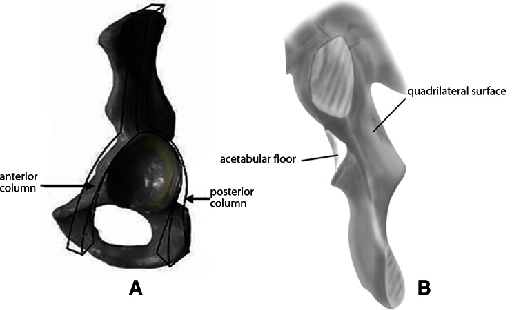
Purpose: Three-dimensional computerised tomography (3DCT) can provide comprehensive patho-anatomy of complex bone on a single image. Though important, the key articular quadrilateral [Q] surface has not been a part of the systems developed for classifying acetabulum fractures. The purpose of the study was to simplify the complexity of classification by the direct sign of the broken Q surface which lies opposite the entire floor of the acetabulum.
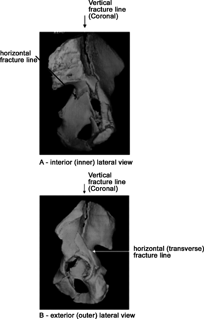
Methods: The study reviewed 84 acetabular fractures using 3DCT images of the interior lateral view (IL) taken between June 2002 to December 2009. Fractures were traditionally classified using the anatomical disruption, plane of the fracture line breaking through or not through the bone column described by Judet and Letournel.
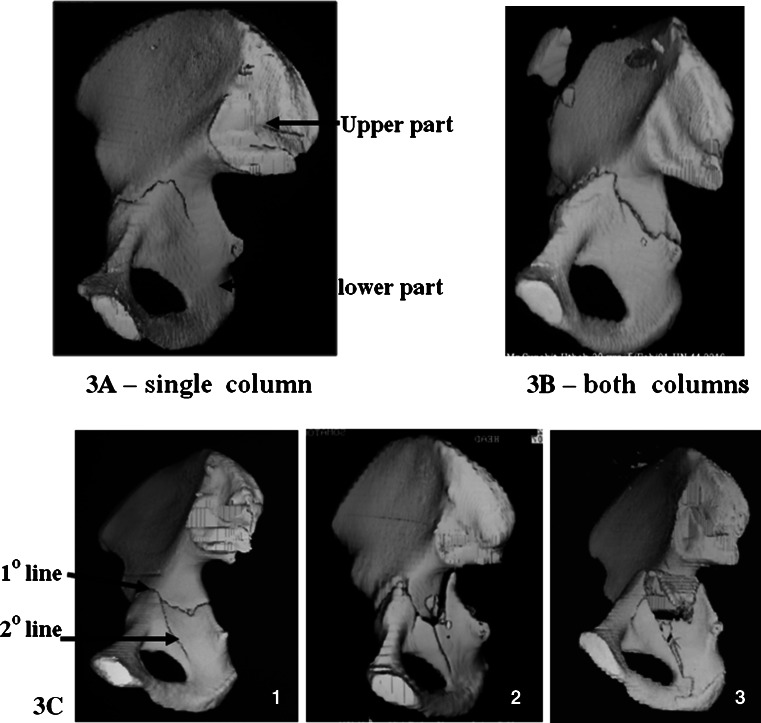
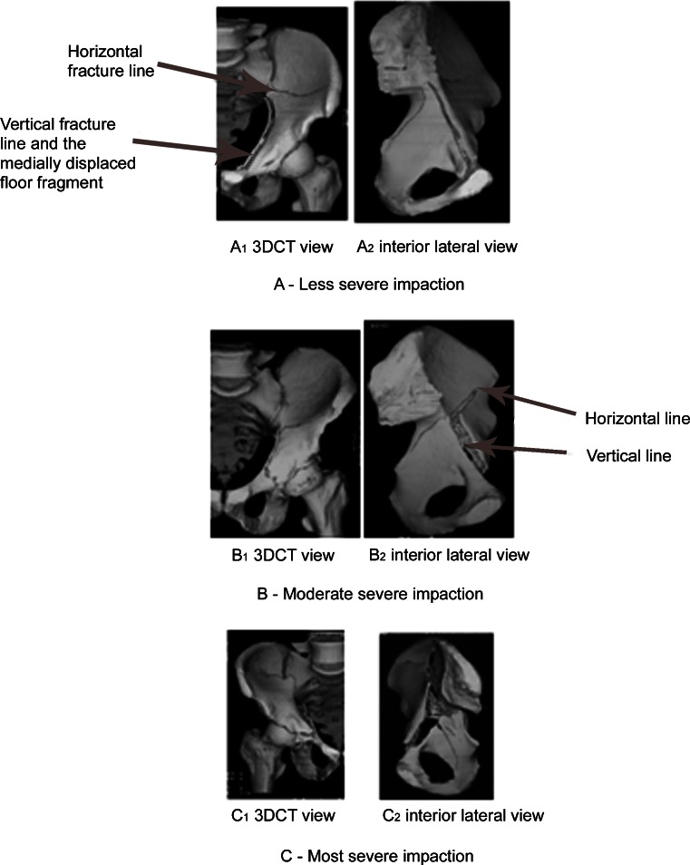
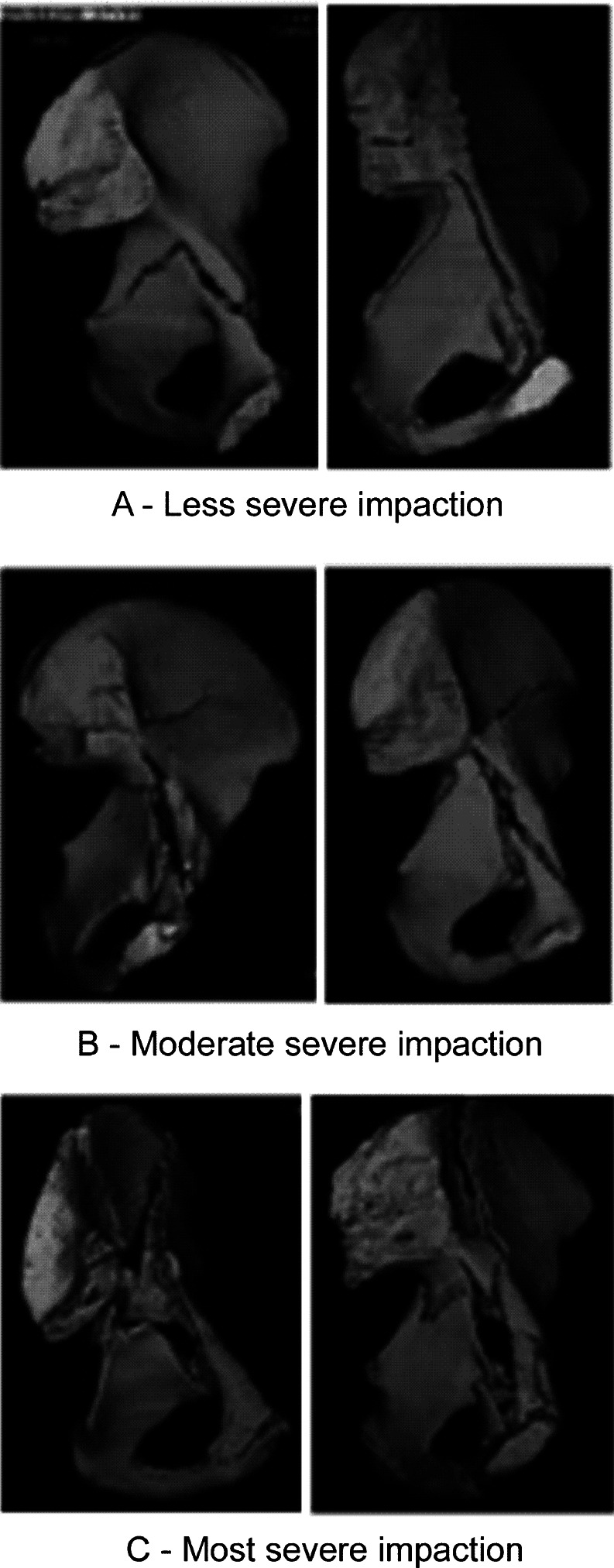
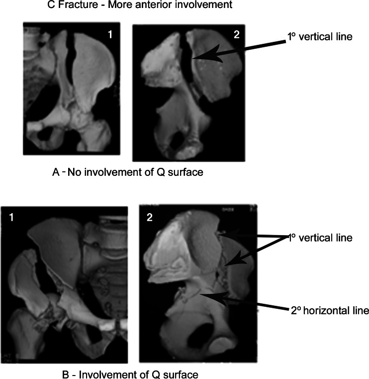
Results: The 3D images clearly show the primary site of impaction acting on the acetabulum and the whole course of fracture. The image could not illustrate disruption of the lips of acetabulum and congruity of hip joints in 20 cases of wall (W) fracture. There were 30 transverse (T) fractures classified when the acetabulum was divided horizontally from front to back into upper and lower parts and 34 cases of column (C) fracture when the main vertical lines run and collide along the anterior and posterior column.
Conclusions: This study showed that the well-known complex fractures can be satisfactorily classified with the broad flat inner plane of the Q surface.
Figures
Similar articles
-
A study on fracture lines of the quadrilateral plate based on fracture mapping.J Orthop Surg Res. 2019 Sep 12;14(1):310. doi: 10.1186/s13018-019-1318-3. J Orthop Surg Res. 2019. PMID: 31514742 Free PMC article.
-
Acetabular fractures: what radiologists should know and how 3D CT can aid classification.Radiographics. 2015 Mar-Apr;35(2):555-77. doi: 10.1148/rg.352140098. Radiographics. 2015. PMID: 25763739 Review.
-
Quadrilateral plate classification program of acetabular fractures based on three-column classification: a three-dimensional fracture mapping study.J Orthop Surg Res. 2024 May 16;19(1):298. doi: 10.1186/s13018-024-04783-z. J Orthop Surg Res. 2024. PMID: 38755648 Free PMC article.
-
[Computer tomography in the diagnosis and therapy of acetabular fractures].Srp Arh Celok Lek. 2001 Jul-Aug;129(7-8):194-8. Srp Arh Celok Lek. 2001. PMID: 11797449 Serbian.
-
[Diagnosis, classification and surgical indications in acetabulum fractures].Orthopade. 1992 Nov;21(6):427-41. Orthopade. 1992. PMID: 1475129 Review. German.
Cited by
-
Establishment of fluoroscopy views and standardized procedure of percutaneous magic screw insertion for acetabulum fractures.BMC Musculoskelet Disord. 2018 Sep 12;19(1):332. doi: 10.1186/s12891-018-2228-y. BMC Musculoskelet Disord. 2018. PMID: 30208885 Free PMC article.
-
Quadrilateral plate fractures of the acetabulum: Classification, approach, implant therapy and related research progress.World J Clin Cases. 2022 Jan 14;10(2):412-425. doi: 10.12998/wjcc.v10.i2.412. World J Clin Cases. 2022. PMID: 35097066 Free PMC article. Review.
-
A New, Easy, Fast, and Reliable Method to Correctly Classify Acetabular Fractures According to the Letournel System.JB JS Open Access. 2018 Feb 16;3(1):e0032. doi: 10.2106/JBJS.OA.17.00032. eCollection 2018 Mar 29. JB JS Open Access. 2018. PMID: 30229234 Free PMC article.
-
A novel navigation template for fixation of acetabular posterior column fractures with antegrade lag screws: design and application.Int Orthop. 2016 Apr;40(4):827-34. doi: 10.1007/s00264-015-2813-8. Epub 2015 Jun 27. Int Orthop. 2016. PMID: 26112873
-
Late surgery for acetabular fractures in a Chinese level I trauma centre: surgical experience and outcomes.Int Orthop. 2015 Sep;39(9):1865-71. doi: 10.1007/s00264-015-2932-2. Epub 2015 Aug 5. Int Orthop. 2015. PMID: 26243451
References
-
- Dorland (1994) Dorland’s Illustrated medical dictionary, 28th edition. Saunders, Philadelphia
-
- Judet R, Judet J, Letournel E. Fractures of the acetabulum: classification and surgical approaches for open reduction, preliminary report. J Bone Joint Surg Am. 1964;46:1615–1646. - PubMed
-
- AO Foundation (2008) A short history of pelvic trauma surgery. http://www.ao-asif.ch/portal/AOFileServer/PortalFiles?FilePath=/Extranet.... Accessed 19 June 2008
-
- Letournel E, Judet R. Fractures of the acetabulum. 2. Berlin: Springer; 1993.
MeSH terms
LinkOut - more resources
Full Text Sources
Other Literature Sources
Medical
