. 2013 Jun 15;19(12):3165-75.
doi: 10.1158/1078-0432.CCR-12-3314. Epub 2013 Apr 23.

Gliomas promote immunosuppression through induction of B7-H1 expression in tumor-associated macrophages

Affiliations

Gliomas promote immunosuppression through induction of B7-H1 expression in tumor-associated macrophages

Orin Bloch et al. Clin Cancer Res. .

Abstract

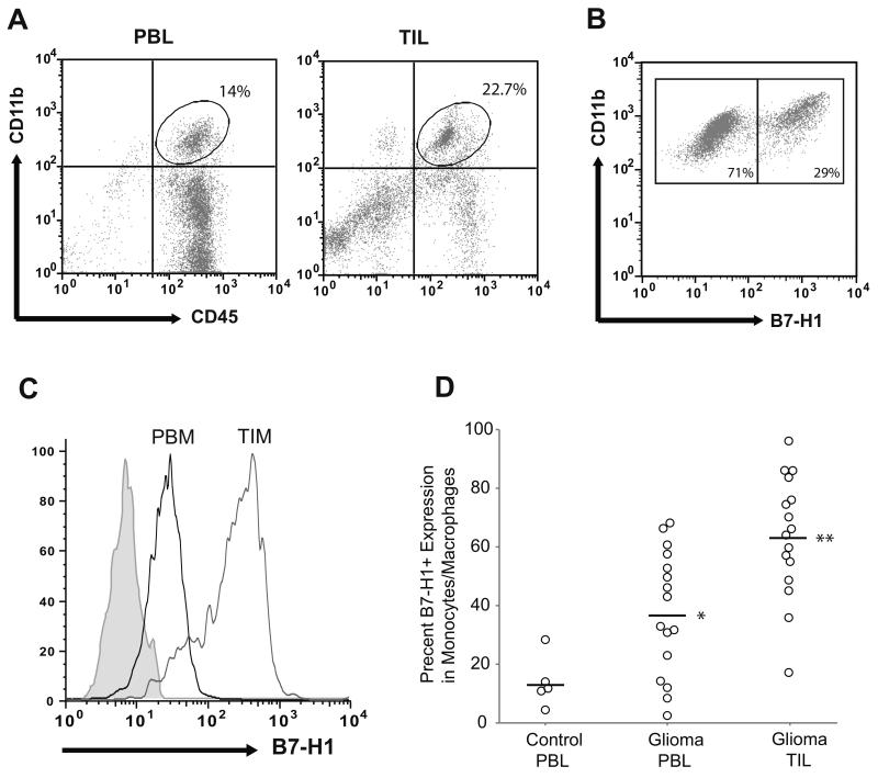
Purpose: Gliomas are known to induce local and systemic immunosuppression, inhibiting T-cell-mediated cytotoxic responses to tumor growth. Tumor-associated macrophages are a significant component of the immune infiltrate in gliomas and may express immunosuppressive surface ligands, such as B7-H1.

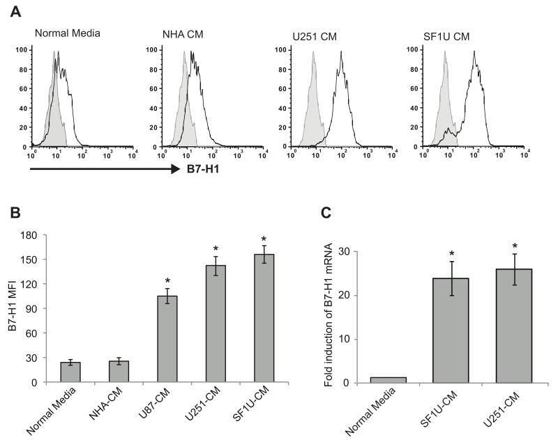
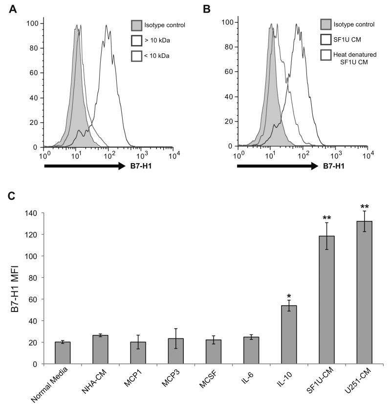
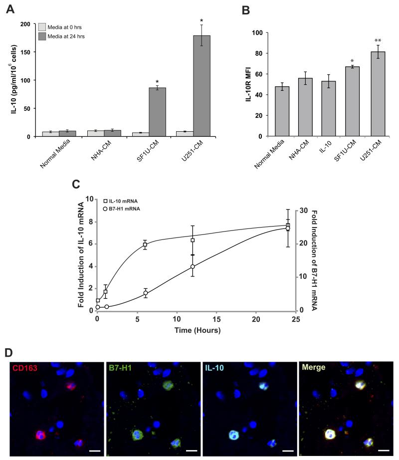
Experimental design: Tumor and peripheral blood samples from patients with glioblastoma (GBM) were analyzed by flow cytometry to evaluate the expression of B7-H1 in circulating and tumor-infiltrating macrophages. Human monocytes from healthy patients were stimulated with conditioned media from glioma cells to evaluate B7-H1 expression. Production of interleukin (IL)-10 by stimulated monocytes was measured by ELISA, and stimulation with IL-10 alone was evaluated for the ability to induce B7-H1 expression. The effect of inhibiting IL-10 and its receptor on glioma-induced B7-H1 expression in monocytes was evaluated.

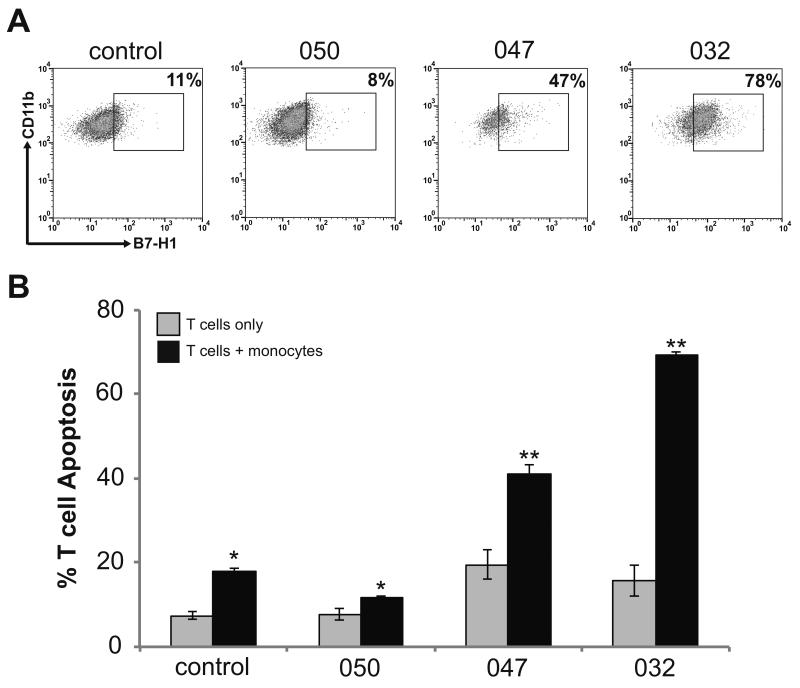
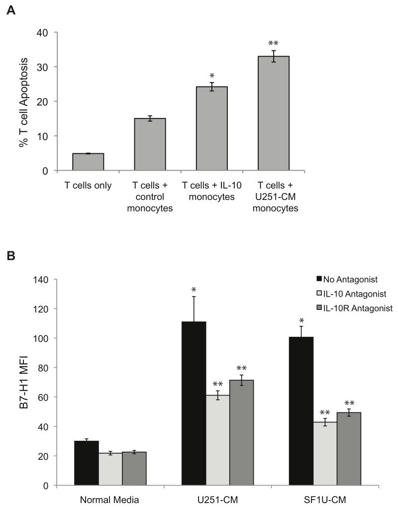
Results: Circulating monocytes in patients with GBM had significantly increased expression of B7-H1 compared with healthy control patients. Tumor-associated macrophages from matched GBM tissue had even greater B7-H1 expression. Treatment of normal monocytes with glioma-conditioned media could significantly increase B7-H1 expression. Stimulation of monocytes with conditioned media resulted in substantial production of IL-10 and upregulation of the IL-10 receptor. Stimulation of monocytes with IL-10 alone could significantly increase B7-H1 expression, sufficient to induce T-cell apoptosis when cocultured with stimulated monocytes. Inhibition of IL-10 and the IL-10 receptor could knock down the effect of glioma media on B7-H1 by more than 50%.

Conclusions: Gliomas can upregulate B7-H1 expression in circulating monocytes and tumor-infiltrative macrophages through modulation of autocrine/paracrine IL-10 signaling, resulting in an immunosuppressive phenotype.


Figures

Figure 1
Figure 1. Peripheral blood monocytes and tumor-infiltrating macrophages in GBM patients express elevated levels of B7-H1
(A) Representative gating of peripheral blood leukocytes (PBL) and tumor-infiltrating leukocytes (TIL) from GBM patients analyzed by flow cytometry is shown. Monocytes/macrophages were identified by co-positive CD45 and CD11b staining. (B) Representative analysis of the percent of B7-H1 positive cells among the CD45+/CD11b+ monocytes gated in panel (A) is shown. (C) Representative analysis of B7-H1 expression intensity in peripheral blood monocytes (PBM) and tumor-infiltrating macrophages (TIM) of a GBM patient is shown. B7-H1 intensity was measured after gating on CD45+/CD11b+ cells. Curves depict the isotype control (gray shaded region), peripheral monocytes (black line), and tumor-infiltrating macrophages (gray line). (D) The fraction of B7-H1 expressing cells as a percentage of the total number of monocytes/macrophages in peripheral blood and tumor from GBM patients (n=16), as well as from the peripheral blood of non-glioma control patients (n=5) is shown. Absolute percentage for each patient and group means are depicted. B7-H1 expression in peripheral monocytes from GBM patients is significantly greater than healthy control patients (* p < 0.05). B7-H1 expression in tumor-associated macrophages is significantly greater than in peripheral monocytes among GBM patients (** p < 0.001).
Figure 2
Figure 2. Peripheral blood monocytes from GBM patients induce autologous T cell apoptosis relative to B7-H1 expression
(A) Gating demonstrating the percentage of B7-H1 positive monocytes isolated from peripheral blood of three GBM patients and a single non-glioma control patient. Monocytes were isolated from total peripheral blood leukocytes by CD14 positive selection. Cells were stained for flow cytometry and monocytes were identified by gating on CD45+/CD11b+ cells. This figure demonstrates the subgate of CD11b+/B7-H1+ cells. The cutoff for B7-H1 positivity was determined based on isotype control staining. (B) The percentage of T cells undergoing apoptosis when monocytes from patients in panel (A) were co-cultured with autologous activated T cells is shown. The percentage of T cell undergoing apoptosis was determined by co-staining cells for CD3 and Annexin V and analyzed by flow cytometry. Columns represent mean apoptotic T cell counts ± SEM from 3 independent trials. T cells co-cultured with monocytes demonstrated significantly increased apoptosis as compared to the same cells in culture alone (* p<0.05, ** p<0.01).
Figure 3
Figure 3. Glioma-dervied soluble factor(s) can induce B7-H1 expression in peripheral monocytes from healthy donors
Monocytes were stimulated for 24 hours with normal media (control) or conditioned media from NHA or glioma cells lines. (A) Representative histograms of B7-H1 expression from stimulated monocytes are depicted (black), as well as isotype controls (gray shaded region). (B) Quantification of mean fluorescence intensity for B7-H1 staining measured by flow cytometry is shown for monocytes stimulated with various conditioned media. B7-H1 intensity was significantly increased for monocytes treated with media from all three glioma cell lines (* p<0.01) but not for media from NHA when compared to unstimulated cells. Columns represent mean fluorescence intensity ± SEM from 5 independent samples. Each sample was tested in triplicate and averaged as a single data point. (C) Quantification of B7-H1 mRNA levels from control monocytes and glioma conditioned monocytes using quantitative RT-PCR. mRNA levels were normalized to 18S rRNA and are reported relative to control monocyte expression. Monocytes treated with glioma-conditioned media have significantly increased B7-H1 mRNA expression (* p <0.05). Columns represent mean relative expression ± SEM from 3 independent samples.
Figure 4
Figure 4. The glioma-derived B7-H1 inducing factor has properties consistent with a soluble protein
(A) B7-H1 expression in normal monocytes treated for 24 hours with conditioned media from SF1U cells filtered to separate solutes with a molecular weight >10 kDa (dark gray) from solutes <10 kDa (light gray). The isotype control (gray shaded region) is also depicted. As shown, B7-H1 expression is significantly increased by a component in the > 10 kDa fraction. (B) B7-H1 expression in normal monocytes treated for 24 hours with unmodified conditioned media from SF1U cells (dark gray) or heat denatured media (light gray), demonstrating that heat denaturation eliminates the stimulatory effect of the conditioned media. (C) Mean fluorescence intensity of B7-H1 staining in normal monocytes treated with glioma-conditioned media or the following cytokines: MCP1 (200 ng/ml), MCP3 (200 ng/ml), M-CSF (50 ng/ml), IL-6 (10 ng/ml), IL-10 (10 ng/ml). Among the cytokines tested, only IL-10 stimulates a significant increase in B7-H1 expression (* p < 0.01). Compared to the response to IL-10, the increase seen with glioma-conditioned media stimulation was significantly greater (** p < 0.05). Columns represent mean fluorescence intensity ± SEM from 4 independent samples. Each sample was tested in triplicate and averaged as a single data point.
Figure 5
Figure 5. Glioma-derived soluble factor(s) enhance IL-10 signaling
(A) Human IL-10 expression measured by ELISA from culture media of normal monocytes stimulated with glioma-conditioned media before and after 24 hours of culture. None of the conditioned media initially contained appreciable levels of IL-10. After 24 hours, IL-10 expression was significantly increased in cells stimulated with glioma-conditioned media compared to normal media (* p<0.01). Columns represent mean total IL-10 ± SEM from 3 independent samples. (B) IL-10 receptor expression was measured by flow cytometry in monocytes stimulated with glioma-conditioned media. Compared to unstimulated cells, treated monocytes had a modest but significant increase in IL-10R mean fluorescence intensity (* p < 0.05, ** p < 0.01). Columns represent mean fluorescence intensity ± SEM from 3 independent samples. Each sample was tested in triplicate and averaged as a single data point. (C) Time course of IL-10 and B7-H1 mRNA expression in normal monocytes stimulated with SF1U conditioned media. Transcript levels were measured at 0, 1, 6, 12, and 24 hours after stimulation using quantitative RT-PCR and normalized to 18S rRNA. Normalized expression is reported as a relative increase over initial expression. Each point represents mean relative expression ± SEM from 3 independent samples. (D) Representative section of GBM tissue with fluorescent immunestaining for CD163 (red), B7-H1 (green), IL-10 (cyan), and DAPI (blue) demonstrating co-localization of all three probes to the same subset of tumor-associated macrophages (scale bar = 10μm).
Figure 6
Figure 6. IL-10 is necessary and sufficient to stimulate an immunosuppressive phenotype in monocytes
(A) The percentage of T cells undergoing apoptosis was measured by Annexin V staining after co-culture with stimulated or unstimulated (control) monocytes. Monocytes stimulated with IL-10 or glioma-conditioned media induce significantly more T cell apoptosis compared to control monocytes (* p < 0.05, ** p < 0.005). Columns represent mean apoptotic T cell counts ± SEM from 3 independent samples. Each sample was tested in triplicate and averaged as a single data point. (B) Soluble IL-10 and binding to the IL-10 receptor were inhibited to evaluate the effect on glioma-induced B7-H1 expression in monocytes. Monocytes were treated with normal media or glioma-conditioned media in the presence of no inhibitor (black bars), a soluble IL-10 neutralizing antibody at 5 μg/ml (light gray bars), or an IL-10 receptor antagonist at 5 μg/ml (dark gray bars) for 24 hours and B7-H1 expression was measured by flow cytometry. As previously shown, treatment with glioma-conditioned media in the presence of no inhibitors significantly increases the expression of B7-H1 (* p<0.01). However, the addition of either an IL-10 or IL-10R antagonist reduces the expression of B7-H1 by greater than 50% (** p<0.05). Columns represent mean fluorescence intensity ± SEM from 3 independent samples. Each sample was tested in triplicate and averaged as a single data point.

References

    1. Surawicz TS, Davis F, Freels S, Laws ER, Jr., Menck HR. Brain tumor survival: results from the National Cancer Data Base. J Neurooncol. 1998;40:151–60. - PubMed
    1. Deorah S, Lynch CF, Sibenaller ZA, Ryken TC. Trends in brain cancer incidence and survival in the United States: Surveillance, Epidemiology, and End Results Program, 1973 to 2001. Neurosurg Focus. 2006;20:E1. - PubMed
    1. Laws ER, Parney IF, Huang W, Anderson F, Morris AM, Asher A, et al. Survival following surgery and prognostic factors for recently diagnosed malignant glioma: data from the Glioma Outcomes Project. J Neurosurg. 2003;99:467–73. - PubMed
    1. Stupp R, Mason WP, van den Bent MJ, Weller M, Fisher B, Taphoorn MJ, et al. Radiotherapy plus concomitant and adjuvant temozolomide for glioblastoma. N Engl J Med. 2005;352:987–96. - PubMed
    1. Gousias K, Markou M, Arzoglou V, Voulgaris S, Vartholomatos G, Kostoula A, et al. Frequent abnormalities of the immune system in gliomas and correlation with the WHO grading system of malignancy. J Neuroimmunol. 2010;226:136–42. - PubMed

Publication types

MeSH terms