Review
. 2013 Jul 1;591(13):3159-65.
doi: 10.1113/jphysiol.2012.248625. Epub 2013 Apr 22.

Organization and function of transmitter release sites at the neuromuscular junction

Affiliations
Review

Organization and function of transmitter release sites at the neuromuscular junction

Stephen D Meriney et al. J Physiol. .

Abstract

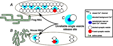
The neuromuscular junction is known as a strong and reliable synapse. It is strong because it releases an excess of chemical transmitter, beyond what is required to bring the postsynaptic muscle cell to threshold. Because the synapse can sustain suprathreshold muscle activation during short trains of action potentials, it is also reliable. The presynaptic mechanisms that lead to reliability during short trains of activity have only recently been elucidated. It appears that there are relatively few calcium channels in individual active zones, that channels open with a low probability during action potential stimulation and that even if channels open the resulting calcium flux only rarely triggers vesicle fusion. Thus, each synaptic vesicle may only associate with a small number of calcium channels, forming an unreliable single vesicle release site. Strength and reliability of the neuromuscular junction emerge as a result of its assembly from thousands of these unreliable single vesicle release sites. Hence, these synapses are strong while at the same time only releasing a small subset of available docked vesicles during each action potential, thus conserving transmitter release resources. This prevents significant depression during short trains of action potential activity and confers reliability.


Figures

Figure 1
Figure 1. Structure and function of the frog neuromuscular junction
A, portion of a large frog neuromuscular junction stained using FITC-labelled peanut lectin to decorate Schwann cell extracellular matrix (green) and Alexa594-labelled α-bungarotoxin to identify the location of postsynaptic receptor folds (red) immediately opposite presynaptic active zones. A single acetylcholine receptor band that represents the predicted position of one active zone is circled. Image obtained by S.D.M. following the protocols of Ko (1987) and Reddy et al. (2003). B, graphical depiction of the spatial organization of docked synaptic vesicles (large white circles) and presynaptic Ca2+ channels (small yellow circles; filled circles represent the fraction of channels that open on average during an AP stimulus) overlaid onto a freeze-fracture replica of about half of a frog neuromuscular junction active zone (Heuser et al. 1979). Graphic adapted from Luo et al. (2011). C, graphical representation of a small portion of the MCell computer model of the frog neuromuscular junction active zone (bottom view). In this graphic, synaptic vesicles are large grey or yellow spheres, synaptotagmin binding sites are represented as an array of black dots at the base of synaptic vesicles (binding sites with bound Ca2+ are coloured according to the Ca2+ channel contributing the ion), triangles represent the position of presynaptic active zone proteins (some of which are occupied by voltage-gated Ca2+ channels; VGCC), and coloured dots represent Ca2+ ions (Ca2+), colour coded based on the voltage-gated Ca2+ channel of origin. D, endplate potential (EPP) recorded from a single frog neuromuscular junction following exposure to 4 μmμ-conotoxin PIIIA to block selectively postsynaptic sodium channels (average of 10 sweeps). Inset, spontaneous miniature endplate potentials (mEPPs) recorded in the absence of nerve stimulation. Data in D obtained by S.D.M. from the cutaneous pectoris nerve-muscle preparation following the protocol of Shon et al. (1998).
Figure 2
Figure 2. Structure and function of the mouse neuromuscular junction
A, an entire single mouse neuromuscular junction stained with Alexa 594 α-bungarotoxin to label postsynaptic acetylcholine receptors (red), and Alexa 488 bassoon antibody to label presynaptic active zones (green spots). Confocal brightest projection image obtained by S.D.M. following the protocol of Nishimune et al. (2004), and processed for deconvolution. Grid lines = 5 μm. B, freeze-fracture replica of a single mouse neuromuscular junction active zone. White circles represent the predicted position of docked synaptic vesicles. Adapted from Nagwaney et al. (2009). Scale bar = 50 nm. C, average endplate potential (EPP) recorded from a single mouse neuromuscular junction following exposure to 1 μmμ-conotoxin GIIIB to block selectively postsynaptic sodium channels (average of 10 sweeps). Inset, spontaneous miniature endplate potentials (mEPPs) recorded in the absence of nerve stimulation. Data in C obtained by S.D.M. from the epitrochleoanconeus nerve-muscle preparation following the protocols of Urbano et al. (2003) and Rogozhin et al. (2008).
Figure 3
Figure 3. Organization of neuromuscular junction active zones based on assembly of unreliable single vesicle release sites
A, each of hundreds of active zones in the frog neuromuscular junction (NMJ) is hypothesized to be constructed using a long linear double array of unreliable single vesicle release sites (C). B, each of hundreds of active zones in the mouse neuromuscular junction are separated from one another by about 500 nm, and are hypothesized to be constructed using a short linear array of only two unreliable single vesicle release sites. C, the basic building block of neuromuscular junctions is hypothesized to be an unreliable single vesicle release site. Graphic adapted from Tarr et al. (2013).

Similar articles

Cited by

References

    1. Bekkers JM. Quantal analysis of synaptic transmission in the central nervous system. Curr Opin Neurobiol. 1994;4:360–365. - PubMed
    1. Bennett MR. Neuromuscular transmission at an active zone: the secretosome hypothesis. J Neurocytol. 1996;25:869–891. - PubMed
    1. Chapman ER. Synaptotagmin: a Ca2+ sensor that triggers exocytosis. Nat Rev Mol Cell Biol. 2002;3:498–508. - PubMed
    1. Chen J, Mizushige T, Nishimune H. Active zone density is conserved during synaptic growth but impaired in aged mice. J Comp Neurol. 2012;520:434–452. - PMC - PubMed
    1. Dittrich M, Pattillo JM, King JD, Cho S, Stiles JR, Meriney SD. 2013. An excess calcium binding site model predicts neurotransmitter release at the NMJ Biophysical J. (in press) - PMC - PubMed

MeSH terms

Substances

LinkOut - more resources