

doi: 10.4070/kcj.2013.43.3.196.
Epub 2013 Mar 31.
Coronary Slow Flow Phenomenon Leads to ST Elevation Myocardial Infarction
Affiliations
- PMID: 23613699
- PMCID: PMC3629248
- DOI: 10.4070/kcj.2013.43.3.196
Item in Clipboard
Coronary Slow Flow Phenomenon Leads to ST Elevation Myocardial Infarction
Korean Circ J.
2013 Mar.
Abstract
The exact etiology of the coronary slow flow phenomenon (CSFP) is not certain. CSFP is not a normal variant as it is an absolutely pathological entity. Furthermore, CSFP not only leads to myocardial ischemia but it can also cause classical acute ST elevation myocardial infarction, which necessitates coronary angiography for a definite diagnosis.
Keywords: Acute anterior wall myocardial infarction; Slow-flow phenomenon.
Conflict of interest statement
The author has no financial conflicts of interest.
Figures
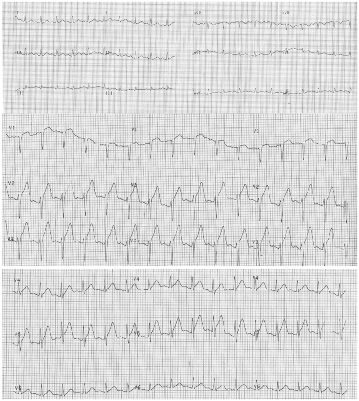
The ECG shows marked ST elevation in the anterior precordial leads. This ECG leads to a diagnosis of hyperacute anterior myocardial infarction. ECG: electrocardiography.
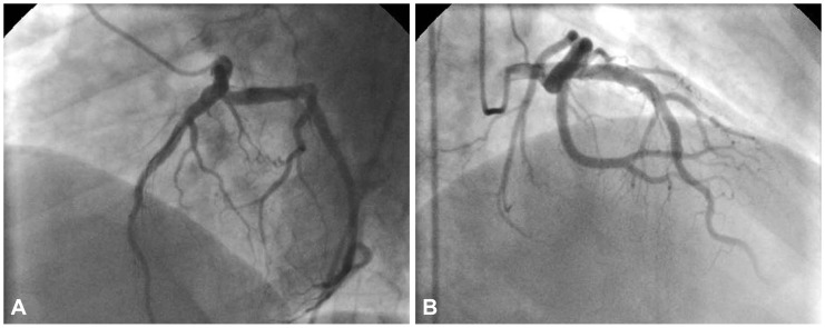
This angiographical image (A: left anterior oblique cranial view, B: right anterior oblique view) reveals left sided normal coronary arteries except slow coronary flow in the left anterior descending artery.
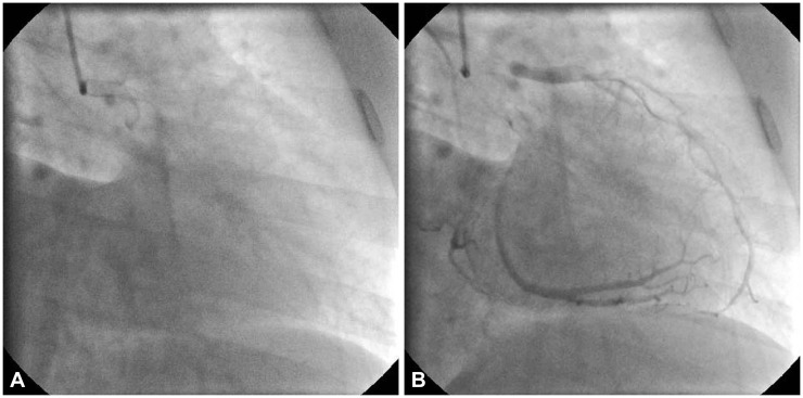
The Thrombolysis in Myocardial Infarction (TIMI) frame count for the left anterior descending coronary artery (A and B) was 67-9=58. The corrected TIMI frame count was calculated as 58/1.7=34.1 (normal range 21±3).
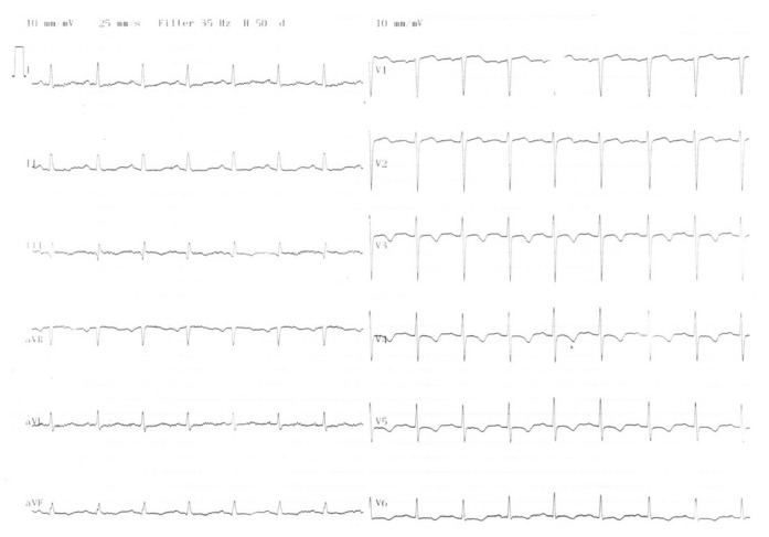
After the chest pain ceased, the electrocardiography showed the resolution ST elevation and negative T waves between V 3-6 leads.
References
-
- Mangieri E, Macchiarelli G, Ciavolella M, et al. Slow coronary flow: clinical and histopathological features in patients with otherwise normal epicardial coronary arteries. Cathet Cardiovasc Diagn. 1996;37:375–381. - PubMed
-
- Mosseri M, Yarom R, Gotsman MS, Hasin Y. Histologic evidence for small-vessel coronary artery disease in patients with angina pectoris and patent large coronary arteries. Circulation. 1986;74:964–972. - PubMed
-
- Singh S, Kothari SS, Bahl VK. Coronary slow flow phenomenon: an angiographic curiosity. Indian Heart J. 2004;56:613–617. - PubMed
-
- Gibson CM, Cannon CP, Daley WL, et al. TIMI frame count: a quantitative method of assessing coronary artery flow. Circulation. 1996;93:879–888. - PubMed
-
- Demirkol MO, Yaymaci B, Mutlu B. Dipyridamole myocardial perfusion single photon emission computed tomography in patients with slow coronary flow. Coron Artery Dis. 2002;13:223–229. - PubMed
LinkOut - more resources
Full Text Sources
Other Literature Sources
