. 2013 Apr 17;8(4):e60926.
doi: 10.1371/journal.pone.0060926. Print 2013.

Bridge-induced chromosome translocation in yeast relies upon a Rad54/Rdh54-dependent, Pol32-independent pathway

Affiliations

Bridge-induced chromosome translocation in yeast relies upon a Rad54/Rdh54-dependent, Pol32-independent pathway

Valentina Tosato et al. PLoS One. .

Abstract

While in mammalian cells the genetic determinism of chromosomal translocation remains unclear, the yeast Saccharomyces cerevisiae has become an ideal model system to generate ad hoc translocations and analyze their cellular and molecular outcome. A linear DNA cassette carrying a selectable marker flanked by perfect homologies to two chromosomes triggers a bridge-induced translocation (BIT) in budding yeast, with variable efficiency. A postulated two-step process to produce BIT translocants is based on the cooperation between the Homologous Recombination System (HRS) and Break-Induced Replication (BIR); however, a clear indication of the molecular factors underlying the genetic mechanism is still missing. In this work we provide evidence that BIT translocation is elicited by the Rad54 helicase and completed by a Pol32-independent replication pathway. Our results demonstrate also that Rdh54 is involved in the stability of the translocants, suggesting a mitotic role in chromosome pairing and segregation. Moreover, when RAD54 is over-expressed, an ensemble of secondary rearrangements between repeated DNA tracts arise after the initial translocation event, leading to severe aneuploidy with loss of genetic material, which prompts the identification of fragile sites within the yeast genome.


Conflict of interest statement

Competing Interests: The authors have declared that no competing interests exist.

Figures

Figure 1
Figure 1. Frequency of recombination events with a BIT cassette in eight different mutants.
The frequency (ν) of each genetic event x (transformation: νt; ectopic integration: νiect; one-end DNA integration: νiadh and νidur3; translocation: νTsl) in the wild type and in eight mutants (see figure legend on the left) divided by the transformability of the strain (νp, Table S1). The frequency values νx/νp and the relative standard errors (here represented as plus bar only for graphical reasons) are summarized in Table S2b. νx values are calculated as total number of transformants obtained on G418 per event divided by the total amount of treated cells (raw data are in Table S2a). For details of the computation see Materials and Methods. To better interpret the data of Table S2a, the ratio distribution of the various integration events for RAD54, RDH54, and POL32 mutants and for the wild type, from the total number of transformants, is plotted graphically as percentage pie charts. For the rad54Δ/rad54Δ mutant no pie has been drawn since only one transformant has been recovered, integrated only at the adh1 locus. A schematic representation of the BIT cassette with the two homologies targeting chromosomes VIII and XV is also shown at the bottom of the figure. As negative control, in rad52Δ/rad52Δ background only three ectopic clones were obtained, one coming from GT and two from BIT. The transformability of rad52Δ/rad52Δ with the centromeric plasmid YCp50 was assessed to be 1/6 of the wild type and therefore does not justify the low number of transformants obtained with a linear DNA cassette (Table S1 and S2). Thus, the deletion of RAD52 completely abolishes the possibility to get any kind of targeted DNA integration confirming that BIT is HRS-dependent.
Figure 2
Figure 2. CHEF and Southern hybridization analysis of translocants unbalanced for Rdh54 and Rad54 amount.
A) A scheme showing the bridge between chromosome VIII and XV and the new hybrid chromosome (Translocant = T). The chromosomal positions of the probes and of the translocation breakpoints (bold and underlined) are reported. B) CHEF (left) and Southern hybridization (right), with a probe against kanamycin, of the translocants in RDH54 and RAD54 deletants. Lanes from left to right: 1, San1ΔMSH2::Kan (it indicates chromosome XV); in rdh54Δ/rdh54Δ background: 2, cl4; 3, cl7; 4, cl16; 5, cl23n; 6, cl23*; 7, cl30; 8, cl47; in CRAD54 background: 9, cl1; 10, cl4; in OeRAD54 background: 11, clw; 12, cl37; 13, cl38; 14, San1ΔDUR3::Kan (chromosome VIII). n means phenotypically normal and * indicates sectored colony (see text for details). C) CHEF and Southern bots of the following samples (from left to right): 1, San1; in rdh54Δ/rdh54Δ background: 2, cl4; 3, cl7; 4, cl16; 5, cl23; 6, cl30 and 7, cl47. In CRAD54 background: 8, cl1 and 9, cl4; in OeRAD54 background: 10, clw; 11, cl37 and 12, cl38; 13, top1Δ/top1Δcl18; 14, xrs2Δ/xrs2Δcl6; 15, San1, hybridized with probes for brx1, hal9 (upper line) and cdc33 (not shown because identical to hal9) for chromosome XV and with crp1, rim4 and ste20 for chromosome VIII (bottom line). Two clones, obtained in TOP1 and XRS2 deletants, were also included in the CHEF (lanes 13 and 14); brx and hal9 hybridization disclosed the complete loss of the native chromosome XV in xrs2Δ/xrs2Δcl6 (lane 14).
Figure 3
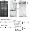
Figure 3. CHEF Southern hybridization using probes against the right arms of chromosomes VIII and XII.
1: wild type San1, 2: rad54Δ/rad54Δ, 3: OeRAD54, 4: OeRAD54clw, 5: OeRAD54cl37, 6: OeRAD54cl38. The hybridization with cdc23 (VIII) points out the unexpected bands (a, b, c) in the translocated strains (lanes 4, 5, and 6) that are also probed with rsc2 (XII). The same pattern obtained with cdc23 was verified with probes against rim4, rps20, ydr213 and arn2 (not shown). A scheme of the recombination between LTRs explaining the bands a, b and c of the hybridizations is provided at the bottom.
Figure 4
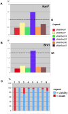
Figure 4. Chromosome stability in rdh54Δ/rdh54Δ translocants.
A) Kanamycin gene copy number ( = N) obtained by quantitative PCR in the six rdh54Δ/rdh54Δ translocants compared with the control (K). The control K is a strain where one copy of kanamycin stably replaces the DUR3 gene. B) Gene copy number ( = N) of BRX1 in rdh54Δ/rdh54Δ translocants compared with the wild type (that has two copies of this gene). BRX1 is located within chromosome XV (see Figure 2A) and on the translocated chromosome XV–VIII. Each histogram is the result of nine independent readings. C) Loss (%) of the chromosome carrying kanamycin, indicated by lack of growth on G418 medium (in red), versus viability (in blue) derived by replica-plating. The strains were grown overnight and plated without selection and then replicated on G418. The results are expressed in percentage. 1: wild type strain, 2: K, 3: rdh54Δ/rdh54Δcl4, 4: rdh54Δ/rdh54Δcl7, 5: rdh54Δ/rdh54Δcl16, 6: rdh54Δ/rdh54Δcl23, 7: rdh54Δ/rdh54Δcl30, 8 rdh54Δ/rdh54Δcl47.
Figure 5
Figure 5. KO and translocation (T) events with short and long homologies between two homologous chromosomes VIII.
The two homologs are labeled here as chrVIII a and b, and are bridged by a DNA cassette carrying kanamycin resistance (KAN). On the top, the molecular event that can give rise to KO (left) and to T (right) as previously reported using a cassette with short homologies (40 nt). F and Y are the two possible chromosome configurations for the KO as reported in the text. The only translocant found was cl77 while the opposite configuration was never detected. At the bottom (framed area), the different result obtained with a homology of 400 nt (reported in this work). On the left the transformants with the KO in the homologous chrVIII a (F); it is shown that spacer sp2 is lost from both chromosomes VIII; on the contrary, in the transformants where the KO happened in the homolog chrVIII b (Y) sp2 is present on both chromosomes VIII. This LOH, with the absence of sp2 on both chromosomes, is maintained also in the two translocants cl94 and cl44 (frame, right) while cl44 only, which is in rdh54Δ/rdh54Δ background, has retained also sp3 as it is shown experimentally in details in Fig. 6C.
Figure 6
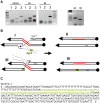
Figure 6. Characterization of the translocants between homologs and proposed model explaining the role of Rdh54.
A) Characterization through colony-PCR of cl44 and cl94. The presence of spacer2 (lane 1 = primers sp2/sp2Fw), spacer3 (lane 2 = primers sp3/Dur3’int) and spacer1 (lane3 = primers Sp1/Dur3R) are shown from left to right in the wild type strain YF123, in the translocant 44 (rdh54Δ/rdh54Δ background) and in the translocant 94 (wt YF123) respectively. In the panel on the right the amplification size of the region surrounding spacer3 in clone 44 and 94 is compared (DurEXT/Dur3’int). The partial sequence of these fragments is shown in details on panel C. B) Two hypothetical models to explain the loss or the retaining of spacer sp3 in the wild type cl 94 (I, II) and in the rdh54Δ/rdh54Δcl 44 respectively (III, IV). C) DNA consensus from the alignment of a partial sequence of cl44 and cl94 around spacer3 confirming that only in the first one the spacer is present (here indicated as green, small letters). The capital letters indicate the nucleotides present in both clones.
Figure 7
Figure 7. Characterization of nine independent pol32Δ/pol32Δ translocants.
A) Southern hybridization of genomic DNA from all the pol32Δ/pol32Δ translocants with probes against kanamycin, rim4 (chromosome VIII) and ndj1 (chromosome XV) loci are shown. Lane 1:wild type San1, 2: pol32Δ/pol32Δcl3, 3: pol32Δ/pol32Δcl22, 4: pol32Δ/pol32Δcl23, 5: pol32Δ/pol32Δcl26, 6 pol32Δ/pol32Δcl31, 7: pol32Δ/pol32Δcl35, 8 pol32Δ/pol32Δcl43, 9 pol32Δ/pol32Δcl44, 10: pol32Δ/pol32Δcl46. T indicates the band of the translocated chromosome. The question mark indicates unknown bands coming from a rearrangement of the left part of chromosome XV on the membrane hybridized with ndj1 B) Scheme of the location of the probes used with respect to the breakpoints (underlined) C) The RIM4 copy number is reported with its own legend on the right. The results are referred to the wild type (here represented by a line with a value of two); each bar is the result of nine independent readings (see Materials & Methods for details).

Similar articles

Cited by

References

    1. Teicher BA, Bagley RG, Rouleau C, Kruger A, Ren Y, et al. (2011) Characteristics of human Ewing/PNET sarcoma models. Ann Saudi Med 31: 174–182. - PMC - PubMed
    1. Poitras JL, Costa D, Kluk MJ, Amrein PC, Stone RM, et al. (2011) Genomic alterations in myeloid neoplasms with novel, apparently balanced translocations. Cancer Genet 204: 68–76. - PubMed
    1. Klemke M, Drieschner N, Laabs A, Rippe V, Belge G, et al. (2011) On the prevalence of the PAX8-PPARG fusion resulting from the chromosomal translocation t(2;3)(q13;p25) in adenomas of the thyroid. Cancer Genet 204: 334–339. - PubMed
    1. Hervé AL, Florence M, Philippe M, Michel A, Thierry F, et al. (2011) Molecular heterogeneity of multiple myeloma: pathogenesis, prognosis, and therapeutic implications. J Clin Oncol 29: 1893–1897. - PubMed
    1. Zhang Y, Gostissa M, Hildebrand DG, Becker MS, Boboila C, et al. (2010) The role of mechanistic factors in promoting chromosomal translocations found in lymphoid and other cancers. Adv Immunol 106: 93–133. - PMC - PubMed

MeSH terms

LinkOut - more resources