

Catechol-O-methyltransferase (COMT) protein expression and activity after dopaminergic and noradrenergic lesions of the rat brain
- PMID: 23613844
- PMCID: PMC3628796
- DOI: 10.1371/journal.pone.0061392
Catechol-O-methyltransferase (COMT) protein expression and activity after dopaminergic and noradrenergic lesions of the rat brain
Abstract
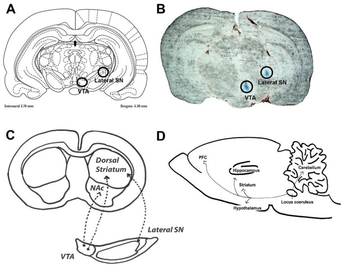
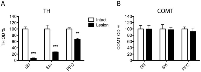
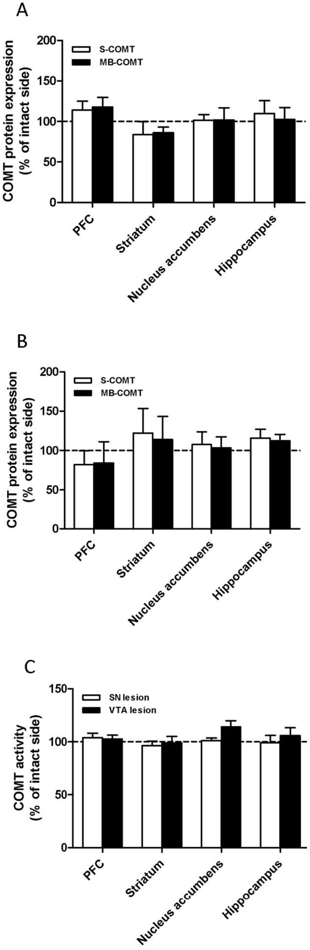
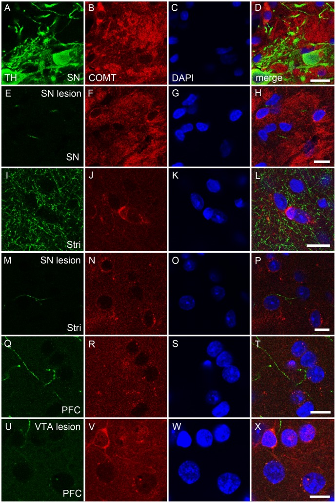
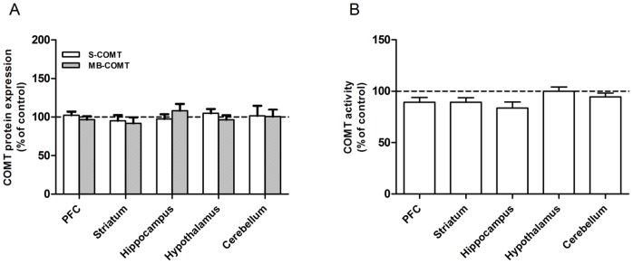
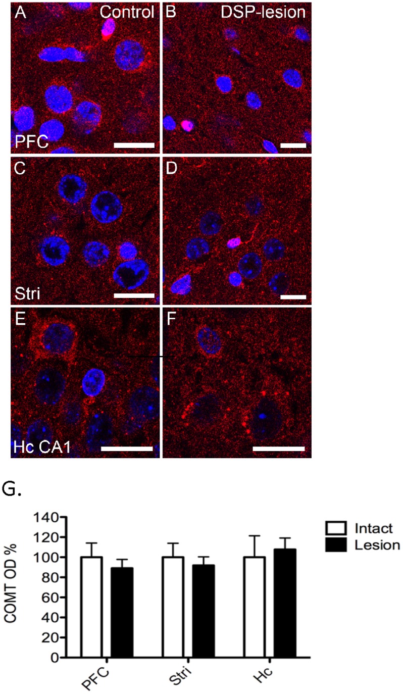
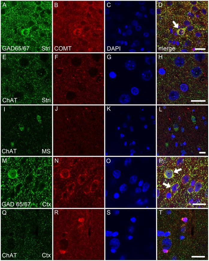
The occurrence of catechol-O-methyltransferase (COMT) in presynaptic neurons remains controversial. This study utilized dopaminergic and noradrenergic toxins to assess the presence of COMT in the presynaptic neurons originating from the substantia nigra, ventral tegmental area or locus coeruleus. Destruction of dopaminergic and noradrenergic neurons was assessed by measuring the dopamine and noradrenaline content in the projection areas of these neurons. Additionally, COMT protein expression and activity were examined in several projection areas to determine whether there are any changes in COMT values. Colocalization studies were done to identify COMT-containing postsynaptic neurons. Despite successful lesioning of dopaminergic and noradrenergic neurons, no changes in COMT protein expression or activity could be noted. These results strongly suggest that COMT is not present in presynaptic dopaminergic and noradrenergic neurons. There was a high colocalization of COMT with the GABAergic marker of short neurons both in the striatum and cortex but only a weak, if any, with the cholinergic marker in the cortex.
Conflict of interest statement
Figures
Similar articles
-
Potentiation of parkinsonian symptoms by depletion of locus coeruleus noradrenaline in 6-hydroxydopamine-induced partial degeneration of substantia nigra in rats.Eur J Neurosci. 2003 Jun;17(12):2586-92. doi: 10.1046/j.1460-9568.2003.02684.x. Eur J Neurosci. 2003. PMID: 12823465
-
Striatal membrane-bound and soluble catechol-O-methyl-transferase after selective neuronal lesions in the rat.J Neural Transm. 1987;69(3-4):221-8. doi: 10.1007/BF01244343. J Neural Transm. 1987. PMID: 3114426
-
Catechol O-methyltransferase mRNA expression in human and rat brain: evidence for a role in cortical neuronal function.Neuroscience. 2003;116(1):127-37. doi: 10.1016/s0306-4522(02)00556-0. Neuroscience. 2003. PMID: 12535946
-
Role of catechol-O-methyltransferase (COMT)-dependent processes in Parkinson's disease and L-DOPA treatment.CNS Neurol Disord Drug Targets. 2012 May;11(3):251-63. doi: 10.2174/187152712800672436. CNS Neurol Disord Drug Targets. 2012. PMID: 22483291 Review.
-
The role of catechol-O-methyltransferase in reward processing and addiction.CNS Neurol Disord Drug Targets. 2012 May;11(3):306-23. doi: 10.2174/187152712800672409. CNS Neurol Disord Drug Targets. 2012. PMID: 22483300 Review.
Cited by
-
Monoaminergic and Histaminergic Strategies and Treatments in Brain Diseases.Front Neurosci. 2016 Nov 24;10:541. doi: 10.3389/fnins.2016.00541. eCollection 2016. Front Neurosci. 2016. PMID: 27932945 Free PMC article. Review.
-
Initial Molecular Mechanisms of the Pathogenesis of Parkinson's Disease in a Mouse Neurotoxic Model of the Earliest Preclinical Stage of This Disease.Int J Mol Sci. 2024 Jan 22;25(2):1354. doi: 10.3390/ijms25021354. Int J Mol Sci. 2024. PMID: 38279354 Free PMC article.
-
Limiting Monoamines Degradation Increases L-DOPA Pro-Locomotor Action in Newborn Rats.Int J Mol Sci. 2023 Sep 29;24(19):14747. doi: 10.3390/ijms241914747. Int J Mol Sci. 2023. PMID: 37834195 Free PMC article.
-
Should "on-demand" treatments for Parkinson's disease OFF episodes be used earlier?Clin Park Relat Disord. 2022 Aug 12;7:100161. doi: 10.1016/j.prdoa.2022.100161. eCollection 2022. Clin Park Relat Disord. 2022. PMID: 36033905 Free PMC article. Review.
-
Associations between catechol-O-methyltransferase (COMT) genotypes at rs4818 and rs4680 and gene expression in human dorsolateral prefrontal cortex.Exp Brain Res. 2020 Feb;238(2):477-486. doi: 10.1007/s00221-020-05730-0. Epub 2020 Jan 20. Exp Brain Res. 2020. PMID: 31960101
References
-
- Männistö PT, Kaakkola S (1999) Catechol-O-methyltransferase (COMT): Biochemistry, molecular biology, pharmacology, and clinical efficacy of the new selective COMT inhibitors. Pharmacol Rev 51: 593–628. - PubMed
-
- Kaakkola S, Männistö PT, Nissinen E (1987) Striatal membrane-bound and soluble catechol-O-methyl-transferase after selective neuronal lesions in the rat. J Neur Transm 69: 221–228. - PubMed
-
- Kaplan GP, Hartman BK, Creveling CR (1981) Localization of catechol-O-methyltransferase in the leptomeninges, choroid plexus and ciliary epithelium: Implications for the separation of central and peripheral catechols. Brain Res 204: 353–360. - PubMed
-
- Rivett AJ, Francis A, Roth JA (1983) Distinct cellular localization of membrane-bound and soluble forms of catechol-O-methyltransferase in brain. J Neurochem 40: 215–219. - PubMed
-
- Karhunen T, Tilgmann C, Ulmanen I, Panula P (1995) Catechol-O-methyltransferase (COMT) in rat brain: Immunoelectron microscopic study with an antiserum against rat recombinant COMT protein. Neurosc Lett 187: 57–60. - PubMed
Publication types
MeSH terms
Substances
LinkOut - more resources
Full Text Sources
Other Literature Sources
Miscellaneous
