Effect of age and cytoskeletal elements on the indentation-dependent mechanical properties of chondrocytes
- PMID: 23613892
- PMCID: PMC3628340
- DOI: 10.1371/journal.pone.0061651
Effect of age and cytoskeletal elements on the indentation-dependent mechanical properties of chondrocytes
Abstract
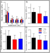
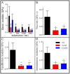
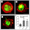
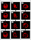
Articular cartilage chondrocytes are responsible for the synthesis, maintenance, and turnover of the extracellular matrix, metabolic processes that contribute to the mechanical properties of these cells. Here, we systematically evaluated the effect of age and cytoskeletal disruptors on the mechanical properties of chondrocytes as a function of deformation. We quantified the indentation-dependent mechanical properties of chondrocytes isolated from neonatal (1-day), adult (5-year) and geriatric (12-year) bovine knees using atomic force microscopy (AFM). We also measured the contribution of the actin and intermediate filaments to the indentation-dependent mechanical properties of chondrocytes. By integrating AFM with confocal fluorescent microscopy, we monitored cytoskeletal and biomechanical deformation in transgenic cells (GFP-vimentin and mCherry-actin) under compression. We found that the elastic modulus of chondrocytes in all age groups decreased with increased indentation (15-2000 nm). The elastic modulus of adult chondrocytes was significantly greater than neonatal cells at indentations greater than 500 nm. Viscoelastic moduli (instantaneous and equilibrium) were comparable in all age groups examined; however, the intrinsic viscosity was lower in geriatric chondrocytes than neonatal. Disrupting the actin or the intermediate filament structures altered the mechanical properties of chondrocytes by decreasing the elastic modulus and viscoelastic properties, resulting in a dramatic loss of indentation-dependent response with treatment. Actin and vimentin cytoskeletal structures were monitored using confocal fluorescent microscopy in transgenic cells treated with disruptors, and both treatments had a profound disruptive effect on the actin filaments. Here we show that disrupting the structure of intermediate filaments indirectly altered the configuration of the actin cytoskeleton. These findings underscore the importance of the cytoskeletal elements in the overall mechanical response of chondrocytes, indicating that intermediate filament integrity is key to the non-linear elastic properties of chondrocytes. This study improves our understanding of the mechanical properties of articular cartilage at the single cell level.
Conflict of interest statement
Figures


References
-
- Loeser RF (2000) Chondrocyte integrin expression and function. Biorheology 37: 109–116. - PubMed
-
- Mow VC, Kuei SC, Lai WM, Armstrong CG (1980) Biphasic creep and stress relaxation of articular cartilage in compression? Theory and experiments. J Biomech Eng 102: 73–84. - PubMed
-
- Maroudas A (1979) Physicochemical properties of articular cartilage. In: Freeman, M AR (Ed) Adult Articular Cartilage Pitman Medical, Kent: 215–290.
-
- Mow VC, Ratcliffe A (1997) Structure and function of articular cartilage and meniscus. In: Mow, VC, Hayes, WC (Eds), Basic Orthopaedic Biomechanics Lippincott-Raven Philadelphia: 113–177.
Publication types
MeSH terms
Grants and funding
LinkOut - more resources
Full Text Sources
Other Literature Sources
Medical
Miscellaneous

