. 2013 Apr 16;8(4):e61878.
doi: 10.1371/journal.pone.0061878. Print 2013.

AMIGO3 is an NgR1/p75 co-receptor signalling axon growth inhibition in the acute phase of adult central nervous system injury

Affiliations

AMIGO3 is an NgR1/p75 co-receptor signalling axon growth inhibition in the acute phase of adult central nervous system injury

Zubair Ahmed et al. PLoS One. .

Abstract

Axon regeneration in the injured adult CNS is reportedly inhibited by myelin-derived inhibitory molecules, after binding to a receptor complex comprised of the Nogo-66 receptor (NgR1) and two transmembrane co-receptors p75/TROY and LINGO-1. However, the post-injury expression pattern for LINGO-1 is inconsistent with its proposed function. We demonstrated that AMIGO3 levels were significantly higher acutely than those of LINGO-1 in dorsal column lesions and reduced in models of dorsal root ganglion neuron (DRGN) axon regeneration. Similarly, AMIGO3 levels were raised in the retina immediately after optic nerve crush, whilst levels were suppressed in regenerating optic nerves, induced by intravitreal peripheral nerve implantation. AMIGO3 interacted functionally with NgR1-p75/TROY in non-neuronal cells and in brain lysates, mediating RhoA activation in response to CNS myelin. Knockdown of AMIGO3 in myelin-inhibited adult primary DRG and retinal cultures promoted disinhibited neurite growth when cells were stimulated with appropriate neurotrophic factors. These findings demonstrate that AMIGO3 substitutes for LINGO-1 in the NgR1-p75/TROY inhibitory signalling complex and suggests that the NgR1-p75/TROY-AMIGO3 receptor complex mediates myelin-induced inhibition of axon growth acutely in the CNS. Thus, antagonizing AMIGO3 rather than LINGO-1 immediately after CNS injury is likely to be a more effective therapeutic strategy for promoting CNS axon regeneration when combined with neurotrophic factor administration.


Conflict of interest statement

Competing Interests: The authors have declared that no competing interests exist.

Figures

Figure 1
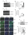
Figure 1. Levels of AMIGO at 1 day after DC injury.
(A) Semi-quantitative RT-PCR and (B) densitometry to show changes in mRNA levels of AMIGO isoforms and LINGO-1 expressed in non-regenerating DRGN compared to intact controls. GAPDH is used as a housekeeping gene. (C) Representative western blots and (D) densitometry to show changes in protein levels of AMIGO isoforms and LINGO-1 in non-regenerating DRGN relative to intact controls. (E–H) Immunohistochemistry in sections from intact controls and 1 d DC injured DRGN shows immunolabelling for AMIGO (E), AMIGO2 (F), AMIGO3 (G) and (H) LINGO-1. Insets in E-H show images from blocking peptide controls to demonstrate specificity of the relevant antibody Scale bars in E–H = 100 µm. *** = P<0.0001, ANOVA.
Figure 2
Figure 2. AMIGO3 levels at 10 days in intact controls, non-regenerating DC, and regenerating SN and pSN+DC models.
(A), (C) Semi-quantitative RT-PCR and (B), (D) densitometry was used to quantify AMIGO3 and LINGO-1 expression levels using total RNA extracted from appropriately treated or untreated DRG. (E–S) Immunohistochemistry, (T) western blotting and (U) densitometry to show high levels of AMIGO3 localisation in DRGN from non-regenerating DC models compared to those in regenerating SN and pSN+DC models. High power insets in (H) and (J) show neuronal and glial localisation of AMIGO3 in sections double stained with Neurofilament 200 and AMIGO3 (arrowhead). Blocking peptide control (Q–S) shows the specificity of AMIGO3 antibodies used. Scale bar = 100 µm. *** = p<0.0001, ANOVA.
Figure 3
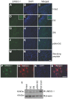
Figure 3. LINGO1 levels in intact, non-regenerating DC and regenerating (SN and pSN+DC lesioned) DRGN models 10 days after injury.
High power insets in (D) and (F) show neuronal and glial localisation of AMIGO3. (P–S) Co-localisation of LINGO-1 with AMIGO3 in DRGN from non-regenerating DC models. (T) Western blotting of LINGO-1 levels in DRG in our experimental model paradigms showing low and unchanged levels of LINGO-1 compared to a positive control lane using rat brain lysates. β-actin was used as a loading control. Scale bars = 100 µm.
Figure 4
Figure 4. Suppressed levels of AMIGO3 correlate with optic nerve regeneration.
(A) Representative western blot of AMIGO3 in NRM and RM at 0, 6, 8 and 20 days after ONC and (B) densitometry to show higher levels of AMIGO3 in NRM at 6, 8 and 20 days after ONC. β-actin was used as a protein loading control. (C) Immunohistochemistry to show that high levels of AMIGO3 is expressed in the majority of RGC in NRM (e.g. arrows) while lower levels are present in RGC from RM retinae. Insets show co-localisation of βIII-tubulin with AMIGO3 (arrowheads). Scale bar in (C) = 100 µm, in insets = 20 µm. *** = P<0.0001, ANOVA.
Figure 5
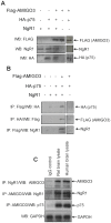
Figure 5. AMIGO3 interacts with NgR1 and p75.
(A) Western blot of COS-7 total cell lysates extracted 48 h after transfection with combinations of FLAG-AMIGO3, HA-p75 and NgR1 genes. (B) Co-immunoprecipitation of AMIGO3, NgR1 and p75 detected after western blotting for relevant anti-HA, anti-FLAG or rabbit anti-NgR1 antibodies from COS-7 cells co-expressing combinations of Flag-AMIGO3, NgR1 and HA-p75. (C) Co-immunoprecipitation of AMIGO3, NgR1 and p75 from adult rat and human brain lysates using combinations of goat anti-AMIGO3, rabbit anti-NgR1 and mouse anti-p75 antibodies for immunoprecipitation (IP) and western blotting (WB). IgG was used as a control antibody for IP and GAPDH was used as a loading control.
Figure 6
Figure 6. Co-transfection of AMIGO3, NgR1 and p75 activates RhoA.
(A) RhoGTP levels detected by a Rho pulldown assay in control untransfected and transfected COS-7 cells expressing NgR1-p75 and NgR1-p75-AMIGO3 genes, in the presence (+) and absence of CME (−). Total Rho levels were also detected by western blotting. (B) Colorimetric Rho activation assay in untransfected control and in COS7 cells transfected with NgR1-p75 and NgR1-p75-AMIGO3 gene combinations in the presence of CME (+). (C) Colorimetric Rho activation assay in untransfected control COS7 cells and cells transfected with NgR1-TROY-AMIGO3, NgR1-p75-AMIGO3 CT1- genes, in the presence of CME (+).
Figure 7
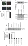
Figure 7. AMIGO3 is localised in DRGN and its suppression using siAMIGO3 in the presence of CME prevents Rho activation.
(A) Representative AMIGO3 localisation in DRGN (arrows = AMIGO3+, arrowhead = AMIGO3) and (B) quantification of the percentage ± SEM of DRGN which are AMIGO3+ and AMIGO3, in DRG cultures. (C) Western blotting and densitometry showing the levels of knockdown, compared to Scr-siAMIGO3 and siGFP after siAMIGO3 treatment. Re-stripped western blots are also shown for AMIGO, AMIGO2 and LINGO-1 to demonstrate that knockdown of AMIGO3 does not affect the levels of these molecules in culture. β-actin is used as a loading control. (D) Rho activation assay in untransfected and siRNA transfected DRG cells in the presence of CME. Total Rho levels were also detected by western blotting. Scale bar in (A) = 50 µm.
Figure 8
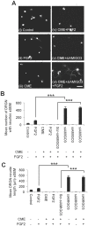
Figure 8. Knockdown of AMIGO3 promotes DRGN neurite outgrowth in the presence of CME.
(A) Representative photomicrographs of βIII-tubulin+ DRGN neurite outgrowth in untransfected and siRNA transfected DRG cells in the presence or absence of CME with or without FGF-2 ± SEM. Quantification of (B) mean number of DRGN with neurites ± SEM and (C) mean neurite length after treatment with AMIGO3 in the presence of CME, with and without FGF-2 ± SEM. *** = P<0.0001, ANOVA. Scale bar in (A) = 100 µm.
Figure 9
Figure 9. Knockdown of AMIGO3 promote RGC neurite outgrowth in the presence of inhibitory concentration of CME.
(A) Immunocytochemistry and (B) quantitation to show that >95% of RGC were AMIGO3+ (arrow) while a small proportion of RGC were AMIGO (arrowheads). (C) Representative photomicrographs of βIII-tubulin+ RGC neurite outgrowth in CNTF and siRNA transfected retinal cells in the presence and absence of CME with or without CNTF. Quantification of (D) mean proportion of RGC with neurites and (E) mean neurite length after treatment with siAMIGO3 in the presence of CME, with and without CNTF. *** = P<0.0001, ANOVA. Scale bars in (A) = 50 µm and (C) = 100 µm.

Similar articles

Cited by

References

    1. Sandvig A, Berry M, Barrett LB, Butt A, Logan A (2004) Myelin-, reactive glia-, and scar-derived CNS axon growth inhibitors: expression, receptor signaling, and correlation with axon regeneration. Glia 46: 225–251. - PubMed
    1. Hunt D, Coffin RS, Anderson PN (2002) The Nogo receptor, its ligands and axonal regeneration in the spinal cord; a review. J Neurocytol 31: 93–120. - PubMed
    1. Berry M, Ahmed Z, Lorber B, Douglas M, Logan A (2008) Regeneration of axons in the visual system. Restor Neurol Neurosci 26: 147–174. - PubMed
    1. Park KK, Liu K, Hu Y, Smith PD, Wang C, et al. (2008) Promoting axon regeneration in the adult CNS by modulation of the PTEN/mTOR pathway. Science 322: 963–966. - PMC - PubMed
    1. Filbin MT (2003) Myelin-associated inhibitors of axonal regeneration in the adult mammalian CNS. Nat Rev Neurosci 4: 703–713. - PubMed

Publication types

LinkOut - more resources