. 2013 Apr 16;8(4):e61980.
doi: 10.1371/journal.pone.0061980. Print 2013.

Paraquat modulates alternative pre-mRNA splicing by modifying the intracellular distribution of SRPK2

Affiliations

Paraquat modulates alternative pre-mRNA splicing by modifying the intracellular distribution of SRPK2

Silvia Vivarelli et al. PLoS One. .

Abstract

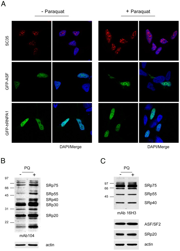
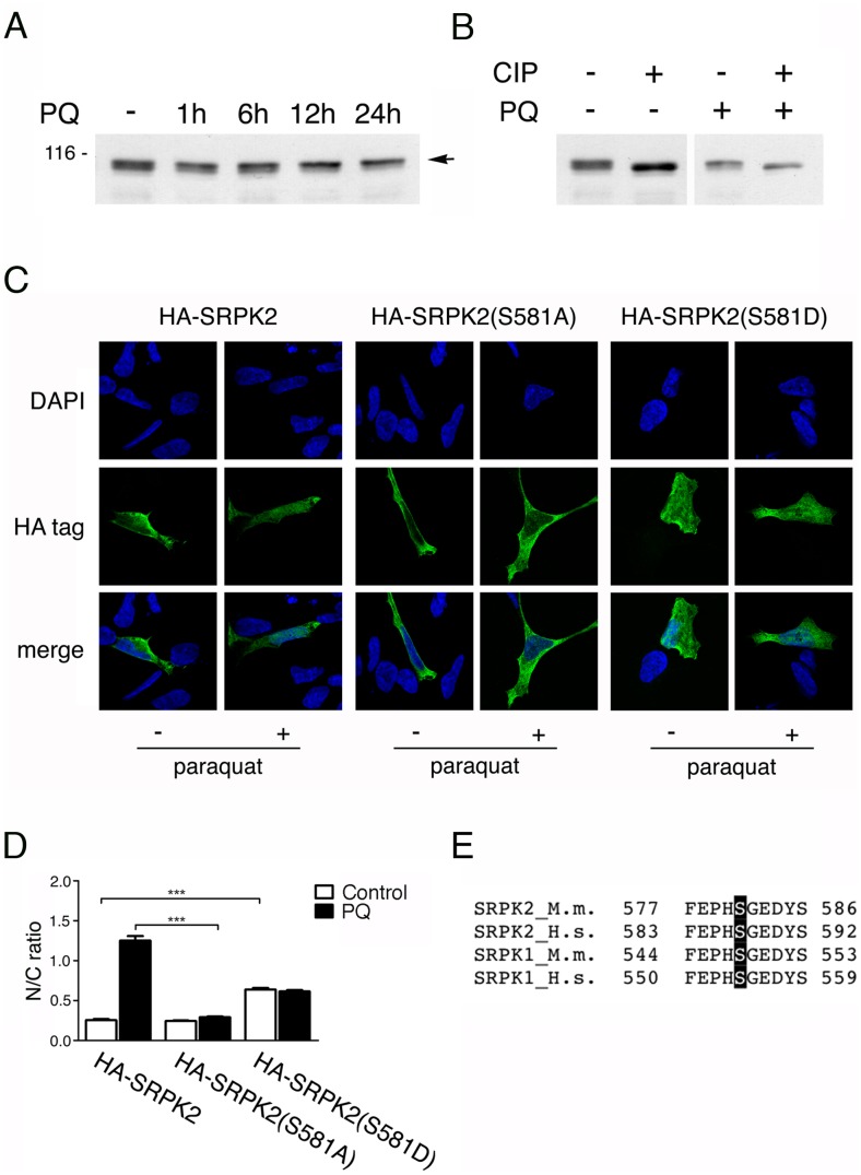
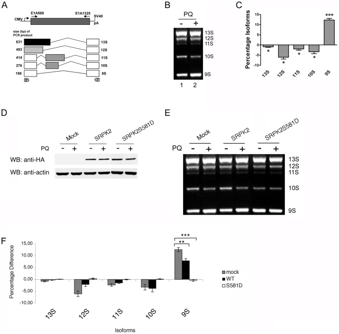
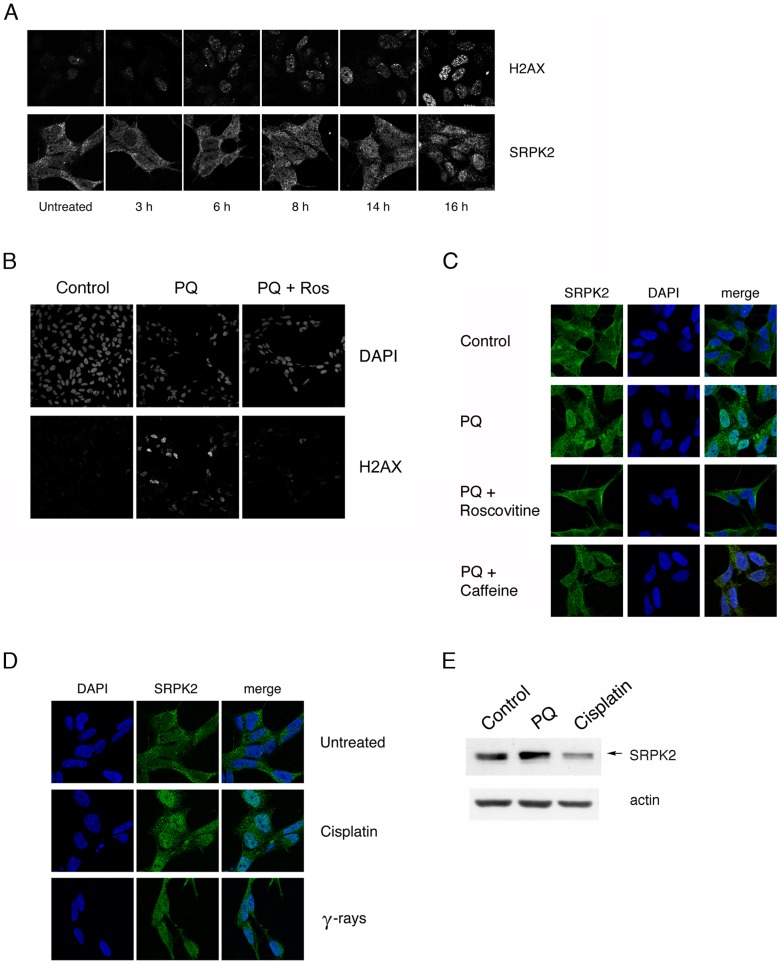
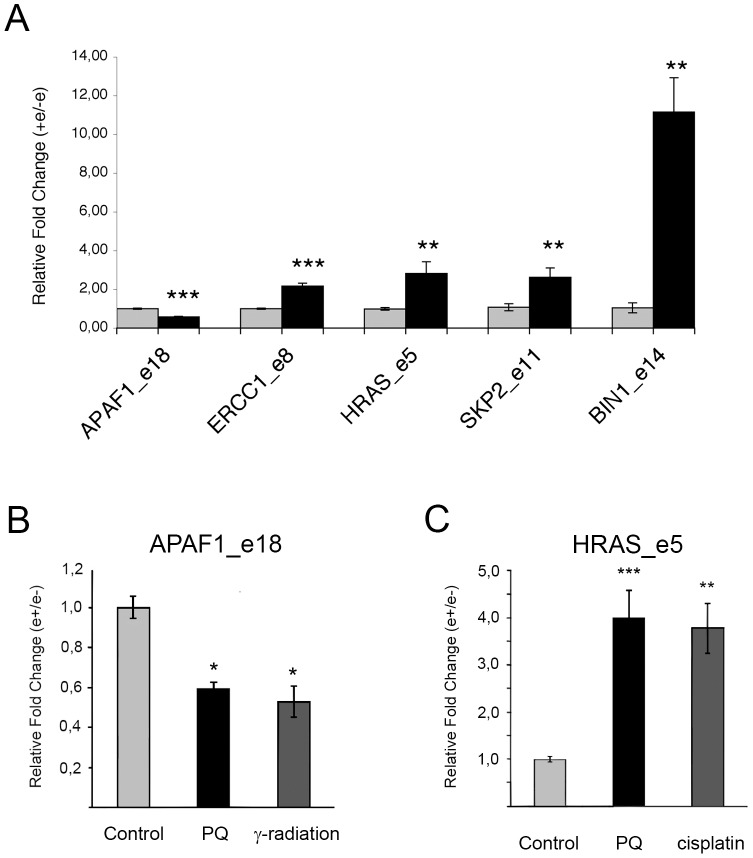
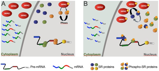
Paraquat (PQ) is a neurotoxic herbicide that induces superoxide formation. Although it is known that its toxic properties are linked to ROS production, the cellular response to PQ is still poorly understood. We reported previously that treatment with PQ induced genome-wide changes in pre-mRNA splicing. Here, we investigated the molecular mechanism underlying PQ-induced pre-mRNA splicing alterations. We show that PQ treatment leads to the phosphorylation and nuclear accumulation of SRPK2, a member of the family of serine/arginine (SR) protein-specific kinases. Concomitantly, we observed increased phosphorylation of SR proteins. Site-specific mutagenesis identified a single serine residue that is necessary and sufficient for nuclear localization of SRPK2. Transfection of a phosphomimetic mutant modified splice site selection of the E1A minigene splicing reporter similar to PQ-treatment. Finally, we found that PQ induces DNA damage and vice versa that genotoxic treatments are also able to promote SRPK2 phosphorylation and nuclear localization. Consistent with these observations, treatment with PQ, cisplatin or γ-radiation promote changes in the splicing pattern of genes involved in DNA repair, cell cycle control, and apoptosis. Altogether, our findings reveal a novel regulatory mechanism that connects PQ to the DNA damage response and to the modulation of alternative splicing via SRPK2 phosphorylation.


Conflict of interest statement

Competing Interests: The authors have declared that no competing interests exist.

Figures

Figure 1
Figure 1. Increased phosphorylation of SR proteins in PQ-treated cells.
A. PQ induced relocalization of SR proteins in the nucleus of treated cells. SH-SY5Y cells treated with vehicle or with 0.75 mM PQ for 18 h were immunostained with an anti-SC35 antibody (upper row), transiently transfected with GFP-ASF/SF2 (middle row), or with GFP-hnRNPA1 (lower row). Nuclei were stained with DAPI. B. Increased phosphorylation of SR proteins in PQ-treated cells. Total extract of control or PQ-treated cells was probed with mAb104 to determine the phosphorylation state of classical SR proteins. C. The same extracts used for the Western blot shown in panel B were probed with the 16H3 monoclonal antibody that detects SR proteins regardless of their phosphorylation status, with anti ASF/SF2 and anti-SRp20 monoclonal antibodies. Actin was used as loading control.
Figure 2
Figure 2. Nuclear translocation of SRPK2 and phosphorylation of SR proteins in response to PQ.
A. Representative confocal micrographs of SH-SY5Y cells stained with an SRPK2-specific antibody. Upper row: cells treated with vehicle; lower row: cells treated with 0.75 mM PQ for 18 h. DAPI was used to identify the nuclei. B. The average nuclear to cytoplasmic ratio (N/C) ratio of SRPK2 fluorescent signal was determined for 50 cells as described in Materials and Methods. *** indicates p<0.001 treated vs. control group by unpaired t-test. C. RNA-mediated silencing strongly reduced SRPK1 and SRPK2 expression. Western blot analysis of the expression level of SRPK proteins in the same extract used for the Western blot shown in figure 2D and 2E. CPSF73K was used as loading control. D. Silencing of SRPK1 and SRPK2 abolishes PQ-mediated phosphorylation of SR proteins. Western blot analysis of nuclear extracts prepared from control SH-SY5Y cells treated with vehicle or with PQ, and from cells depleted of both SRPK1 and SRPK2. Phosphorylated SR proteins were detected with mab104. E. To control for equal loading of the samples SR proteins were also detected with mab16H3.
Figure 3
Figure 3. Phosphorylation of Ser581 is required for the nuclear localization of SRPK2.
A. Western blots of total cell extract prepared from untreated SH-SY5Y cells or from cells treated with PQ for the indicated times. The blot was probed with an anti-SRPK2 antibody. Upon PQ treatment an increase of the slower migrating band (indicated by an arrowhead) could be observed. B. SRPK2 phosphorylation was confirmed by CIP treatment. Total cell lysates prepared from untreated cells and from cells treated with PQ were incubated with calf intestinal phosphatase (CIP) as described in Material and methods. C. Representative confocal micrographs of SH-SY5Y cells transfected with a construct expressing HA-tagged SRPK2, HA-SRPK2(S581A), or HA-SRPK2(S581D). Upper row: DAPI; middle row: cells stained with an HA-specific antibody; lower row: merge of the DAPI and antibody signals. D. The nuclear to cytoplasmic ratio (N/C) of the fluorescence signal was determined for 50 transfected cells. The graph shows the average N/C ratio of SRPK2 signal measured as described in Materials and Methods. E. *** indicates p<0.0001 of groups compared by one-way ANOVA and Tukey post-test analysis. F. Sequence alignment of the 577–586 aa region in SRPK2 highlighting conservation of CK2 consensus sites across species. A black, vertical bar indicates the conserved serine.
Figure 4
Figure 4. PQ treatment or nuclear SRPK2 affect splice site selection of the E1A minigene transcript.
A. Schematic diagram illustrating the structure of the construct and the splicing pattern of the E1A minigene reporter. Arrows mark the position of the primers used for PCR analysis. The alternative 5′ splice site and splicing events that generate the different mRNA variants are indicated. B. Representative agarose gel of the splicing assay. SH-SY5Y cells were transiently transfected with the E1A reporter plasmid. 24 h after transfection cells were treated PQ. Total RNA was then isolated and the alternative splicing pattern of the E1A transcripts was determined by RT-PCR. The treatment with PQ induced an increase of the production of the 9 S transcript variant with respect to the other isoforms. C. Densitometric analysis of the E1A splicing products (mean ± S.E., n = 3) in cells treated with vehicle or with PQ. *** indicates p<0.001, and * indicates D. p<0.05, compared with control by paired, two-tailed Student's t test. E. Western blot analysis of the expression level of HA-tagged wild type and the S581D mutant in untreated and in PQ-treated SH-SY5Y cells. Blots were probed with an anti-HA antibody. Actin was used as loading control. F. SH-SY5Y cells were transiently transfected with the E1A splicing reporter minigene alone or together with expression plasmids coding for HA-tagged wild type SRPK2 or with the S581D mutant. RNA and protein fractions were simultaneously prepared. The alternative splicing pattern of the E1A transcripts was determined by RT-PCR. G. Densitometric analysis of the splicing products (mean ± S.E., n = 3) in untreated and PQ-treated cells. The relative levels of 13 S, 12 S, and 9 S mRNAs were quantitated as described in Materials and Methods. *** indicates p<0.001, * * indicates p<0.01, by one-way ANOVA and Dunnett's post test.
Figure 5
Figure 5. Genotoxic stress induces nuclear accumulation and phosphorylation of SRPK2.
A. PQ induces DNA damage. Representative confocal micrographs of SH-SY5Y cells treated with PQ and fixed at the indicated time-points. Cells were stained with an anti-γH2AX antibody (upper row) or with an antibody specific for SRPK2 (lower row). B. Inhibition of PQ-induced H2AX phosphorylation by roscovitine. Representative confocal micrographs of SH-SY5Y cells incubated with 10 µM roscovitine and PQ treatment. ãH2AX was detected by immunocytochemistry; nuclei were stained with DAPI. C. Inhibition of the DDR blocks nuclear localization of SRPK2. Representative confocal micrographs of control SH-SY5Y cells (first row), or SH-SY5Y cells incubated with PQ alone (second row), or with PQ and with 10 µM roscovitine (third row), or with PQ and 10 mM caffeine (forth row). SRPK2 was detected by immunocytochemistry; nuclei were stained with DAPI. D. Genotoxic treatments induce nuclear localization of SRPK2. Representative confocal micrographs of untreated SH-SY5Y cells (upper row) or cells treated with 20 µM cisplatin for 18 h (middle row) or irradiated with 10 Gy (lower row) were stained with DAPI and with an anti-SRPK2 antibody. E. Genotoxic treatments induce hyperphosphorylation of SRPK2. Western blots of total cell extract prepared from untreated SH-SY5Y cells or from cells treated with PQ, or with cisplatin for the indicated times. The blot was probed with an anti-SRPK2 antibody. Actin was used as loading control. The slower migrating band is indicated by an arrow.
Figure 6
Figure 6. Genotoxic stress modifies alternative splicing of endogenous genes.
A. SH-SY5Y cells were incubated with vehicle or with PQ as described in Material and Methods. The bar graph represents the quantification of the RT-PCR splicing analysis of the alternatively spliced regions of the APAF1 (exon 18, e18), H-RAS (exon 5, e 5), ERCC1 (exon 8, e 8), SKP2 (exon 11, e 11), and BIN1 (exon 14, e14) transcripts. The indicated splice forms were subcloned and sequenced to verify their identity. The inclusion or the skipping of variable exons after PQ treatment (black bars) was normalized relative to that observed in the respective controls (light grey bars). Error bars indicate the standard error three biological replicates. The asterisks represents the result of two-tailed t-test: *** indicates p<0.001, ** indicates p<0.01. B. RT-PCR splicing analysis of the alternatively spliced exon 18 (e18) of the APAF1 transcript in SH-SY5Y cells treated with vehicle (control), PQ, or γ-radiation. The asterisks represents the result of one-way ANOVA and Tukey's post test: * indicates p<0.05. C. RT-PCR splicing analysis of the alternatively spliced exon 5 (e5) of the H-RAS transcript in SH-SY5Y cells treated with vehicle, PQ, or cisplatin as described in Material and Methods. The asterisks represents the result of one-way ANOVA and Dunnett's post test: *** indicates p<0.001, ** indicates p<0.01.
Figure 7
Figure 7. Model of SRPK2 action inthe modulation of stress-dependent splicing.
A. Under normal conditions SRPK2 is mainly located in the cytoplasm. Its low level in the nucleus is counteracted by phosphatases that keep SR proteins in a hypophosphorylated state. B. Upon activation of stress-dependent signalling (f. ex. induced by DNA damage) SRPK2 is phosphorlyated in serine 581 and translocates to the nucleus. Here, the increased SRPK2 concentration overrides dephosphorlyation by PPases and leads to hyperphosphorylation of SR proteins. Since hyperphosphorylation of the RS domain is detrimental for splicing activity, this results in the reduction of the active pool of SR proteins and to a change in the balance between SR proteins and hnRNP proteins thus modifying the choice of alternative splice sites.

Similar articles

Cited by

References

    1. Cocheme HM, Murphy MP (2008) Complex I is the major site of mitochondrial superoxide production by paraquat. J Biol Chem 283: 1786–1798. - PubMed
    1. Lenzken SC, Romeo V, Zolezzi F, Cordero F, Lamorte G, et al. (2011) Mutant SOD1 and mitochondrial damage alter expression and splicing of genes controlling neuritogenesis in models of neurodegeneration. Hum Mutat 32: 168–182. - PubMed
    1. Schwerk C, Schulze-Osthoff K (2005) Regulation of apoptosis by alternative pre-mRNA splicing. Mol Cell 19: 1–13. - PubMed
    1. Shin C, Manley JL (2004) Cell signalling and the control of pre-mRNA splicing. Nat Rev Mol Cell Biol 5: 727–738. - PubMed
    1. Cooper TA, Wan L, Dreyfuss G (2009) RNA and disease. Cell 136: 777–793. - PMC - PubMed

Publication types

MeSH terms