. 2013 Apr 17;8(4):e62025.
doi: 10.1371/journal.pone.0062025. Print 2013.

Anatomic pathways of peripancreatic fluid draining to mediastinum in recurrent acute pancreatitis: visible human project and CT study

Affiliations

Anatomic pathways of peripancreatic fluid draining to mediastinum in recurrent acute pancreatitis: visible human project and CT study

Haotong Xu et al. PLoS One. .

Abstract

Background: In past reports, researchers have seldom attached importance to achievements in transforming digital anatomy to radiological diagnosis. However, investigators have been able to illustrate communication relationships in the retroperitoneal space by drawing potential routes in computerized tomography (CT) images or a virtual anatomical atlas. We established a new imaging anatomy research method for comparisons of the communication relationships of the retroperitoneal space in combination with the Visible Human Project and CT images. Specifically, the anatomic pathways of peripancreatic fluid extension to the mediastinum that may potentially transform into fistulas were studied.

Methods: We explored potential pathways to the mediastinum based on American and Chinese Visible Human Project datasets. These drainage pathways to the mediastinum were confirmed or corrected in CT images of 51 patients with recurrent acute pancreatitis in 2011. We also investigated whether additional routes to the mediastinum were displayed in CT images that were not in Visible Human Project images.

Principal findings: All hypothesized routes to the mediastinum displayed in Visible Human Project images, except for routes from the retromesenteric plane to the bilateral retrorenal plane across the bilateral fascial trifurcation and further to the retrocrural space via the aortic hiatus, were confirmed in CT images. In addition, route 13 via the narrow space between the left costal and crural diaphragm into the retrocrural space was demonstrated for the first time in CT images.

Conclusion: This type of exploration model related to imaging anatomy may be used to support research on the communication relationships of abdominal spaces, mediastinal spaces, cervical fascial spaces and other areas of the body.


Conflict of interest statement

Competing Interests: The authors have declared that no competing interests exist.

Figures

Figure 1
Figure 1. Visualization of pathways into the retrocrural space across the esophageal hiatus.
(A) The oblique sagittal plane shows Route 1, Route 2 and Route 3 track together from the left extraperitoneal space to the retrocrural space across the esophageal hiatus (white arrowheads) from the lateral superior view on CVH2. Route 1 (black open arrows) is displayed as the path from the peripancreatic space to the left extraperitoneal space. Route 2 (crooked arrows) presents the conduit in the retromesenteric plane (blue lines) spreading to the left extraperitoneal space. Route 3 (white long arrows) shows the pathway from the retromesenteric plane to the retrorenal plane (green lines) and further to the left extraperitoneal space. (B) Case 1, a 47-year-old man with recurrent acute pancreatitis. Route 1 to Route 3 on the reformed CT plane corresponds well to the paths on CVH2. The thickened bridging septa distributes on Route 3 (white short arrows). A: left extraperitoneal space; B: pancreas; C: left kidney; D: left adrenal gland; E: esophageal hiatus; F: retrocrural space; G: pericapsular fluid collections. (C) Route 4 (white open arrows) from the retromesenteric plane to the left retrorenal plane across the left fascial trifurcation and further into the retrocrural space via the esophageal hiatus is displayed on the dataset of VHPA. (D) Case 2, a 60-year-old woman with recurrent acute pancreatitis. Route 4 is present in the CT image.
Figure 2
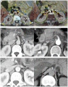
Figure 2. Manifestation of pathways into the retrocrural space across the aortic hiatus.
(A, B) Route 5 to Route 11 into the retrocrural space across the aortic hiatus is displayed on CVH2. (A) The potential Route 5 to Route 9 is shown in the superior mesenteric artery section. Route 5 (white arrows) is from the retromesenteric plane to the left retrorenal plane via the left bridging septa, and further into the retrocrural space across the aortic hiatus; Route 6 is present as the similar conduit on the opposite side. Route 7 (arrowheads) is from the retromesenteric plane to the left retrorenal plane through the left fascial trifurcation and additionally into the retrocrural space; Route 8 is the contralateral pathway into the retrocrural space. Route 9 (black arrows) is the channel into the retrocrural space along the superior mesenteric artery. (B) Route 10 and Route 11 (crooked arrows) are the pathways along the left renal vein and right renal artery into the retrocrural space across the aortic hiatus on the left renal vein section. A: pancreas; B: superior mesenteric artery; C: inferior vena cava; D: right renal artery; E: left renal vein. (C, E) Case 1, a 34-year-old woman with recurrent acute pancreatitis. Route 5, Route 9 and Route 10 are present as fluid spreading along the left bridging septa (white arrows), superior mesenteric artery (black arrows) and left renal vein (crooked arrows) into the retrocrural space, respectively. (D) Case 2, a 47-year-old woman with recurrent acute pancreatitis. Route 6 is displayed as the fluid extending to the retrocrural space via the right bridging septa (white arrows). (F) Case 3, a 42-year-old man with recurrent acute pancreatitis. Route 11 is manifested as the fluid flowing along the right renal vein (crooked arrows) to the retrocrural space across the aortic hiatus.
Figure 3
Figure 3. Presentation of the pathways into the right mediastinum via the inferior vena cava hiatus.
(A) The oblique sagittal plane shows the potential, cranial Route 12 (long arrow) from the retromesenteric plane (blue lines) and retrorenal plane (green lines) to the bare area of the liver, and further into the right mediastinum via the inferior vena cava hiatus from the medial inferior view on CVH2. (B, C) A 72-year-old woman with recurrent acute pancreatitis. Sagittal plane shows Route 12, in which the fluid drains from the bare area of the liver to the right mediastinum across the inferior vena cava hiatus (long arrow). The peripancreatic fluid in the mediastinum further develops to the pleural effusion and the right lower lobar lung consolidation. The axial CT image depicts the pancreatic fluid flows from the retromesenteric plane to the right retrorenal plane through the right fascial trifurcation (arrowheads) and the bridging septa (short arrow). A: inferior vena cava, B: right lung, C: right kidney, D: inferior vena cava hiatus.
Figure 4
Figure 4. Visualization of the pathway into the retrocrural space by penetrating the diaphragm.
(A) A 89-year-old woman with recurrent acute pancreatitis. The CT image manifests Route 13, in which the pancreatic fluid extends from the left perirenal space to the retrocrural space via the narrow space on the left diaphragm (white arrow). (B) The axial CVH5 image illustrates the potential pathway of Route 13 from the left perirenal space to the retrocrural space through the narrow space between the left costal diaphragm and left crural diaphragm. A: crural diaphragm, B: costal diaphragm, C: retromesenteric plane.
Figure 5
Figure 5. Displayed conduits across the left bridging septa to the subcapsular space.
(A) The left perirenal bridging septa may provide a latent channel for renal subcapsular fluid to spread to the fascial planes (white arrows). Conversely, these pathways may be recruited to drain pancreatic fluid from the fascial planes to the subcapsular space in acute pancreatitis (black arrows). (B) A 61-year-old man with carcinoma at the pancreatic head, which led to secondary pancreatitis. The pancreatic fluid extends along the left bridging septa to the subcapsular space (black arrows).

References

    1. Mediero A, Frenkel SR, Wilder T, He W, Mazumder A, et al. (2012) Adenosine A2A Receptor Activation Prevents Wear Particle–Induced Osteolysis. Science Translational Medicine 4: 135ra165–135ra165. - PMC - PubMed
    1. Goldstein LE, Fisher AM, Tagge CA, Zhang X-L, Velisek L, et al. (2012) Chronic Traumatic Encephalopathy in Blast-Exposed Military Veterans and a Blast Neurotrauma Mouse Model. Science Translational Medicine 4: 134ra160–134ra160. - PMC - PubMed
    1. Zhang SX, Heng PA, Liu ZJ (2006) Chinese visible human project. Clin Anat 19: 204–215. - PubMed
    1. Spitzer VM (1995) The Visible Human Project. Available: http://www.nlm.nih.gov/research/visible/visible_human.html via the Internet. Accessed 11 Sep 2003.
    1. Lee SL, Ku YM, Rha SE (2010) Comprehensive reviews of the interfascial plane of the retroperitoneum: normal anatomy and pathologic entities. Emerg Radiol 17: 3–11. - PubMed

Publication types