. 2013 Apr 16;8(4):e62046.
doi: 10.1371/journal.pone.0062046. Print 2013.

Continued expression of GATA3 is necessary for cochlear neurosensory development

Affiliations

Continued expression of GATA3 is necessary for cochlear neurosensory development

Jeremy S Duncan et al. PLoS One. .

Abstract

Hair cells of the developing mammalian inner ear are progressively defined through cell fate restriction. This process culminates in the expression of the bHLH transcription factor Atoh1, which is necessary for differentiation of hair cells, but not for their specification. Loss of several genes will disrupt ear morphogenesis or arrest of neurosensory epithelia development. We previously showed in null mutants that the loss of the transcription factor, Gata3, results specifically in the loss of all cochlear neurosensory development. Temporal expression of Gata3 is broad from the otic placode stage through the postnatal ear. It therefore remains unclear at which stage in development Gata3 exerts its effect. To better understand the stage specific effects of Gata3, we investigated the role of Gata3 in cochlear neurosensory specification and differentiation utilizing a LoxP targeted Gata3 line and two Cre lines. Foxg1(Cre)∶Gata3(f/f) mice show recombination of Gata3 around E8.5 but continue to develop a cochlear duct without differentiated hair cells and spiral ganglion neurons. qRT-PCR data show that Atoh1 was down-regulated but not absent in the duct whereas other hair cell specific genes such as Pou4f3 were completely absent. In addition, while Sox2 levels were lower in the Foxg1(Cre):Gata3(f/f) cochlea, Eya1 levels remained normal. We conclude that Eya1 is unable to fully upregulate Atoh1 or Pou4f3, and drive differentiation of hair cells without Gata3. Pax2-Cre∶Gata3(f/f) mice show a delayed recombination of Gata3 in the ear relative to Foxg1(Cre):Gata3(f/f) . These mice exhibited a cochlear duct containing patches of partially differentiated hair cells and developed only few and incorrectly projecting spiral ganglion neurons. Our conditional deletion studies reveal a major role of Gata3 in the signaling of prosensory genes and in the differentiation of cochlear neurosenory cells. We suggest that Gata3 may act in combination with Eya1, Six1, and Sox2 in cochlear prosensory gene signaling.


Conflict of interest statement

Competing Interests: The authors have declared that no competing interests exist.

Figures

Figure 1
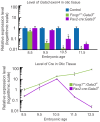
Figure 1. Gata3 recombination.
The top panel, using qRT-PCR, shows the relative level of recombined Gata3 exon4 in the inner ear of both Cre lines compared to control (set to 1, for each age group). There is an earlier and more effective recombination of the Gata3 floxed allele in Foxg1Cre ears than in Pax2-Cre. Pax2-Cre ears first show a significant reduction in Gata3 exon4 at E10.5 two days after Foxg1Cre. This is correlated with the timing of sensory vs non-sensory specification in the ear. The lower panel shows a comparison of Cre expression levels, using qRT-PCR, between Foxg1Cre: Gata3f/f and Pax2-Cre:Gata3f/f ears. Cre expression levels are relative to the level of Cre mRNA in Foxg1Cre: Gata3f/f ears at E8.5 (set to 1). The scale on the left is logarithmic. Pax2-Cre mRNA expression levels are 8-fold less at E8.5 than Foxg1Cre. All data represent 3 biological replicates and 3 technical replicates per-time point as per MIQE guidelines . *p<.01, students t-test.
Figure 2
Figure 2. Morphological development of Foxg1Cre∶ Gata3f/f ears.
3D reconstruction of Foxg1Cre:Gata3f/f inner ears compared to control; utilizing confocal microscopy and Amira software. A-C) At E9.5 there is very little difference in size between the two genotypes. By E10.5 there is a slight reduction in the mutant ear dorsally. At E16.5 there is morphologic development of a cochlear duct (red) in the mutant ear, although it is truncated compared to control. There is also morphologic development of the vestibular portion of the mutant ear (green) although it is highly abnormal. While there is a noticeable saccular out-pouching, none of the other vestibular structures are easily identifiable. The endolymphatic duct is present in the mutant ear (purple) and extends dorsally. Dorsal is up anterior is to the right in all images. All scale bars represent 100 µm.
Figure 3
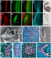
Figure 3. Foxg1CreGata3f/f mice show loss of cochlear neurosensory epithelia.
A) E16.5 Control ear showing lipophilic dye tracing of normal wild-type distribution of efferent, VII, and GP nerves. A portion of the posterior projection of the VII nerve was removed in order to visualize the cochlear (C) afferents. Injection of dye was in ipsilateral rhombomere 4. B,C) Injection of lipophilic dye in E16.5 Foxg1Cre:Gata3f/f hindbrain to label fibers to the ear as in (A), except in addition red lipophilic dye was injected to label the cochlear and vestibular afferents. The facial nerve (VII) shows a normal course around the ear. No fibers are projecting towards the cochlear duct (c). Blue arrow indicates fibers projecting to a non-existent horizontal canal. White arrow indicates fibers initially projecting to the location of where a posterior canal should reside, but in its absence these fibers diverge and project to the remaining epithelia. D) Foxg1Cre: Gata3f/f ear labeled with Myo7a immunohistochemistry shown in red. Positive cells are located in the saccule, utricle, and one canal crista. There are no Myo7a positive cells in the cochlear duct. In the center of the single canal crista there is a lack of non-sensory epithelia (blue arrow). D′′) Low magnification of entire mutant ear. E) SEM of E16.5 control organ of Corti, showing the normal stereotyped pattern of four rows of hair cells (lilac). The early tectorial membrane could be visualized during preparation. F) SEM showing a flat epithelium in the Foxg1Cre: Gata3f/f ear. No hair cells could be identified in the cochlear duct. No tectorial membrane was observed during dissection. There was also a lack of microvilli on the remaining epithelia. G,H) Inner ear efferents and FBM were both labeled with red dye on the same side simultaneously where the facial nerve wraps around the ear. The contralateral FBM were labeled with green dye. G) Efferent cell bodies can be seen (as marked by the white oval). These cells were labeled from the contralateral ear. H) Efferent axons can be seen (arrow) as they diverge from the facial nerve. All aspects of efferent cell body location are in concordance with wild type phenotype. I and J) Showing Atoh1 in situ hybridization (ISH) at E14.5. Insets show positive expression remains in the vestibular portion of the ear correlating with the presence of Myo7a positive cells, and absence within the mutant cochlear duct. K and L) Showing Sox2 ISH at e12.5. Sox2 is necessary for specification of sensory epithelia in the ear. In the control ear (K) all sensory epithelia show positive expression. In the Foxg1Cre: Gata3f/f ear (L) the vestibular portion is positive for Sox2 expression while the organ of Corti shows no expression. M and N) Showing Jag1 ISH at E14.5. Jag1 is a known marker of the prosensory domain and necessary for sensory cell development in the inner ear. Jag1 expression appears more highly upregulated with respect to topology and intensity in the mutant (N) compared to control (M). Anterior is to the right and dorsal is up in all images. Scale bars C and D indicate 10 µm; G,H, K-N indicate 100 µm, I and J indicate 200 µm. A-F, dorsal is up and anterior is to the right. G,H rostral is to the right. c, cochlea; p, posterior crista; hc, horizontal crista; ac, anterior crista; VII, facial nerve; gp, greater petrosal nerve; u, uturicle; s, saccule; FBM, facial branchial motorneurons; IN, intermediate nerve; oc, organ of Corti.
Figure 4
Figure 4. Many genes known in cochlear neurosensory development show altered expression levels in the absence of Gata3.
A) qRT-PCR analysis comparing gene expression of E16.5 control (set to1) and Foxg1Cre: Gata3f/f microdissected cochleas. Gene comparisons were normalized with Actb. Fgf10 has previously been shown to be regulated by Gata3 in the early developing otic vesicle. Shown here is that Fgf10 is specifically affected by levels of Gata3 in the cochlea, and at a later time point than shown previously. Fgf10 was the most affected by loss of Gata3 compared to the other mRNA levels assessed. Tecta is not expressed in Gata3 positive cells and is necessary for normal formation of the tectorial membrane that covers the organ of Corti. Its levels show significant reduction in the mutant. Atoh1 continues to be expressed (albeit at a significantly reduced level) even though there is no indication of hair cells or organ of Corti. Genes downstream to Atoh1 are undetectable by qRT-PCR. Islet1, Prox1, and Bmp4, are necessary for prosensory specification and patterning of the organ of Corti. In the absence of Gata3 all of these genes are significantly down-regulated. Notch, Smo, and Eya1 all thought to be needed to drive prosensory specification are not altered in the absence of Gata3. Axin2 is a marker of non-sensory tissue of the growing cochlear duct and is not affected. Fgfr2b and Pax2 are necessary for sensory specification, and are significantly upregulated. The overexpression of these genes is not sufficient to drive sensory development. *p<.01, students t-test. Eight ears were pooled together for one biological replicate. Three biological replicates with technical replicates were assessed for each stage.
Figure 5
Figure 5. Pax2-Cre: Gata3f/f ears have altered neurosensory development.
A and B) 3D reconstruction of a Pax2-Cre: Gata3f/f ear compared to control at P0. Colors correspond to figure 2. A) P0 control ear yellow and blue correspond to scala tympani and scala vestibule which are absent in the mutant (B). Only the base of the endolymphatic duct has been recsonstructed in both images. C,D,F,G) E18.5 Gata3 LacZ expression in Gata3 heterozygotes with a wildtype allele (C,D) and with a conditional deletion of the second floxed allele with Pax2-Cre (F,G). In the mutant there is a reduction in the length of the cochlea. The mutant spiral ganglion (SG) is not adjacent to the cochlear duct, rather all spiral ganglion cells are coalesced in a single area. E,H-K) Lipophilic dye tracing as in Fig. 3. In contrast to the Foxg1Cre mutant spiral ganglion cells are present and project to the cochlea. In contrast to control, (E), innervation to the cochlea is patchy and is targeted to specific areas. Afferent radial fibers project a straight and short distance in the control in contrast to the mutant where the radial fibers project a long distance from a single location of the spiral ganglion neurons to the patches of sensory epithelia. I-K) Show higher magnification of innervation to sensory epithelia. L) Immunohistochemistry for α-tubulin (green) and Myo7a (red) of mutant cochlear duct. Tubulin staining also shows patchy innervation to the cochlear duct. The remaining innervation targets patches of hair cells that remain along the length of the cochlear duct. Scale bars represent 100 µm. c, cochlea; pc, posterior crista; hc, horizontal crista; ac, anterior crista; VII, facial nerve; gg, geniculate ganglion; u, uturicle; s, saccule; sg, spiral ganglion; oc, organ of Corti.
Figure 6
Figure 6. Pax2-Cre: Gata3f/f ears have altered organ of Corti topology.
A-D) High magnification of organ of Corti showing immunohistochemistry for BDNF (red, hair cells), Sox2 (green, supporting cells), α-tubulin (cyan, neurons). In the mutant patch of sensory epithelia there is one row of inner hair cells and three rows of outer hair cells as in the control. However there are some ectopic BDNF positive cells which are always paired with a Sox2 positive cell (arrows B′). A major difference between the mutant and control is where the hair cells are located within the Sox2 expressing domain. In the control there are very few Sox2 expressing cells lateral to the hair cells and a larger portion medially. However, this setup is reversed in the mutant; indicating that there is a shift in the relative topology of hair cells and the Sox2 positive supporting cell domain. The neurons of the mutant are overshooting the hair cells into the ectopic Sox2 domain then looping. E and E′) Fgf8 in situ hybridization in control and mutant cochlea. Even though BDNF staining indicated specification of inner versus outer hair cells, the inner hair cell marker Fgf8 was absent. F) Low magnification SEM of the torus like vestibular structure. The saccular pouch is not shown. G) The utricular stereocilia lack connections between each other and are splayed out in a random fashion. Stereocilia from a single hair cell have been false-colored violet. H) Low magnification of cochlear duct. The sensory patch apical-basal extent is marked by the white bracket. The abnormal tectorial membrane can be seen as a web like structure that partially covers the sensory patch. There is no spiral limbus in which the tectorial membrane is attached. I) A single Pax2-Cre: Gata3f/f hair cell in the location of an inner hair cell. The thickness of the stereocilia is different than in the wt (J) and does not tapper at the bottom. K) Hair cell in the location of an outer hair cell. There is no polarity with respect to the stereocilia, and no staircase pattern as in the control (L). All images are of E18.5 cochlea except J and L (P5).
Figure 7
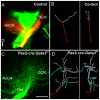
Figure 7. Inner ear central projection.
Lipophilic dye was placed into the cochlea of E18.5 mice. In the control mouse red and green dyes were placed into the base and apex. A and C) Confocal image of cochlear nerve (Cne) projection into the hindbrain (cochlear nucleus). In the control there is a single bifurcation of each afferent axon to the dorsal cochlear nucleus (DCN) and antero-ventral cochlear nucleus (AVCN). In the mutant the cochlear fibers are bifurcating at several branch points with the terminal fibers looping and misdirected. B and D) Amira software was used with the confocal images from A and C to trace individual axons. A single axon was reconstructed, and the root of this axon is labeled in red, and its branches in gray. B) A representative control axon. Each axon analyzed had a single bifurcation. D) In the mutant blue branches are neurites looping back toward the entry point. The single neuron has many bifurcations compared to the control. The right panel of B and D) shows the fibers rotated 90 degrees. The control shows the fibers staying within a single plane. The mutant fibers do not show this restriction in 2D space and branch throughout the dorsal-ventral extent of the hindbrain.
Figure 8
Figure 8. Timing of Gata3 loss reveals different phenotypes.
The loss of Gata3 during different stages of development reveals changing roles of Gata3 in cochlear neurosensory development. In this diagram the solid arrow indicates normal embryonic progression while the dashed arrows indicate the aproximate embryonic age of Gata3 loss from the ear and subsequent altered development. In the Gata3 null mutant the inner ear was shown to contain only a single patch of sensory epithelia (saccule) and corresponding afferent neurons. The cochlear duct was completely devoid of all neurosensory cell types. In the Foxg1Cre conditional Gata3 mutant there is formation of saccule, utricule, and anterior canal neurosensory cell types. The cochlear sensory epithelia is not properly specified, some prosensory genes remain, and many sensory differentiation genes are absent or downregulated. Only in the Pax2-Cre mutant where recombination occurs later than the other two indicated muants (between E8.5 and E10.5) are some cochlear neurosensory cells present, but this later loss of Gata3 results in improper differentiation of these cell types.

Similar articles

Cited by

References

    1. Grocott T, Tambalo M, Streit A (2012) The peripheral sensory nervous system in the vertebrate head: a gene regulatory perspective. Dev Biol 370: 3–23. - PubMed
    1. Fritzsch B, Beisel KW, Hansen LA (2006) The molecular basis of neurosensory cell formation in ear development: a blueprint for hair cell and sensory neuron regeneration? Bioessays 28: 1181–1193. - PMC - PubMed
    1. Fekete DM, Wu DK (2002) Revisiting cell fate specification in the inner ear. Curr Opin Neurobiol 12: 35–42. - PubMed
    1. Brigande JV, Kiernan AE, Gao X, Iten LE, Fekete DM (2000) Molecular genetics of pattern formation in the inner ear: do compartment boundaries play a role? Proc Natl Acad Sci U S A 97: 11700–11706. - PMC - PubMed
    1. Fritzsch B, Beisel KW, Bermingham NA (2000) Developmental evolutionary biology of the vertebrate ear: conserving mechanoelectric transduction and developmental pathways in diverging morphologies. Neuroreport 11: R35–44. - PubMed

Publication types

MeSH terms

Substances

LinkOut - more resources