Clinical Trial
. 2013 Apr 15;8(4):e62056.
doi: 10.1371/journal.pone.0062056. Print 2013.

Re-evaluation of sarcolemma injury and muscle swelling in human skeletal muscles after eccentric exercise

Affiliations
Clinical Trial

Re-evaluation of sarcolemma injury and muscle swelling in human skeletal muscles after eccentric exercise

Ji-Guo Yu et al. PLoS One. .

Abstract

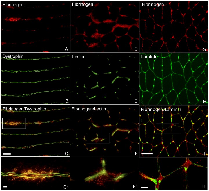
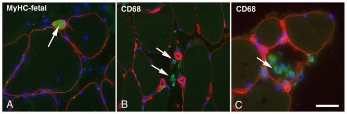
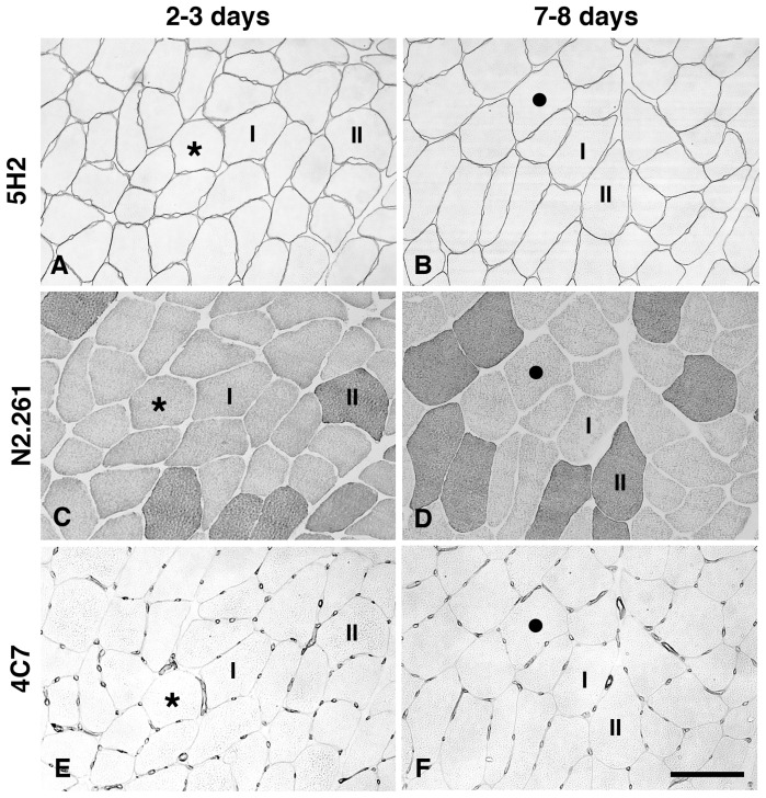
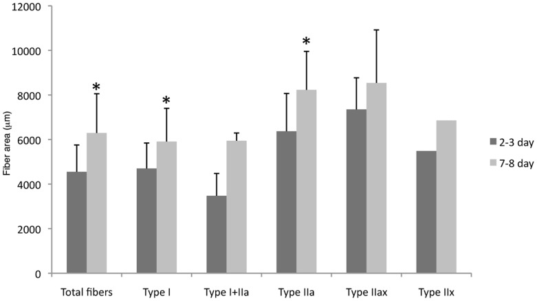
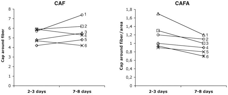
The results regarding the effects of unaccustomed eccentric exercise on muscle tissue are often conflicting and the aetiology of delayed onset muscle soreness (DOMS) induced by eccentric exercise is still unclear. This study aimed to re-evaluate the paradigm of muscular alterations with regard to muscle sarcolemma integrity and fibre swelling in human muscles after voluntary eccentric exercise leading to DOMS. Ten young males performed eccentric exercise by downstairs running. Biopsies from the soleus muscle were obtained from 6 non-exercising controls, 4 exercised subjects within 1 hour and 6 exercised subjects at 2-3 days and 7-8 days after the exercise. Muscle fibre sarcolemma integrity, infiltration of inflammatory cells and changes in fibre size and fibre phenotype composition as well as capillary supply were examined with specific antibodies using enzyme histochemistry and immunohistochemistry. Although all exercised subjects experienced DOMS which peaked between 1.5 to 2.5 days post exercise, no significant sarcolemma injury or inflammation was detected in any post exercise group. The results do not support the prevailing hypothesis that eccentric exercise causes an initial sarcolemma injury which leads to subsequent inflammation after eccentric exercise. The fibre size was 24% larger at 7-8 days than at 2-3 days post exercise (p<0.05). In contrast, the value of capillary number per fibre area tended to decrease from 2-3 days to 7-8 days post exercise (lower in 5 of the 6 subjects at 7-8 days than at 2-3 days; p<0.05). Thus, the increased fibre size at 7-8 days post exercise was interpreted to reflect fibre swelling. Because the fibre swelling did not appear at the time that DOMS peaked (between 1.5 to 2.5 days post exercise), we concluded that fibre swelling in the soleus muscle is not directly associated with the symptom of DOMS.


Conflict of interest statement

Competing Interests: The authors have declared that no competing interests exist.

Figures

Figure 1
Figure 1. Scheme of fibre phenotype classification.
The staining intensity of the four different antibodies in all fibres were briefly classified into “unstained”, “weak”, “Moderate” and “Strong”. An individual fibre was classified into a specific fibre type according to its staining intensities with the four different antibodies against different MyHC isoforms.
Figure 2
Figure 2. Immunofluorescence staining for visualization of muscle fibre sarcolemma integrity.
A–C, one longitudinal section from a biopsy of 1 h group double stained for fibrinogen (A) and dystrophin (B). C is merged images of A and B, and C1 is enlargement of boxed areas in C. Several sites of intracellular staining for fibrinogen outlined by dystrophin were seen along the length of a fibre, whereas staining for dystrophin did not reveal distinct disruption of plasma membrane. D–F, one transverse section from a biopsy of 1 h group double stained for fibrinogen (D) and lectin (E). F is merged images of D and E, and F1 is enlargement of boxed areas in F. Intracellular staining for fibrinogen was seen close to capillaries revealed by lectin staining. G–I, one transverse section from a biopsy of control group double stained for fibrinogen (G) and laminin (H). I is merged images of G and H, and I1 is enlargement of boxed areas in I. Several sites of distinct extracellular staining for fibrinogen outlined by laminin were seen. Bars 50 µm
Figure 3
Figure 3. Immunofluorescence staining for visualization of developmental and inflammatory cells.
A, one transverse sections from biopsy of 7–8 day group double stained with antibodies against MyHC-fetal (green) and dystrophin (red). One small neonatal fibre was seen between normal sized fibres (arrow). B and C, transvers sections from biopsies of 2–3 day group double stained with antibodies against CD68 (green) and laminin (red). Several extracellular monocytes/macrophages (arrows) were seen in B whereas in C monocytes/macrophages were seen inside a necrotic fiber. Nuclei (blue) were visualized in all sections by using mounting medium containing DAPI. Bar 100 µm
Figure 4
Figure 4. Immunohistochemical staining for visualization of muscle fibre size, fibre phenotype and capillary supply.
Serial transverse sections from one biopsy in the 2–3 day group (A, C, E) and one from the 7–8 day group (B, D, F) stained with antibodies 5H2 for fibre profile (A, B), N2.261 for fibre phenotypes (C, D) and 4C7 for capillaries (E, F). Most fibres are larger in size in the 7–8 days group (B) compared to the 2–3 days group (A). Type I and type II fibres are marked (C, D). There was no distinct difference in number of capillaries around each fibre between the two groups (E, F) despite the fibre size was larger in the 7–8 day group (F). Stars and black dots mark same fibre in series sections. Bar 100 µm
Figure 5
Figure 5. Comparison in fibre size between 2–3 day and 7–8 day groups.
Fibre size in 7–8 day group was significantly larger than in 2–3 day group. Among the five different fibre phenotypes, only type I and type IIa fibres presented larger fibre size in 7–8 day group than in 2–3 day group (*p<0.05).
Figure 6
Figure 6. Comparison of individual values in CAF and CAFA from the same subjects in 2–3 day and 7–8 day groups.
Compared to 2–3 day group, CAF was higher in three subjects (1, 3, 5) and lower in two subjects (4, 6), whereas CAFA was lower in five subjects (1, 3, 4, 5, 6) in 7–8 day group (p<0.05).

References

    1. Paulsen G, Mikkelsen UR, Raastad T, Peake JM (2012) Leucocytes, cytokines and satellite cells: what role do they play in muscle damage and regeneration following eccentric exercise? Exerc Immunol Rev 18: 42–97. - PubMed
    1. Lieber RL, Friden J (2002) Morphologic and mechanical basis of delayed-onset muscle soreness. J Am Acad Orthop Surg 10: 67–73. - PubMed
    1. Clarkson PM, Hubal MJ (2002) Exercise-induced muscle damage in humans. Am J Phys Med Rehabil/Association of Academic Physiatrists 81: S52–69. - PubMed
    1. Lewis PB, Ruby D, Bush-Joseph CA (2012) Muscle soreness and delayed-onset muscle soreness. Clin Sports Med 31: 255–262. - PubMed
    1. Crameri RM, Aagaard P, Qvortrup K, Langberg H, Olesen J, et al. (2007) Myofibre damage in human skeletal muscle: effects of electrical stimulation versus voluntary contraction. J Physiol 583: 365–380. - PMC - PubMed

Publication types

MeSH terms

Substances