. 2013 May 21;52(20):3588-600.
doi: 10.1021/bi301616c. Epub 2013 May 7.

Kinetic mechanism of human histidine triad nucleotide binding protein 1

Affiliations

Kinetic mechanism of human histidine triad nucleotide binding protein 1

Xin Zhou et al. Biochemistry. .

Abstract

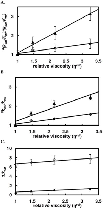
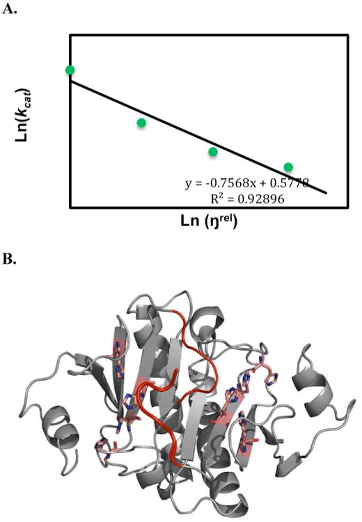
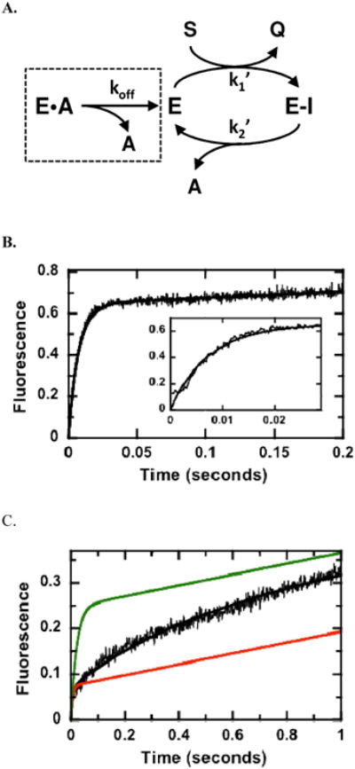
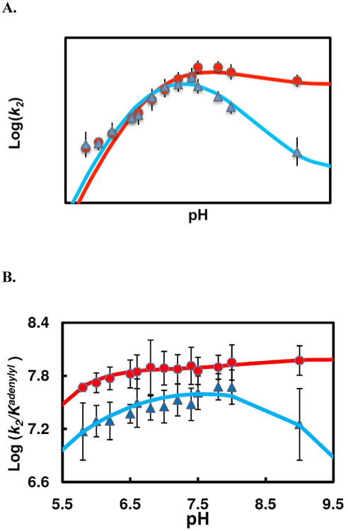
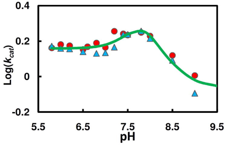
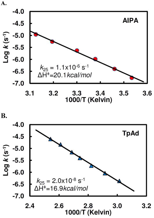
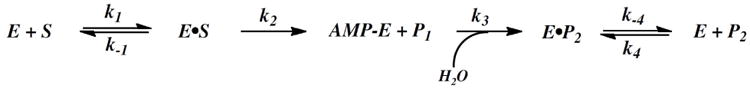
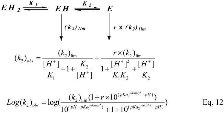
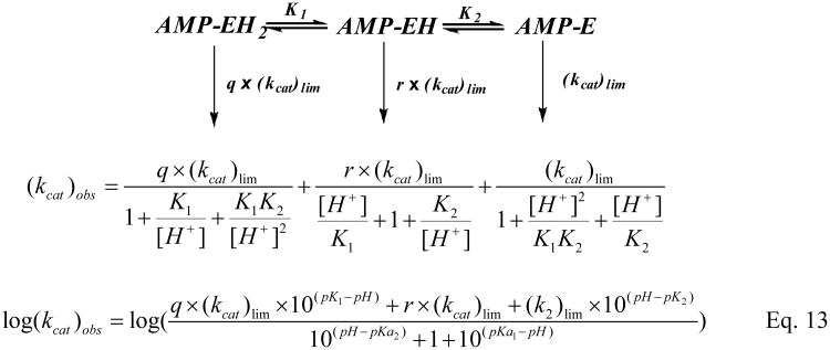
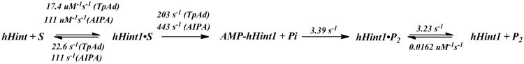
Human histidine triad nucleotide binding protein 1 (hHint1) is a member of a ubiquitous and ancient branch of the histidine triad protein superfamily. hHint1 is a homodimeric protein that catalyzes the hydrolysis of model substrates, phosphoramidate and acyl adenylate, with a high efficiency. Recently, catalytically inactive hHint1 has been identified as the cause of inherited peripheral neuropathy [Zimon, M., et al. (2012) Nat. Genet. 44, 1080-1083]. We have conducted the first detailed kinetic mechanistic studies of hHint1 and have found that the reaction mechanism is consistent with a double-displacement mechanism, in which the active site nucleophile His112 is first adenylylated by the substrate, followed by hydrolysis of the AMP-enzyme intermediate. A transient burst phase followed by a linear phase from the stopped-flow fluorescence assay indicated that enzyme adenylylation was faster than the subsequent intermediate hydrolysis and product release. Solvent viscosity experiments suggested that both chemical transformation and diffusion-sensitive events (product release or protein conformational change) limit the overall turnover. The catalytic trapping experiments and data simulation indicated that the true koff rate of the final product AMP is unlikely to control the overall kcat. Therefore, a protein conformational change associated with product release is likely rate-limiting. In addition, the rate of Hint1 adenylylation was found to be dependent on two residues with pKa values of 6.5 and 8, with the former pKa agreeing well with the nuclear magnetic resonance titration results for the pKa of the active site nucleophile His112. In comparison to the uncatalyzed rates, hHint1 was shown to enhance acyl-AMP and AMP phosphoramidate hydrolysis by 10(6)-10(8)-fold. Taken together, our analysis indicates that hHint1 catalyzes the hydrolysis of phosphoramidate and acyl adenylate with high efficiency, through a mechanism that relies on rapid adenylylation of the active residue, His112, while being partially rate-limited by intermediate hydrolysis and product release associated with a conformational change. Given the high degree of sequence homology of Hint proteins across all kingdoms of life, it is likely that their kinetic and catalytic mechanisms will be similar to those elucidated for hHint1.


Figures

Figure 1
Figure 1
(A) The active site of hHint1 with AMP binding (PDB: 1KPF). (B) Structure of substrates.
Figure 2
Figure 2
Active site titration. (A) The WT human Hint1 (1, 2, 3, 4, 5μM) was mixed with AIPA (10 μM) in stopped flow cell and the time course of IPA production was monitored at 360nm. (B) The amplitude of “burst” vs. human Hint1 monomer concentration was plotted. A slope of 0.77 with R2 =0.955 indicated that 77% of the active site catalyzes the reaction in the first catalytic cycle.
Figure 3
Figure 3
Solvent viscosity effects on the steady state hydrolysis of TpAd (▲) and TrAd (◊) in HEPES buffer at pH 7.2. (A) plot of the relative second-order rate constant as a function of relative viscosity (ŋrel); (B) plot of the relative kcat vs. relative viscosity (ŋrel); (C) plot of 1/kcat vs. relative viscosity (ŋrel).
Figure 4
Figure 4
(A) Data from viscosity experiment were fit to Kramer's model (eq 16), ln(kcat) is linearly dependent on ln (ηrel) with a slope of -0.76 (R2 =0.93), indicating a strong coupling of product release with the active site conformational change. (B) The C-termini of hH1 homodimer that is proposed to undergo conformational change coupled with AMP release. The active sites are shown in pink and the C-termini are shown in red.
Figure 5
Figure 5
Catalytic Trapping of AMP.(A) Kinetic mechanism. Data in the absence and presence of AMP are simulated using the program DynaFit and a two-step acylation/deacylation mechanism. (B) Kinetic transient in the absence of AMP. Hint1 (5 μM) is mixed with TpAd (20μM) in the stopped flow instrument and the fluorescence changes are simulated using the kinetic scheme in panel A where k1′ and k2′ are 7.2 μM−1s−1 and 0.5 s−1 and the output for Q is 0.26 volts/mM. (C) Kinetic transient in the presence of 2500 mM AMP. Conditions are the same as in panel B except 2500 mM AMP is preequilbrated with the enzyme prior to mixing. The fluorescence changes are simulated using fixed values for k1 ′ and k2′ from panel B, 0.7 and 1.8 mM E and E•A, an output for Q of 0.1 volts/mM and a koff of 2.7 s−1 (black line). The data were also simulated using koff values of 50 (green line) and 0.5 s−1 (red line).
Figure 6
Figure 6
pH dependence of pre-steady state kinetic parameters of the enzyme adenylylation by TpAd (▲) and AIPA(●). (A) Plots of adenylylation rate (k2) vs. pH. The data fitting (Scheme 2, eq 12) for AIPA reaction (red line) yields following values of the characterizing parameters: pK1= 6.68 ± 0.09, pK2= 8.03 ± 0.16, r= 0.72 ± 0.11, (k2)lim= 673±50 s−1; for TpAd reaction (blue line), pK1= 6.56 ± 0.13, pK2= 7.90 ± 0.24, r= 0.16 ± 0.08, (k2)lim= 634 ± 86 s−1. (B) Plots of k2/Kadenylyl vs. pH. The pH profiles for AIPA reaction (red line) and for TpAd (blue line) were relatively flat, indicating substrates are sticky.
Figure 7
Figure 7
Comparison of pH (▲, TpAd; ●, AIPA) dependence of steady state kinetic parameters (kcat). The pH profile data were fit by eq 13 (Scheme 3), yielding pK1= 7.52 ± 0.18, pK2= 8.15 ± 0.10, r= 7.3 ± 2.0, q= 1.68 ± 0.16, and (kcat)lim for the three active forms (AMP-EH2, AMP-EH, and AMP-E) are 1.44±0.08 s−1, 6.28±0.56 s−1, and 0.85±0.05 s−1, respectively.
Figure 8
Figure 8
Arrhenius plots for hydrolysis of AIPA (A) and TpAd (B) at 25°C at pH 6.8.
Scheme 1
Scheme 1
Proposed minimal 4-step mechanism of hHint1.
Scheme 2
Scheme 2
Scheme 3
Scheme 3
Scheme 4
Scheme 4
Proposed hHint1 kinetic mechanism with the substrates AIPA and TpAd.

References

    1. Zimon M, Baets J, Almeida-Souza L, De Vriendt E, Nikodinovic J, Parman Y, Battaloglu E, Matur Z, Guergueltcheva V, Tournev I, Auer-Grumbach M, De Rijk P, Petersen BS, Muller T, Fransen E, Van Damme P, Loscher WN, Barisic N, Mitrovic Z, Previtali SC, Topaloglu H, Bernert G, Beleza-Meireles A, Todorovic S, Savic-Pavicevic D, Ishpekova B, Lechner S, Peeters K, Ooms T, Hahn AF, Zuchner S, Timmerman V, Van Dijck P, Rasic VM, Janecke AR, De Jonghe P, Jordanova A. Loss-of-function mutations in HINT1 cause axonal neuropathy with neuromyotonia. Nat Genet. 2012;44:1080–1083. - PubMed
    1. Brenner C, Garrison P, Gilmour J, Peisach D, Ringe D, Petsko GA, Lowenstein JM. Crystal structures of HINT demonstrate that histidine triad proteins are GalT-related nucleotide-binding proteins. Nat Struct Bio. 1997;l4:231–238. - PMC - PubMed
    1. Mozier NM, Walsh MP, Pearson JD. Characterization of a novel zinc binding site of protein kinase C inhibitor-1. FEBS Lett. 1991;279:14–18. - PubMed
    1. Lima CD, Klein MG, Weinstein IB, Hendrickson WA. Three-dimensional structure of human protein kinase C interacting protein 1, a member of the HIT family of proteins. Proc Natl Acad Sci U S A. 1996;93:5357–5362. - PMC - PubMed
    1. Rodriguez-Munoz M, de la Torre-Madrid E, Sanchez-Blazquez P, Wang JB, Garzon J. NMDAR-nNOS generated zinc recruits PKCgamma to the HINT1-RGS17 complex bound to the C terminus of Mu-opioid receptors. Cell Signal. 2008;20:1855–1864. - PubMed

Publication types