

Cells behave distinctly within sponges and hydrogels due to differences of internal structure
- PMID: 23614637
- PMCID: PMC3761434
- DOI: 10.1089/ten.TEA.2012.0393
Cells behave distinctly within sponges and hydrogels due to differences of internal structure
Abstract
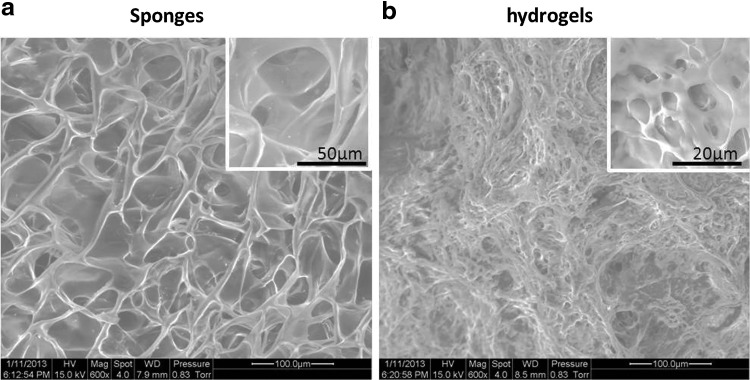
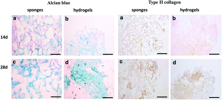
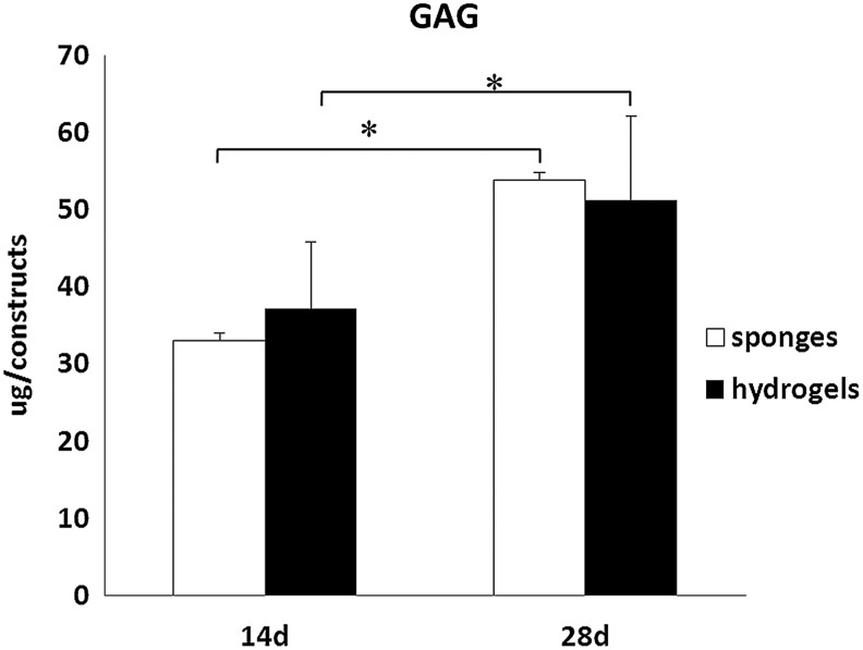
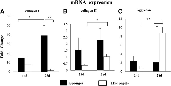
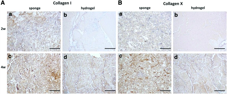
Different forms of biomaterials, including microspheres, sponges, hydrogels, and nanofibers, have been broadly used in cartilage regeneration; however, effects of internal structures of the biomaterials on cells and chondrogenesis remain largely unexplored. We hypothesized that different internal structures of sponges and hydrogels led to phenotypic disparity of the cells and may lead to disparate chondrogenesis. In the current study, the chondrocytes in sponges and hydrogels of chitosan were compared with regard to cell distribution, morphology, gene expression, and production of extracellular matrix. The chondrocytes clustered or attached to the materials with spindle morphologies in the sponges, while they distributed evenly with spherical morphologies in the hydrogels. The chondrocytes proliferated faster with elevated gene expression of collagen type I and down-regulated gene expression of aggracan in sponges, when compared with those in the hydrogels. However, there was no significant difference of the expression of collagen type II between these two scaffolds. Excretion of both glycosaminoglycan (GAG) and collagen type II increased with time in vitro, but there was no significant difference between the sponges and the hydrogels. There was no significant difference in secretion of GAG and collagen type II in the two scaffolds, while the levels of collagen type I and collagen type X were much higher in sponges compared with those in hydrogels during an in vivo study. Though the chondrocytes displayed different phenotypes in the sponges and hydrogels, they led to comparable chondrogenesis. An optimized design of the biomaterials could further improve chondrogenesis through enhancing functionalities of the chondrocytes.
Figures


Similar articles
-
The influence of scaffold microstructure on chondrogenic differentiation of mesenchymal stem cells.Biomed Mater. 2014 Jun;9(3):035011. doi: 10.1088/1748-6041/9/3/035011. Epub 2014 May 12. Biomed Mater. 2014. PMID: 24818859
-
Effect of three-dimensional expansion and cell seeding density on the cartilage-forming capacity of human articular chondrocytes in type II collagen sponges.J Biomed Mater Res A. 2010 Dec 1;95(3):924-31. doi: 10.1002/jbm.a.32917. J Biomed Mater Res A. 2010. PMID: 20845491
-
Comparative phenotypic analysis of articular chondrocytes cultured within type I or type II collagen scaffolds.Tissue Eng Part A. 2009 Jun;15(6):1233-45. doi: 10.1089/ten.tea.2008.0114. Tissue Eng Part A. 2009. PMID: 18950259
-
Animal Models Used for Testing Hydrogels in Cartilage Regeneration.Curr Stem Cell Res Ther. 2018;13(7):517-525. doi: 10.2174/1574888X13666180514123103. Curr Stem Cell Res Ther. 2018. PMID: 29756585 Review.
-
The Molecular Interaction of Collagen with Cell Receptors for Biological Function.Polymers (Basel). 2022 Feb 23;14(5):876. doi: 10.3390/polym14050876. Polymers (Basel). 2022. PMID: 35267698 Free PMC article. Review.
Cited by
-
Single-step laser-based fabrication and patterning of cell-encapsulated alginate microbeads.Biofabrication. 2013 Dec;5(4):045006. doi: 10.1088/1758-5082/5/4/045006. Epub 2013 Nov 6. Biofabrication. 2013. PMID: 24192221 Free PMC article.
-
Charting a quarter-century of commercial cartilage regeneration products.J Orthop Translat. 2025 Jan 31;50:354-363. doi: 10.1016/j.jot.2024.10.009. eCollection 2025 Jan. J Orthop Translat. 2025. PMID: 39968336 Free PMC article. Review.
-
Articular cartilage and osteochondral tissue engineering techniques: Recent advances and challenges.Bioact Mater. 2021 May 28;6(12):4830-4855. doi: 10.1016/j.bioactmat.2021.05.011. eCollection 2021 Dec. Bioact Mater. 2021. PMID: 34136726 Free PMC article. Review.
-
Mechanotransducive Biomimetic Systems for Chondrogenic Differentiation In Vitro.Int J Mol Sci. 2021 Sep 7;22(18):9690. doi: 10.3390/ijms22189690. Int J Mol Sci. 2021. PMID: 34575847 Free PMC article. Review.
-
Collagen Scaffolds in Cartilage Tissue Engineering and Relevant Approaches for Future Development.Tissue Eng Regen Med. 2018 Jul 25;15(6):673-697. doi: 10.1007/s13770-018-0135-9. eCollection 2018 Dec. Tissue Eng Regen Med. 2018. PMID: 30603588 Free PMC article. Review.
References
-
- Ge Z.G., et al. Osteoarthritis and therapy. Arthritis Rheum-Arthritis Care Res. 2006;55:493. - PubMed
-
- Alford J.W. Cartilage restoration, part 1: basic science, historical perspective, patient evaluation, and treatment options. Am J Sports Med. 2005;33:295. - PubMed
-
- Alford J.W. Cartilage restoration, part 2: techniques, outcomes, and future directions. Am J Sports Med. 2005;33:443. - PubMed
-
- Liumin H., et al. Microstructure and properties of nano-fibrous PCL-b-PLLA scaffolds for cartilage tissue engineering. Eur Cells Mater. 2009;18:63. - PubMed
Publication types
MeSH terms
Substances
LinkOut - more resources
Full Text Sources
Other Literature Sources
