. 2013 May;22(5):342-8.
doi: 10.1111/exd.12140.

Delphinidin, a dietary antioxidant, induces human epidermal keratinocyte differentiation but not apoptosis: studies in submerged and three-dimensional epidermal equivalent models

Affiliations

Delphinidin, a dietary antioxidant, induces human epidermal keratinocyte differentiation but not apoptosis: studies in submerged and three-dimensional epidermal equivalent models

Jean Christopher Chamcheu et al. Exp Dermatol. 2013 May.

Abstract

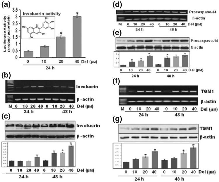
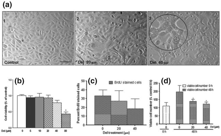
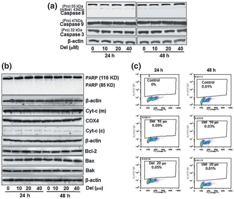
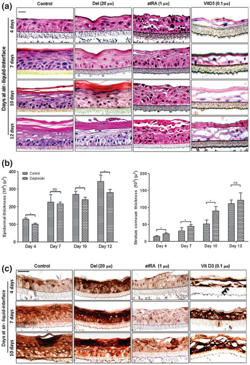
Delphinidin (Del), [3,5,7,3'-,4'-,5'-hexahydroxyflavylium], an anthocyanidin and a potent antioxidant abundantly found in pigmented fruits and vegetables exhibits proapoptotic effects in many cancer cells. Here, we determined the effect of Del on growth, apoptosis and differentiation of normal human epidermal keratinocytes (NHEKs) in vitro in submerged cultures and examined its effects in a three-dimensional (3D) epidermal equivalent (EE) model that permits complete differentiation reminiscent of in vivo skin. Treatment of NHEKs with Del (10-40 μm; 24-48 h) significantly enhanced keratinocyte differentiation. In Del-treated cells, there was marked increase in human involucrin (hINV) promoter activity with simultaneous increase in the mRNA and protein expressions of involucrin and other epidermal differentiation markers including procaspase-14 and transglutaminase-1 (TGM1), but without any effect on TGM2. Del treatment of NHEKs was associated with minimal decrease in cell viability, which was not associated with apoptosis as evident by lack of modulation of caspases, apoptosis-related proteins including Bcl-2 family of proteins and poly(ADP-ribose) polymerase cleavage. To establish the in vivo relevance of our observations in submerged cultures, we then validated these effects in a 3D EE model, where Del was found to significantly enhance cornification and increase the protein expression of cornification markers including caspase-14 and keratin 1. For the first time, we show that Del induces epidermal differentiation using an experimental system that closely mimics in vivo human skin. These observations suggest that Del could be a useful agent for dermatoses associated with epidermal barrier defects including aberrant keratinization, hyperproliferation or inflammation observed in skin diseases like psoriasis and ichthyoses.


Conflict of interest statement

Conflict of interests: The authors have declared no conflicting interests.

Figures

Figure 1
Figure 1
Del treatment of normal human epidermal keratinocyte (NHEK) induces the gene and protein expression of epidermal differentiation-related proteins Normal human epidermal keratinocytes prepared from neonatal foreskin were treated with Del (0–40 μm) for 24–48 h, and mRNA and protein extracts were prepared as described. (a) Histograms represent the human involucrin promoter activity normalized to Renilla activity in cells treated with Del for 24 h, inset represents the molecular structure of Del (2-(3,4,5-trihydroxyphenyl) chromenylium-3,5,7-triol). (b) Involucrin mRNA expression by reverse transcription-polymerase chain reaction (RT-PCR). (c) Involucrin protein expression by immunoblotting. (d–e) Procaspase-14 mRNA expression by RT-PCR and protein expression by western blotting (f–g) TGM1 mRNA expression by RT-PCR and protein expression by western blotting. Blots are representative of three independent experiments for each treatment group. The blots were stripped and reprobed for β-actin to determine equal protein loading, and the histograms are semiquantitative densitometric results and represent the ratio between target protein and β-actin and is the mean ± SD (n = 3). Each film was scanned on an Epson Perfection V350 photo Scanner (Epson America Inc, Long Beach, CA, USA), and the intensities were quantified using ImageJ (NIH). All assays were performed in duplicate, and each experiment was repeated a minimum of three times. Statistica analysis were performed using ANOVA (Bonferroni's multiple comparison test; *P < 0.05).
Figure 2
Figure 2
Del treatment of normal human epidermal keratinocyte (NHEK) modulates morphology, growth, proliferation and viable cell number. (a) Phase contrast photomicrographs of control NHEK (a1) and NHEKs treated with Del 20 μm (a2) and 40 μm (a3). Near-confluent NHEKs were treated for 48 h with or without Del (0–40 μm). Inset (a3). Magnified image showing multivacuolated and stretched adherent cells. (b) Histograms representing effect of Del treatment on cell growth by MTT assay. Normal human epidermal keratinocytes were treated with Del (inset; 0–80 μm) for 48 h, (*P < 0.05). (c) Histograms representing effect of Del treatment on cell proliferation by 5-bromo-2-deoxyuridine (BrdU) labelling. Cells were treated with Del (0–40 μm) for 48 h and then the medium was supplemented with 10 μm BrdU labelling reagent for 6 h. (d) Histograms representing effect of Del treatment on viable cell number using the trypan blue dye exclusion assay. Normal human epidermal keratinocytes were treated with Del (0–40 μm) for 48 h and viable cell number determined and expressed as percentage cell number harvested (48 h) in relation to the cell number present at the time of initiation of treatment (0 h), (*P < 0.05). The Del concentration of 80 μm was used as a positive control for cytotoxicity and apoptosis.
Figure 3
Figure 3
Del treatment of normal human epidermal keratinocyte (NHEK) does not induce apoptosis nor alter cell cycle profile. Normal human epidermal keratinocytes were seeded, grown to near confluence and treated with or without Del (10–40 μm) for 24 or 48 h. (a) Immunoblot analysis of cell lysates probed with antibodies specific for human caspases -3, -8 & -9 (b) Immunoblot analysis of cell lysates probed with antibodies specific for human poly(ADP)-ribose polymerase, cytosolic and mitochondrial cytochrome c, Bcl2, Bax and Bak. The β-actin or COX4 antibodies were used to reprobe to confirm equal protein loading. (c) Flow cytometric analysis of propidium iodide stained NHEKs treated with or without Del (10–20 μm) for 48 h to show the percentage of sub-G1, cells. These experiments were repeated at least three times and only one representative data is presented.
Figure 4
Figure 4
Del treatment enhances cornification and induces caspase-14 expression in the developing 3D epidermal equivalent (EE) model. Normal human epidermal keratinocyte were grown on Millipore PCF cell culture inserts at air–liquid interface to generate a 3D EE treated with or without test compounds as described above, and harvested after 4, 7, 10 and 12 days for histomorphometry and thickness of the EE. (a) Representative H&E photomicrographs of histology of organotypic epidermis untreated controls (left panel) or treated with Del (second panel from left), all-trans retinoic acid (atRA)-treated (third panel from left) or 1, 25(OH)2 vitamin D3 (vitD3)-treated (right panel). (b) Histograms represent the thicknesses of the total epidermis (left panel) and stratum corneum (right panel) of 4, 7, 10 and 12-days-old organotypic epidermis treated with or without Del and analysed as described in Materials and methods. Data represent means ± SD of three independent experiments each performed in triplicate. Statistical analysis was performed using ANOVA (Bonferroni's multiple comparison test; *P < 0.05). (c) Representative micrographs of immunostaining showing caspase-14 expression in developing 3D EE at 4, 7 and 10 days after air-liquid interface. Untreated controls (left panel) or treated with Del (second panel from left) or atRA-treated (third panel from left) and vitD3 (right panel). Bar = 20 μm.

Similar articles

Cited by

References

    1. Elias PM. Curr Allergy Asthma Rep. 2008;8:299–305. - PMC - PubMed
    1. Feingold KR, Schmuth M, Elias PM. J Invest Dermatol. 2007;127:1574–1576. - PubMed
    1. Presland RB, Coulombe PA, Eckert RL, et al. J Invest Dermatol. 2004;123:603–606. - PubMed
    1. Sandilands A, Sutherland C, Irvine AD, et al. J Cell Sci. 2009;122:1285–1294. - PMC - PubMed
    1. Segre JA. J Clin Invest. 2006;116:1150–1158. - PMC - PubMed

Publication types

MeSH terms